Veterinary Science
http://dx.doi.org/10.4142/jvs.2014.15.1.61
Received: 7 Feb. 2013, Revised: 11 Sep. 2013, Accepted: 6 Oct. 2013
Original Article
*Corresponding author: Tel: +82-31-467-1837; Fax: +82-31-467-1845; E-mail: [email protected]
ⓒ 2014 The Korean Society of Veterinary Science.
This is an Open Access article distributed under the terms of the Creative Commons Attribution Non-Commercial License (http://creativecommons.org/licenses/by-nc/3.0) which permits
unrestricted non-commercial use, distribution, and reproduction in any medium, provided the original work is properly cited.
Toxic effects of methylmercury, arsanilic acid and danofloxacin on the differentiation of mouse embryonic stem cells into neural cells
Seok-Jin Kang
1, Sang-Hee Jeong
2, Eun-Joo Kim
1, Young-Il Park
1, Sung-Won Park
1, Hyo-Sook Shin
1, Seong-Wan Son
1, Hwan-Goo Kang
1,*
1
Toxicology and Residue Chemistry Division, Animal and Plant Quarantine Agency, Anyang 430-824, Korea
2
GLP Research Center, College of Natural Sciences, Hoseo University, Industrial-academy Corporation Center I, Asan 336-795, Korea
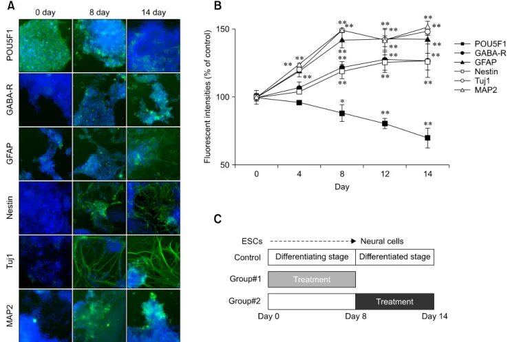
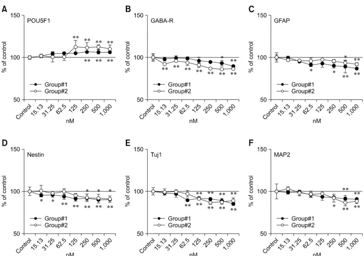
This study was performed to assess the neurotoxic effects of methylmercury, arsanilic acid and danofloxacin by quantification of neural-specific proteins in vitro. Quantitation of the protein markers during 14 days of differentiation indicated that the mouse ESCs were completely differentiated into neural cells by Day 8. The cells were treated with non-cytotoxic concentrations of three chemicals during differentiation. Low levels of exposure to methylmercury decreased the expression of GABA
A-R and Nestin during the differentiating stage, and Nestin during the differentiated stage. In contrast, GFAP, Tuj1, and MAP2 expression was affected only by relatively high doses during both stages.
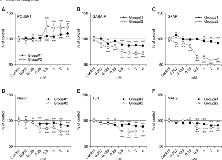
Arsanilic acid affected the levels of GABA
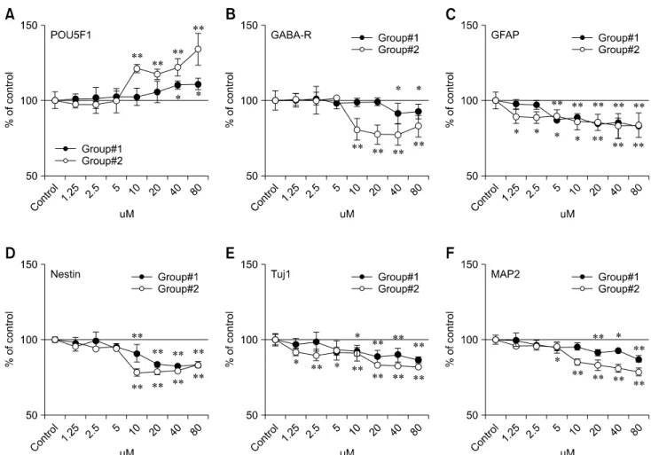
A-R and GFAP during the differentiated stage while the changes of Nestin and Tuj1 were greater during the differentiating stage. For the neural markers (except Nestin) expressed during both stages, danofloxacin affected protein levels at lower concentrations in the differentiated stage than the differentiating stage.
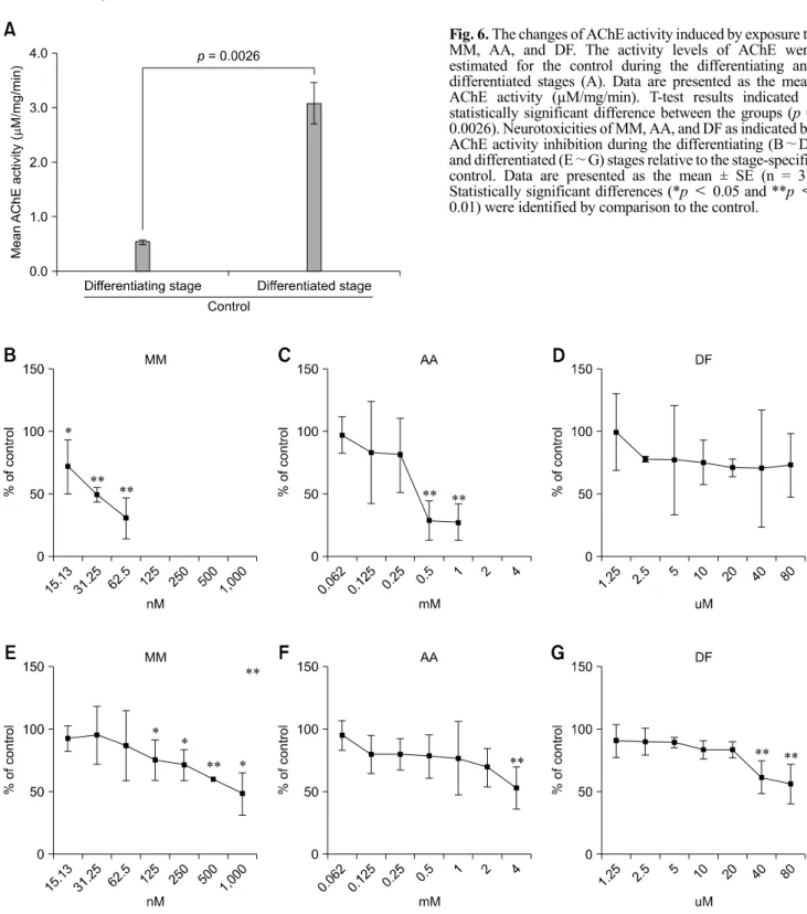
Acetylcholinesterase activity was inhibited by relatively low concentrations of methylmercury and arsanilic acid during the differentiating stage while this activity was inhibited only by more than 40 μM of danofloxacin in the differentiated stage.
Our results provide useful information about the different toxicities of chemicals and the impact on neural development.
Keywords: arsanilic acid, danofloxacin, embryonic stem cell test, methylmercury, neural cell
Introduction
Embryonic stem cells (ESCs) have the unique potential to differentiate into any cell type. A number of studies
have demonstrated their pluripotency in vitro including the ability to differentiate into hepatocytes [24], cardiomyocytes [34], and neurons [18]. These cells are therefore potentially important for cell therapy in the field of regenerative medicine, and are a useful model for assessing the toxicity of new drugs and pharmacological chemicals.
Recently, the embryonic stem cell test (EST) has been used as an in vitro alternative to animal testing and a new predictive model for risk assessment. An EST using ESCs and 3T3 fibroblasts has been validated for use in assessing the embryotoxic potential of chemicals. An inhibition assay involving ESC-derived cardiomyocyte differentiation has also been developed [12,13]. Finally, an EST using functional neuron-like cells derived from ESCs has been established as a neurotoxicity test [30,31]. The European Centre for the Validation of Alternative Methods (ECVAM) has encouraged further optimization of EST-based methods as novel toxicological prediction models.
In vitro neural development models have recently been established for mouse and human cells. ESCs can efficiently differentiate into neural progenitor cells and neural lineage cells. Neural progenitor cells derived from the neuroectoderm can also differentiate into neuronal- and glial-restricted precursor cells [22]. Various approaches have been used to promote the neural differentiation of ESCs [3,17,38]. Basically, all methods are designed to recreate the multistep differentiation process that occurs during in vivo neurogenesis.
During embryogenesis, a large number of tissue-specific
genes systemically interact within the normal
microenvironment to direct specific cell fates. During
ESC-derived neurulation, the expression of neural-specific