Effects of Transient Treatment with Rotenone, a Mitochondrial Inhibitor, on Mouse Subventricular Zone Neural Stem Cells
Ki-Youb Park1* and Man Su Kim2
1Korea Science Academy of KAIST, 105-47 Baegyanggwanmun-ro, Busanjin-Gu, Busan 614-100, Korea
2College of Pharmacy, Inje University, Gimhae 50834, Korea
Received November 20, 2019 /Revised December 5, 2019 /Accepted December 11, 2019
Subventricular zone (SVZ) in the brain contains neural stem cells (NSCs) which self-renew and differ- entiate to neurons and glial cells during postnatal period and throughout adulthood. Since fate deci- sion to either proliferation or differentiation has to respond to intracellular and extracellular con- ditions, many intrinsic and extrinsic factors are involved. Among them, mitochondria have been re- ported to participate in fate decision of NSCs. In our previous report, we showed that long-term treat- ment of a mitochondrial inhibitor rotenone greatly inhibited neurogenesis. In this study, we examined the effects of short-term treatment of rotenone on SVZ NSCs. We found that (1) even one-day treat- ment of rotenone significantly reduced neurogenesis and earlier time points seemed to be more sensi- tive to rotenone, (2) a number of Mash1+ transit amplifying cells was decreased by one-day treatment of rotenone, (3) short-term treatment of rotenone eliminated most of the differentiated Tuj1+ neurons and Olig2+ oligodendrocytes, while glial fibrillary acidic protein (GFAP)+ astrocytes were not affected, and (4) sulfiredoxin 1 (Srxn1) gene expression was increased after one-day treatment of rotenone, in- dicating activation of nuclear factor (erythroid-derived 2)-like 2 (Nrf2) pathway. All these results con- firm that functional mitochondria are necessary during differentiation to neurons or oligodendrocytes as well as maintenance of neurons after differentiation. Also, these data suggest that temporary ex- posure to mitochondrial inhibitor such as rotenone might have long-term effects on neurogenic poten- tial of NSCs.
Key words : Mitochondria, neural stem cells (NSCs), neuronal differentiation, rotenone, subventricular zone (SVZ)
*Corresponding author
*Tel : +82-51-606-2225, Fax : +82-51-606-2226
*E-mail : [email protected]
This is an Open-Access article distributed under the terms of the Creative Commons Attribution Non-Commercial License (http://creativecommons.org/licenses/by-nc/3.0) which permits unrestricted non-commercial use, distribution, and reproduction in any medium, provided the original work is properly cited.
Journal of Life Science 2019 Vol. 29. No. 12. 1329~1336 DOI : https://doi.org/10.5352/JLS.2019.29.12.1329
Introduction
Various aspects of normal function of mitochondria have been reported to be important in NSCs. Disruption of mi- tochondrial fission using an inhibitor of dynamin-related protein 1 reduced neuronal migration and neurogenesis in cultured SVZ NSCs [10]. When NDUFS2 gene that is essen- tial in mitochondrial complex I function was conditionally knocked out, SVZ NSCs did not properly proliferate and differentiate [2]. Mitochondrial DNA repair impairment by amyloid-beta42 oligomers disrupted neuronal differentiation and induced astrocytic differentiation in human NSC cul- tures [14]. During neuronal differentiation from NSCs, mi-
tochondria fuse together and become elongated [23]. We also previously reported that an inhibitor of mitochondrial com- plex I, rotenone, inhibited neurogenesis and oligodendro- genesis in cultured SVZ NSCs [8]. In addition to their role in normal NSC biology, mitochondrial dysfunction has been observed in NSCs from patients with neurological disease.
NSCs from Parkinson’s disease patients had abnormal mi- tochondrial function and shape [25]. Interestingly, boosting mitochondrial function through overexpression of pro-neu- ral transcriptional factor Neurod1 in Alzheimer’s disease mouse model increased neurogenesis [21]. All these reports emphasize the important role of mitochondria in NSC func- tions either in physiologically normal or abnormal condition.
Mash1 is proneural transcription factor during embryo- genesis [1]. In the postnatal brain, Mash1 is critical in fate decision of SVZ NSC to neurons and oligodendrocytes [18].
Also, Mash1 promotes oligodendrocyte development and myelination in postnatal brain [16]. In the adult brain, a high level of Mash1 expression was observed in type C cells of SVZ [9]. In SVZ, true NSCs are called as type B cells, which become type C transit-amplifying cells through cell division
[13]. Then, type C cells differentiate to type A neuroblast cells, which will further fully differentiate to neurons.
Therefore, Mash1 seems to be important in neuronal fate decision at the early phase of differentation.
Nuclear factor (erythroid-derived 2)-like 2 (Nrf2) is a tran- scription factor that activates various genes as a defense mechanisme against cellular oxidative stress [22]. Recently, a role of Nrf2 in NSCs has been reported. Nrf2 expression was observed in normal SVZ NSCs and its expression level decreased with aging [6]. Similarly, expression of Nrf2 in hippocampal NSCs was decreased with aging of the mice [20]. Besides its physiological role in NSCs, Nrf2 is involved in NSCs under cellular stress such as rotenone treatment [19 ]. Many genes were identified as downstream target genes of Nrf2 and their proteins function as detoxification proteins and antioxidant enzymes [7].
Previously, we showed that 4-day long treatment with ro- tenone inhibited neuronal differentiation of cultured SVZ NSCs [8]. In this study, we investigated about effects of tran- sient 1-day long treatment of rotenone on SVZ NSCs and found early time points of neurogenesis were sensitive to inhibitory effects of rotenone. We also examined that early decrease of Mash1+ cell population by rotenone might be involved. In addition, we showed that just two days of treat- ment with rotenone eliminated most of fully differentiated neurons in the SVZ NSC cultures. Finally, we explored the Nrf2 pathway activation upon rotenone. Expectedly, these results provide better understanding about normal processes of neuronal and glial differentiation of SVZ NSCs as well as abnormal processes under mitochondrial malfunction caused by rotenone.
Materials and Methods
Mouse SVZ NSC culture
Mouse pups of 5 days old (CD1 (ICR) from Orient Bio, Sungnam, Korea) were sacrificed after being euthanized us- ing carbon dioxide to obtain brain. All the procedures of animal work followed the national guideline and performed under approval from Inje University Animal Care and Use Committee (approval ID number: Inje 2017-020). Culturing method followed the procedure as previously reported [8].
From slices of brain, SVZ tissues were obtained and dis- sociated using trypsin (Gibco, ThermoFisher, Waltham, MA, USA). Then, cells were plated and incubated at 37℃ with 5% carbon dioxide. Cells from one mouse pup’s SVZ were
plated onto one well of 6-well culture dish. After one-week incubation of P0 cells, cells were trypsinized and plated to new culture dish with 1:2 or 1:3 ratio. For this current study, cells with passage 5 were used.
Cells were maintained and grown in N5 proliferation me- dium which consists of DMEM/F12-GlutaMAXTM supple- mented with 5% fetal bovine serum, 10% N2 supplement, 35 µg/ml bovine pituitary extract, 20 ng/ml epidermal growth factor (EGF), 20 ng/ml basic fibroblast growth factor (bFGF), and 10% antibiotic/antimycotic. All components of N5 medium were purchased from Gibco (ThermoFisher, Waltham, MA, USA) except for fetal bovine serum that was purchased from GenDEPOT (Texas, USA).
Neuronal differentiation
To induce neurogenesis, NSCs were transferred to 8-well CC2 chamberslide (Nunc, ThermoFisher, Waltham, MA, USA) coated with laminin (Invitrogen, ThermoFisher, Wal- tham, MA, USA). Chamberslide was covered with 5 μg/ml laminin solution for 4 hr or overnight in the incubator at 37℃ with 5% carbon dioxide. Chamberslide was rinsed with phosphate-buffered saline (PBS) before transferring cells on- to the chamberslide. After 1 day of transferring, N5 medium was removed, cells were briefly rinsed with N6 differ- entiation medium and then, maintained in N6 medium.
Main ingredients of N6 medium are the same as in N5 me- dium except that N6 medium does not contain EGF, bFGF, and fetal bovine serum. For immunochemistry of Mash1 in cells at proliferating state, cells were fixed after 1 day of transferring to the chamberslide without changing the me- dium to N6.
In experiments testing effects of rotenone, NSCs were in- cubated in N6 medium containing 50 nM rotenone (Sigma, St. Louis, MO, USA) or dimethyl sulfoxide (DMSO) (Sigma, St. Louis, MO, USA) as a vehicle control.
Immunocytochemistry
For immunocytochemistry, cells were fixed in 4% paraf- ormaldehyde (Sigma, St. Louis, MO, USA) for 30 min. After rinsing with PBS, cells were incubated in blocking solution containing 10% normal goat serum (Cell Signaling, Danvers, MA, USA) with 0.1~0.3% triton X-100 (Sigma, St. Louis, MO, USA) for 30 min. Then, primary antibodies diluted in block- ing solution were added to the cells for 2 hr. After rinsing with PBS, secondary antibodies and 4’,6-diamidino-2-phe- nylindole (DAPI, Sigma, St. Louis, MO, USA) diluted in PBS
were added to the cells for 1 hr. Secondary antibodies were diluted at 1:500 and DAPI was at 1:1,000 dilution. After rins- ing with PBS, cells were mounted using Aqua-Poly/Mount (Polysciences, Warrington, PA, USA).
All antibodies and their dilutions used in this study are as follows: mouse anti-Tuj1 (BioLegend, San Diego, CA, USA) at 1:500, rabbit anti-Olig2 (Millipore, Billerica, MA, USA) at 1:500, mouse anti-glial fibrillary acidic protein (GFAP) (Millipore, Billerica, MA, USA) at 1:500, rabbit an- ti-Mash1 (Abcam, Cambridge, UK) at 1:50, Alexa-488-con- jugated anti-mouse, Alexa-488-conjugated anti-rabbit and Alexa-594-conjugated anti-rabbit. All secondary antibodies were purchased from Jackson ImmunoResearch, West Grove, PA, USA.
Fluorescence imagining and analysis of images Immunostained cells were imaged using a fluorescence microscope (Olympus, Tokyo, Japan). For each condition, 3~4 wells of cells were imaged for analysis. When cells were imaged with 40x objective lens, at least four fields of view were imaged for each well. When cells were imaged with 20x objective lens, two fields of view were imaged for each well. The total number of cells stained with DAPI, number of Mash1+ cells, and number of Olig2+ cells were counted using a cell count macro in iSolution software (IMT i-Solu- tion Inc., Vancouver, BC, Canada). The number of Tuj1+ cells and GFAP+ cells were counted manually.
Real-time reverse-transcription PCR
RNA was extracted from SVZ NSCs using QIAshredder column and RNeasy mini kit (borht from Qiagen, Hilden, Germany). During that process, RNase-free DNase (Qiagen, Hilden, Germany) was added to the RNeasy column to re- move genomic DNA. The cDNA was synthesized from those RNA using GoScript reverse transcriptase (Promega, Madi- son, WI, USA) with oligo (dT) primer following the manu- facturers’ protocols. For quantitative real-time PCR (qPCR), cDNA equivalent to 50 ng of RNA was used for each re- action containing master mix from Real Helix qPCR kit (Nanohelix, Daejeon, Korea). The PCR program was 95℃, 15 min; then 40 cycles of 95℃, 20 s; 58℃, 30s; 72℃, 3s. Primer sequences were as following: Nqo1 (forward: 5’-GGC TGGTTTGAGAGAGTGCT-3’, reverse: 5’-GAGTACATGGA GCCGCTACC-3’), Srxn1 (forward: 5’-TTCGTAGTCGCTGT TGCTGT-3’, reverse: 5’-GGTGACAATGGTGGCTAGCT-3’), Hmox1 (forward: 5’-GCCGAGAATGCTGAGTTCAT-3’, re-
verse: 5’-CTGCTTGTTGCGCTCTATCT-3’), Gsr (forward: 5’- AGCAGTGCACTCGGAATTCA-3’, reverse: 5’-CGAATGTT GCATAGCCGTGG-3’), GAPDH (forward: 5’-CAAGGCTGT GGGCAAGGT-3’, reverse: 5’-TCACCACCTTCTTGATGTCA TCA-3’).
Statistical analysis
All the quantitative data were from three independent sets of experiments. Statistical significance was analyzed us- ing Student’s T-test (two-tailed distribution and two-sample equal variance). When p value was below 0.05 or 0.01, the difference was considered as statistically significant.
Results
Inhibition of neurogenesis by transient treatment of rotenone
In the previous report [8], we observed almost complete inhibition of neurogenesis in SVZ NSCs treated with rote- none for 4 days. In this study, we added rotenone onto the SVZ NSCs for shorter time, 1 day. In addition, we wanted to test which time point is critical in neuronal fate determination. Therefore, NSCs were treated with rotenone during 1st (condition “a”), 2nd (condition “b”), 3rd (condition
“c”), or 4th (condition “d”) day of the 4 days of N6 differ- entiation condition as shown in figure 1A. In condition “a”, Tuj1+ cells were 35%, while Tuj1+ cells were only 7.2% in NSCs treated with rotenone (Fig. 1B). In condition “b”, Tuj1+
cells were 32% in control and 11% in rotenone-treated cells.
Similarly, conditions “c” and “d” had less Tuj1+ cells in rote- none, compared to vehicle controls. Tuj1+ cells were 28%
in control and 4.8% in rotenone in condition “c”. Tuj1+ cells were 22% in control and 7.8% in rotenone in condition “d”.
Interestingly, the differences between vehicle control and ro- tenone treatment seemed to decrease from condition “a” to
“d”. This led us think that the early time points of neuro- genesis might be more susceptible to rotenone treatment than the later ones.
Another notable thing is the morphology of Tuj1+ cells in conditions “c” and “d” (Fig. 1C). While Tuj1+ cells in rote- none-treated cells in conditions “a” and “b” looked similar to Tuj1+ cells of Veh, Tuj1+ cells in rotenone-treated cells in conditions “c” and “d” looked very immature with only short neurites. The effects seemed more pronounced in con- dition “d” than in condition “c”. This might be caused by inhibition of neurite formation by rotenone or removal of
A B
C
Fig. 1. Inhibition of neurogenesis by short-term treatment of rotenone. A. Experimental scheme for slides a, b, c, and d. Cultured SVZ NSCs were incubated in N6 differentiation medium for 4 days for all slides. For each slide, cells were incubated in N6 with DMSO (Veh) or 50 nM rotenone (Rot) for only one day as in the scheme. B. Immunocytochemistry was performed to stain Tuj1. Data are mean and standard error from three independent sets of experiment. Each experiment set had three to four different wells of cells. For each of slides a~d, data from Veh and Rot conditions were compared using Student’s T-test (*p<0.05, ** p<0.01, N.S.=not significant). C. Representative images of cells immunostained with antibodies against Tuj1 and DAPI for nuclear staining for each slide. Scale bar=20 μm.
already-formed neurites by rotenone.
Premature decrease of Mash1 by rotenone Since early time points of neuronal differentiation seemed to be more sensitive to rotenone treatment as shown in Fig.
1, we examined whether the early marker of neurogenesis might be changed by rotenone. Since a neurogenic tran- scription marker, Mash1 has been used as a marker for trans- it-amplifying cells [18], NSCs were stained for Mash1 (Fig.
2A). As expected, Mash1 was observed in the nucleus and Mash1+ cells were 63% in NSCs matained in N5 pro- liferation medium (Fig. 2B). When NSCs were incubated in N6 differentiation medium for 1 day or 2 days, Mash1+ cells decreased to 58% and 22%, respectively, in vehicle controls.
This result is consistent with the role of Mash1 in early stage of neurogenesis. On the other hand, Mash1+ cells were greatly decreased to 12% and 10%, when NSCs were in-
cubated in rotenone-containing N6 medium for 1 day or 2 days, respectively. Therefore, rotenone seemed to decrease Mash1+ cells too early during neuronal differentiation.
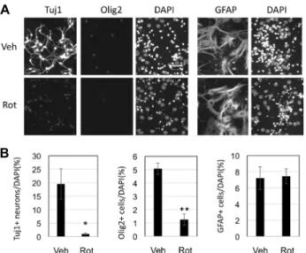
Removal of neurons and oligodendrocytes by rotenone Previously, we showed that both neurogenesis and oligo- dendrogenesis were almost completely blocked by rotenone treatment during 4 days of differentiation [8]. Here, we test- ed whether rotenone has any effects on differentiated neu- rons and glial cells. For this purpose, SVZ NSCs were main- tained in N6 differentiation medium for 4 days, then, me- dium was changed to new N6 medium containing DMSO as a vehicle or 50 nM rotenone for the another 2 days. To our surprise, Tuj1+ cells were only 1% in rotenone-treated cells, while Tuj1+ cells were 20% in control (Fig. 3A, Fig.
3B). After total 6 days of differentiation, control cells had relatively long neurites and cells were distributed more
A B
Fig. 2. Effects of rotenone on Mash1 expression. A. Representative images after immunocytochemistry with antibodies against Mash1 and nuclear staining with DAPI. N5 condition is cells that are maintained in N5 proliferating medium. Others were incubated with N6 differentiation medium containing DMSO (Veh) or 50 nM rotenone (Rot) for 1 or 2 days. Scale bar=100 μm. B.
Immunostained cells were imaged and Mash1+ cells were counted for each condition. Data are average and standard error from three independent sets of experiment. Each experiment set had three different wells of cells. For N6_1d and N6_2d condition, data from Veh and Rot conditions were compared using Student’s T-test (*p<0.05).
A
B
Fig. 3. Effects of rotenone on differentiated SVZ NSCs. A. SVZ NSCs were incubated in N6 differentiation medium for 4 days, and then, new N6 medium containing DMSO (Veh) or 50 nM rotenone (Rot) was applied to them.
Immunocytochemistry was performed with antibodies against Tuj1 and Olig2 together. Separate slides were subject to immunostaining for GFAP. DAPI was used for nuclear staining. B. Number of Tuj1+ cells, Olig2+
cells, and GFAP+ cells were counted. Data are average and standard error from three independent sets of ex- periment. Each experiment set had three to four different wells of cells. Statistical significance between Veh and Rot condition was examined with Student’s T-test (*p<
0.05, **p<0.01).
evenly, compared to clustered pattern as seen in vehicle con- trol after 4 days of differentiation (Fig. 1C). Similarly, Olig2+
cells were 1.3% in rotenone-treated cells, while Olig2+ cells were 5.1% in control (Fig. 3A, Fig. 3B). Yet, astrocytes were not affected by rotenone. Both control and rotenone had sim-
ilar level of GFAP+ cells relative to total number of cells (Fig. 3A, Fig. 3B). Therefore, rotenone seemed to remove Tuj1+ neurons and Olig2+ oligodendrocytes, while GFAP+
astrocytes were resistant to rotenone.
Nrf2 pathway activation by rotenone
Finally, we investigated about effects of rotenone on an expression profile of the Nrf2 target genes. Nrf2 becomes active upon reactive oxygen species in the cells [15].
Rotenone was reported to activate Nrf2 in human induced pluripotent stem cells-derived NSCs [19]. Since mRNA ex- pression can be relatively fast, we examined the gene ex- pression of Nrf2 target genes after short-tem treatment of rotenone such as 5 hr and 1d in N6 differentiation medium.
Interestingly, two genes, heme oxygenase-1 (Hmox1) and sulfiredoxin 1 (Srxn1) were highly expressed only in 1 day treatment of rotenone (Fig. 4). NAD(P)H:quinone oxidor- eductase 1 (Nqo1) mRNA expression increased by 1 day of differentiation in both control and rotenone-treated cells, but glutathione reductase (Gsr) mRNA expression level stayed almost same throughout the different conditions. Both genes, Nqo1 and Gsr, were not affected by rotenone, significantly.
These data suggest that rotenone activates some down stream target genes of Nrf2 pathway.
Discussion
To investigate time window of inhibitory effect of rote- none on neurogenesis, daily treatment of rotenone was per- formed as in Fig. 1. To our much surprise, only 1-day treat- ment rotenone significantly decreased neurogenesis, even though cells were maintained in normal differentiation me-
Fig. 4. Activation of Nrf2 pathway by rotenone. Cultured SVZ NSCs were incubated in N5 proliferation medium (0 hr), N6 differentiation medium containing DMSO (Veh) or 50 nM rotenone (Rot) for 5 hours (5 hr) or 1 day (1 d).
Real-time PCR was done for four genes that are down- stream targets of Nrf2: Srxn1, Hmox1, Nqo1, and Gsr.
Relative quantity of each transcript normalized to GAPDH transcript was plotted. All the values were relative ones to 0 hour differentiation condition. Data are average and standard error from three independent sets of experiment.
Statistical significance between Veh and Rot condition at each time point was examined with Student’s T-test (*p<0.05).
dium for the other 3 days during total 4 days of differ- entiation. This inhibitory effect seemed to be strongest dur- ing the first 1~3 days of differentiation and there was no significant difference between vehicle and rotenone when rotenone was treated for the 4th day of differentiation.
Therefore, rotenone seems to inhibit neuronal fate decision specifically at early time points of differentiation, not just having general toxic effects on the cells. Another interesting phenomenon is the morphology of Tuj1+ cells in rote- none-treated cells at later time points. They had minimal neurites even though cell bodies were strongly positive for the neuronal marker Tuj1. This suggests that later time points of differentiation are more resistant to rotenone, but neurite growth might be still sensitive to rotenone. Our ob- servation is consistent with several previous reports.
Rotenone inhibited neurite growth in human LUHMES cells [11]. In dopaminergic neurons derived from tooth-derived pulp stem cells form autistic children, both neurite develop- ment impairment and mitochondrial dysfunction were ob-
served [17]. Neurite growth of chicken motor neuron was perturbed by rotenone [5]. All these reports and our results suggest that mitochondrial dysfunction can lead to in- hibition of neurite outgrowth.
To test possible mechanism why the early time points of differentiation are more susceptible to rotenone, we checked the Mash1 expression. Mash1 level was higher at the first day of differentiation and the level was much lower at the second day of differentiation. Since Mash1 is a transcription factor for transit-amplifying cells that will become neurons, Mash1 level was expected to decrease throughout the neuro- nal differentiation. Under rotenone condition, there were much less Mash1+ cells even at the first day of differ- entiation. This observation is consistent with the high sensi- tivity of SVZ NSCs to rotenone at the early time points of differentiation as mentioned above. Even though causality has to be further researched, these results support that rote- none decreases Mash1+ cell population and then reducing cells with neurogenic fate.
Rotenone has been used to make a Parkinson’s disease model due to its neurotoxic effects on dopaminergic neurons [24]. In our SVZ cultured NSCs, rotenone also had neuro- toxic effects with only two days of treatment (Fig. 3). Since Tuj1+ or Olig2+ cell numbers were significantly decreased by rotenone, neurons and oligodendrocytes seemed to be highly vulnerable to rotenone. On the other hand, GFAP+
astrocytes were not affected by rotenone. Recent reports showed that platelet-derived growth factor, subtype BB (PDGF-BB) protected astrocytes and astrocytic-like model of T98G human glioblastoma cell line from oxidative stress [3, 4]. Therefore, it might be interesting whether PDGF-BB can provide similar protective effects on neurons and oligoden- drocytes of SVZ NSCs from rotenone.
Finally, we observed that Nrf2 pathway is activated by rotenone at least partially. Among the four genes, Srxn1 gene expression was significantly upregulated by rotenone after 1 day of treatment. Srxn1 was reported to have neuro- protective effect against oxidative stress in rat brains [26].
In astrocytes, Srxn1 reduced oxidative stress caused by oxy- gen-glucose deprivation [27]. Since our SVZ NSC cultures are heterogenous with multiple types of cells containing as- trocytes, the increase of Srxn1 level might have originated from astrocytes. Besides Srxn1, Hmox1 showed a trend of increased expression by rotenone even though it was not statistically significant. In the previous report, Hmox1 was upregulated in SVZ by mitochondrial complex I inhibitor
1-methyl-4-phenyl-1,2,3,6-tetrahydropyridine through Nrf2 pathway in vivo [12]. Therefore, it is worth to check the ex- pression after longer treatment with rotenone. Interestingly, Nqo1 expression was increased by both vehicle and rotenone.
This suggests that some target genes of Nrf2 might be acti- vated during normal neurogenic process.
In summary, we observed various aspects of transient treatment of rotenone on SVZ NSC differentiation. Through- out the 4 days of differentiation and post-differentiation time periods, NSCs were all affected by rotenone, supporting the critical roles of mitochondria during and post neurogenesis.
Also, we observed that some of target genes downstream of Nrf2 were activated by acute treatment of rotenone. This suggests that cells respond to oxidative stress by increasing expression of antioxidant proteins. As a future work, it might be interesting to investigate any of those antioxidant proteins might affect neuronal differentiation.
Acknowledgments
This work was supported by the Korea Science Academy of KAIST with funds from the Ministry of Science and ICT and by Basic Science Research Program through the National Research Foundation of Korea (NRF) funded by the Ministry of Science and ICT (NRF-2018R1A2A3074889).
References
1. Bertrand, N., Castro, D. S. and Guillemot, F. 2002. Proneural genes and the specification of neural cell types. Nat. Rev.
Neurosci. 3, 517-530.
2. Cabello-Rivera, D., Sarmiento-Soto, H., Lopez-Barneo, J. and Munoz-Cabello, A. M. 2019. Mitochondrial complex i func- tion is essential for neural stem/progenitor cells pro- liferation and differentiation. Front. Neurosci. 13, 664.
3. Cabezas, R., Avila, M. F., Gonzalez, J., El-Bacha, R. S. and Barreto, G. E. 2015. Pdgf-bb protects mitochondria from ro- tenone in t98g cells. Neurotox. Res. 27, 355-367.
4. Cabezas, R., Vega-Vela, N. E., Gonzalez-Sanmiguel, J., Gon- zalez, J., Esquinas, P., Echeverria, V. and Barreto, G. E. 2018.
Pdgf-bb preserves mitochondrial morphology, attenuates ros production, and upregulates neuroglobin in an astrocytic model under rotenone insult. Mol. Neurobiol. 55, 3085-3095.
5. Choi, S. Y., Kim, J. Y., Kim, H. W., Cho, B., Cho, H. M., Oppenheim, R. W., Kim, H., Rhyu, I. J. and Sun, W. 2013.
Drp1-mediated mitochondrial dynamics and survival of de- veloping chick motoneurons during the period of normal programmed cell death. FASEB J. 27, 51-62.
6. Corenblum, M. J., Ray, S., Remley, Q. W., Long, M., Harder, B., Zhang, D. D., Barnes, C. A. and Madhavan, L. 2016.
Reduced nrf2 expression mediates the decline in neural stem cell function during a critical middle-age period. Aging Cell 15, 725-736.
7. Hayes, J. D., McMahon, M., Chowdhry, S. and Dinkova- Kostova, A. T. 2010. Cancer chemoprevention mechanisms mediated through the keap1-nrf2 pathway. Antioxid. Redox Signal. 13, 1713-1748.
8. Park, K. Y. and Kim, M. S. 2018. Inhibition of proliferation and neurogenesis of mouse subventricular zone neural stem cells by a mitochondrial inhibitor rotenone. J. Life Sci. 28, 1397-1405.
9. Kim, E. J., Ables, J. L., Dickel, L. K., Eisch, A. J. and Johnson, J. E. 2011. Ascl1 (mash1) defines cells with long-term neuro- genic potential in subgranular and subventricular zones in adult mouse brain. PLoS One 6, e18472.
10. Kim, H. J., Shaker, M. R., Cho, B., Cho, H. M., Kim, H., Kim, J. Y. and Sun, W. 2015. Dynamin-related protein 1 con- trols the migration and neuronal differentiation of sub- ventricular zone-derived neural progenitor cells. Sci. Rep. 5, 15962.
11. Krug, A. K., Balmer, N. V., Matt, F., Schonenberger, F., Merhof, D. and Leist, M. 2013. Evaluation of a human neu- rite growth assay as specific screen for developmental neu- rotoxicants. Arch. Toxicol. 87, 2215-2231.
12. L'Episcopo, F., Tirolo, C., Testa, N., Caniglia, S., Morale, M.
C., Impagnatiello, F., Pluchino, S. and Marchetti, B. 2013.
Aging-induced nrf2-are pathway disruption in the subven- tricular zone drives neurogenic impairment in parkinsonian mice via pi3k-wnt/beta-catenin dysregulation. J. Neurosci.
33, 1462-1485.
13. Lim, D. A. and Alvarez-Buylla, A. 2016. The adult ven- tricular-subventricular zone (v-svz) and olfactory bulb (ob) neurogenesis. Cold Spring Harb. Perspect. Biol. 8, a018820.
14. Lu, J., Li, Y., Mollinari, C., Garaci, E., Merlo, D. and Pei, G. 2019. Amyloid-beta oligomers-induced mitochondrial DNA repair impairment contributes to altered human neu- ral stem cell differentiation. Curr. Alzheimer Res. 16, 934-949.
15. Ma, Q. 2013. Role of nrf2 in oxidative stress and toxicity.
Annu. Rev. Pharmacol. Toxicol. 53, 401-426.
16. Nakatani, H., Martin, E., Hassani, H., Clavairoly, A., Maire, C. L., Viadieu, A., Kerninon, C., Delmasure, A., Frah, M., Weber, M., Nakafuku, M., Zalc, B., Thomas, J. L., Guillemot, F., Nait-Oumesmar, B. and Parras, C. 2013. Ascl1/mash1 promotes brain oligodendrogenesis during myelination and remyelination. J. Neurosci. 33, 9752-9768.
17. Nguyen, H. T. N., Kato, H., Masuda, K., Yamaza, H., Hirofuji, Y., Sato, H., Pham, T. T. M., Takayama, F., Sakai, Y., Ohga, S., Taguchi, T. and Nonaka, K. 2018. Impaired neurite devel- opment associated with mitochondrial dysfunction in dop- aminergic neurons differentiated from exfoliated deciduous tooth-derived pulp stem cells of children with autism spec- trum disorder. Biochem. Biophys. Rep. 16, 24-31.
18. Parras, C. M., Galli, R., Britz, O., Soares, S., Galichet, C., Battiste, J., Johnson, J. E., Nakafuku, M., Vescovi, A. and Guillemot, F. 2004. Mash1 specifies neurons and oligoden- drocytes in the postnatal brain. EMBO J. 23, 4495-4505.
초록:미토콘드리아 저해제인 rotenone의 일시적 처리가 쥐의 뇌실 하 영역 신경 줄기 세포에 미치는 영향
박기엽1*․김만수2
(1KAIST 부설 한국과학영재학교, 2인제대학교 약학대학)
뇌에서 뇌실하 영역은 자가 복제 및 신경세포와 교세포로 분화하는 신경줄기세포가 위치한 곳이다. 이러한 신 경줄기세포는 태어난 직후 뿐만 아니라, 성인기까지 존재한다. 세포 증식과 분화에 대한 결정은 세포 안과 밖의 상황에 따라 조절될 필요가 있기에, 많은 세포 내부 또는 세포 외부의 인자들이 이러한 결정에 관여한다. 이러한 인자들 중에서 미토콘드리아는 신경줄기세포의 운명 결정에 관여함이 보고된 바 있다. 본 저자들의 이전 논문에 서, 미토콘드리아 저해제인 rotenone을 장시간 처리했을 때, 신경세포로의 분화가 거의 일어나지 않았음을 보여주 었다. 이번 연구에서, rotenone을 뇌실하 영역 신경줄기세포에 단기간 처리했을 때의 영향에 대해 조사하였다. 이 를 통해 다음과 같은 결과를 관찰하였다. (1) 하루 동안 rotenone을 처리하자 신경세포로의 분화가 크게 감소하였 고, 특히 분화 초기 단계가 더 민감하게 억제되었다. (2) 일시적 증식세포인 Mash1+ 세포의 수가 rotenone을 하루 처리한 후 감소하였다. (3) 분화가 된 Tuj1+ 신경세포와 Olig2+ 희소 돌기 아교 세포 (oligodendrocytes) 모두 rote- none을 단기간 처리하자 감소하였다. 반면, glial fibrillary acidic protein (GFAP)+성상 세포 (astrocytes)의 수는 변화하지 않았다. (4) sulfiredoxin 1 (Srxn1) 유전자 발현이 rotenone을 하루 처리한 후 증가하였는데, 이는 nu- clear factor (erythroid-derived 2)-like 2 (Nrf2) 신호전달 경로가 활성화 되었음을 말해준다. 이러한 실험 결과는 기능을 갖춘 미토콘드리아가 신경세포 또는 희소 돌기 아교 세포로의 분화 뿐 아니라, 이미 분화가 끝난 신경세포 의 유지에도 필요함을 확인해 주었다. 또한, 이러한 결과는 rotenone과 같은 미토콘드리아의 저해제에 짧은 시간 노출 되더라도 신경줄기세포의 신경세포로의 분화 가능성에 장기적인 영향을 미칠 수 있음을 시사한다.
19. Pistollato, F., Canovas-Jorda, D., Zagoura, D. and Bal-Price, A. 2017. Nrf2 pathway activation upon rotenone treatment in human ipsc-derived neural stem cells undergoing differ- entiation towards neurons and astrocytes. Neurochem. Int.
108, 457-471.
20. Ray, S., Corenblum, M. J., Anandhan, A., Reed, A., Ortiz, F. O., Zhang, D. D., Barnes, C. A. and Madhavan, L. 2018.
A role for nrf2 expression in defining the aging of hippo- campal neural stem cells. Cell Transplant. 27, 589-606.
21. Richetin, K., Moulis, M., Millet, A., Arrazola, M. S., Andraini, T., Hua, J., Davezac, N., Roybon, L., Belenguer, P., Miquel, M. C. and Rampon, C. 2017. Amplifying mitochondrial function rescues adult neurogenesis in a mouse model of alzheimer's disease. Neurobiol. Dis. 102, 113-124.
22. Shaw, P. and Chattopadhyay, A. 2019. Nrf2-ARE signaling in cellular protection: Mechanism of action and the regu- latory mechanisms. J. Cell. Physiol. doi:10.1002/jcp.29219.
23. Son, G. and Han, J. 2018. Roles of mitochondria in neuronal development. BMB Rep. 51, 549-556.
24. von Wrangel, C., Schwabe, K., John, N., Krauss, J. K. and
Alam, M. 2015. The rotenone-induced rat model of parkin- son's disease: Behavioral and electrophysiological findings.
Behav. Brain Res. 279, 52-61.
25. Walter, J., Bolognin, S., Antony, P. M. A., Nickels, S. L., Poovathingal, S. K., Salamanca, L., Magni, S., Perfeito, R., Hoel, F., Qing, X., Jarazo, J., Arias-Fuenzalida, J., Ignac, T., Monzel, A. S., Gonzalez-Cano, L., Pereira de Almeida, L., Skupin, A., Tronstad, K. J. and Schwamborn, J. C. 2019.
Neural stem cells of parkinson's disease patients exhibit aberrant mitochondrial morphology and functionality. Stem Cell Reports 12, 878-889.
26. Wu, J., Chen, Y., Yu, S., Li, L., Zhao, X., Li, Q., Zhao, J.
and Zhao, Y. 2017. Neuroprotective effects of sulfiredoxin-1 during cerebral ischemia/reperfusion oxidative stress injury in rats. Brain Res. Bull. 132, 99-108.
27. Zhou, Y., Duan, S., Zhou, Y., Yu, S., Wu, J., Wu, X., Zhao, J. and Zhao, Y. 2015. Sulfiredoxin-1 attenuates oxidative stress via nrf2/are pathway and 2-cys prdxs after oxygen- glucose deprivation in astrocytes. J. Mol. Neurosci. 55, 941- 950.