46(1) : 52∼ 58 (2015)
52
오미자 70% 에탄올 추출물의 신경줄기세포 증식과 분화에 미치는 영향
바랄삼랏1,2,3,4·파리야르라메시2,4·윤치수2,5·윤종민6·장석오6·김성연2·
오현철2,3,4,5·김윤철1,2,3,4,5·서정원1,2,3,4*
1BK21 PLUS Team, 2원광대학교 약학대학 약품연구소, 3원광대학교 한의학전문대학원
4원광대학교 한방체액조절센터, 5원광대학교 천연물신소재은행, 6원광대학교 한의과대학 내과학교실
Effects of Schisandrae Fructus 70% Ethanol Extract on Proliferation and Differentiation of Human Embryonic Neural Stem Cells
Samrat Baral1,2,3,4, Ramesh Pariyar2,4, Chi-Su Yoon2,5, Jong-Min Yun6, Seok O Jang6, Sung Yeon Kim2, Hyuncheol Oh2,3,4,5, Youn-Chul Kim1,2,3,4,5, and Jungwon Seo1,2,3,4*
1From the BK21 Plus team,
2Institute of Pharmaceutical Research and Development, College of Pharmacy, Wonkwang University, Iksan 570-749, Korea
3Professional Graduate School of Oriental Medicine, Wonkwang University, Iksan 570-749, Korea
4Hanbang Body-Fluid Research Center, Wonkwang University, Iksan 570-749, Korea
5Standardized Material Bank for New Botanical Drugs, College of Pharmacy, Wonkwang University, Iksan 570-749, Korea
6Department of Internal Medicine, College of Korean Medicine, Wonkwang University, Iksan 570-749, Korea
Abstract − Neural stem cells (NSCs), with self-renewal and neuronal differentiation capacity, are a feasible resource in cell- based therapies for various neurodegenerative diseases and neural tissue injuries. In this study, we investigated the effects of Schisandrae Fructus (SF) on proliferation and differentiation of human embryonic NSCs. Treatment with 70% ethanol extract of SF increased the viability of NSCs derived from human embryonic stem cells, which was accompanied by increased mRNA expression of cyclin D1. Whereas 70% ethanol extract of SF also decreased the mRNA expression of nestin, it increased class III β-tublin (Tuj-1) and MAP2 in both growth and differentiation media. Lastly, we found increased mRNA expression of BDNF in SF-treated NSCs. In conclusion, our study demonstrates for the first time that SF induced proliferation and neuronal differentiation of NSCs and increased mRNA expression of BDNF, suggesting its potential as a regulator of NSC fate in NSC- based therapy for neuronal injuries from various diseases.
Key words − Schisandrae Fructus, Human Embryonic Neural Stem Cells, Proliferation, Neuronal Differentiation, BDNF
파킨슨병, 알츠하이머병과 같은 퇴행성 뇌질환이나 뇌졸 중은 공통적으로 신경세포의 사멸에 의해 유발된다. 뇌는 손상된 신경세포에 대한 재생능력이 제한적이어서 치료에 많은 어려움이 존재한다. 신경줄기세포는 자기복제능과 신 경세포로의 분화능을 가진 세포1)로서, 이러한 손상된 신경 세포를 대체할 수 있는 세포치료제로서 활발한 연구가 진 행되고 있다.2,3) 예를 들어, 파킨슨 질환 모델 쥐에 배아줄 기세포4)나 유도만능줄기세포5)에서 유래한 도파민 신경을 이식하면 회전실험에서 회전율이 감소함이 보고되었다. 또 한, 알츠하이머질환 모델 쥐에서 배아줄기세포 유래 신경
전구 세포의 이식은 수중 미로 시험과 공간 기억 시험 결과 를 향상시켰다.6)따라서, 이식하는 신경줄기세포의 증식과 신경세포 분화 유도는 신경줄기세포 기반 치료법의 치료 효 과와 안전성을 높이는데 크게 기여할 수 있을 것으로 기대 된다. 신경줄기세포는 신경 발달 과정에서만 존재하는 것이 아니라, 성체의 뇌에서도 subependymal ventricle zone (SVZ)7)과 hippocampal subgranular zone(SGZ)8)에 존재하고 있다. 성체신경줄기세포는 자기재생 능력과 신경세포나 성 상세포로의 분화 능력을 가지고 있으나, 신경세포로의 분화 비율은 상당히 낮다. 따라서, 성체 신경줄기세포의 증식을 유도하거나 신경세포로의 분화를 촉진할 수 있다면, 신경퇴 행성 질환이나 뇌졸중의 효과적인 치료법이 될 수 있을 것
*교신저자(E-mail):[email protected] (Tel): +82-63-850-6819
이다.
오미자(五味子, Schisandrae Fructus)는 목련과(Magnoliaceae) 의 낙엽덩굴식물인 오미자(Schisandra chinensis Baill.)의 열 매를 건조한 것으로, 당뇨,9,10) 간질환,11) 심혈관 질환,12) 신 경학적 증상13)의 치료에 사용되어 왔다. 특히, 선행 동물 실 험 결과들에 의하면 오미자는 기억력 감퇴를 개선14)하고 신 경독성을 감소15)시킨다고 알려져 있다. 주요 성분은 schisandrin, deoxyschisandrin, schisandrol, gomisin, citric acid, and β-sitosterol 등이 있다.16) 특히 schisandrin A나 schisandrin B와 같은 dibenzocyclooctadiene-type lignan류는 오미자에서 보여지는 신경보호효과,17,18)항신경염증효과,19) 기억력 개선20,21)에 기여하는 중요한 구성성분으로 알려져 있다. 선행연구 결과에 따르면 schisandrin A와 schisandrin B는 쥐의 subventricular zone에서 신경 생성을 증가22)시킨 다고 한다. 이러한 결과는 오미자의 신경 재생의 가능성을 시사하고 있으나, 아직 오미자가 신경줄기세포에 미치는 영 향에 관하여는 연구된 바가 없다. 따라서 본 논문에서는 인 간 배아줄기세포 유래의 신경줄기세포를 이용하여 오미자 의 70% 에탄올 추출물이 신경줄기세포의 증식과 분화에 미 치는 영향을 연구하였다. 신경줄기세포의 분화를 확인하기 위하여, 미분화 신경 줄기세포의 마커인 Nestin, 신경세포 특이적으로 발현하는 단백질인 Tuj1과 MAP2의 mRNA 발 현을 real-time PCR을 이용하여 측정하였으며, 신경줄기세 포의 증식과 분화를 유도하는 성장인자인 BDNF의 mRNA 발현 또한 확인하였다.
재료 및 방법
실험재료 − 본 실험에 사용한 오미자는 2013년 8월 전북 익산시 소재 대학한약국에서 구입하였으며, 원광대학교 약 학과 김윤철 교수가 형태학적 평가를 통하여 동정하였고 표 본시료(NNMB-13-39)는 원광대학교 약학대학 천연물신약 표준화소재은행에 보관되어 있다.
시료제조 − 분말화한 오미자 50 g을 70% 에탄올 200 ml 로 40oC에서 2시간 동안 초음파추출을 한 뒤 여과하고, 2차 추출을 1차 추출과 같은 조건으로 한 뒤 여과한 다음 여액 을 합한 후 감압농축하여 70% 에탄올 추출물(4.39 g; 8.8 w/
w%)을 얻었다. 오미자 70% 에탄올 추출물의 기준시료 (NNMBS48)는 원광대학교 천연물신약표준화소재은행에 보 관되어 있다.
시약 및 기기 − KnockOut DMEM/F-12 배지, Cellstart, StemPro, Glutamax, EGF, FGF를 Life Technologies사에서 구입하였다. L-glutamate와 3'-(4,5-dimethylthiazol-2-yl)-2,5- diphenyltetrazolium bromide(MTT)는 Sigma 사에서 구입하 였다. 96-Well tissue culture plates와 기타 tissue culture dishes는 Nunc사 제품을 이용하였다. 흡광도는 BioRad사의
Microplate Reader 를 이용하여 측정하였다.
세포 배양 − 배아줄기세포 유래 신경줄기세포는 Life Technologies사에서 구입하여 사용하였다(N7800-200). 세포 배양 전dish는 CellStat를 이용하여 1시간 동안 코팅하였다.
신경줄기세포를 2% StemPro Neural Supplement, 2 mM Glutamax, 20 ng/mL recombinant Human EGF, 20 ng/mL recombinant Human bFGF(Invitrogen)를 함유한 KnockOut DMEM/F-12 배지(growth media)를 첨가하여 5% CO2 배양 기 내에서 37oC의 온도로 배양하였다. 신경세포 분화는 EGF, FGF를 넣지 않고, B-27 supplement, 2 mM Glutamax 을 함 유한 Neurobasal 배지(differentiation media)를 첨가하여 4일 동안 분화를 유도하였다.
면역형광 염색법 − 신경줄기세포를 cover-glass bottom dish(SPL101350)에 24시간 배양한 후, 4% paraformaldehyde 로 고정하고 0.25% Triton X-100을 10분간 처리하였다. 1%
BSA/PBST로 blocking하고, 1차 항체인 mouse Nestin monoclonal antibody(Abcam) 단독 혹은 MAP2 polyclonal antibody(Abcam)과 같이 처리 후, 2차 항체인 Alexa Fluor 488의 goat anti-mouse antibody 단독 혹은 Alexa Fluor 568의 goat anti-rabbit antibody를 동시에 처리하여 confocal microscopy(Olympus)로 관찰하였다.
MTT Assay − 신경줄기세포에 대한 세포 생존율을 측정 하기 위해 MTT assay를 사용하였다. 간단히 기술하면 먼저 96 well plate에 1×10⁴ cells/well로 동일하게 분주하고 24시 간 동안 배양하였다. 기존의 배지를 제거하고 새로운 배지 를 넣어준 후 DMSO에 녹인 시료를 다양한 농도로 KnockOut DMEM/F-12 배지에 희석하여 첨가하였다.
DMSO의 처리 농도는 배지 대비 0.1% 이하가 되도록 하였 다. 이를 다시 24 시간 배양한 후에 배지를 제거하고 MTT 시약(5 mg/mL)을 넣고, 4 시간 동안 방치한 후 상등액을 제 거하였다. 형성된 formazan의 각 well에 DMSO 20 µL를 첨 가한 후 orbital shaker를 이용하여 녹이고, 30 분 후 595 nm 에서 흡광도를 측정하였다. 실험은 3회 반복 실시하여 평균 값을 구하였으며, control의 흡광도 값을 기준으로 세포 생 존율을 비교하였다.
Real Time-PCR Analysis − 세포를 60 mm dish에 3×10⁵ cells/well 밀도로 24 시간 배양한 후 각각의 시료를 농도별로 처리하였다. 72시간 또는 96시간 후 세포에 Trizol reagent(Invitrogen)을 처리하여 RNA를 추출한 후, SuperScript III system(Invitrogen)을 이용하여 2 µg의 RNA로부터 oligo- dT primer로 cDNA를 제작하였다. 사용한 primer는 Nestin 5'-CAG CGT TGG AAC AGA GGT TGG-3', 5'-TGG CAC AGG TGT CTC AAG GGT AG-3'; MAP2 5'-AAT GGG ATC AAC GGA GAG CT-3', 5'-TCT TCA GCT GCT AAA GGC AG-3'; Tuj1 5'-AAC AGC ACG GCC ATC CAG GA-3', 5'-CTT GGG GCC CTG GGC CTC
CGA-3'; BDNF 5'-GGC GGC AGA CAA AAA GAC TG-3', 5'-CGT ACG ACT GGG TAG TTC GG-3';
GAPDH 5'-TGC ACC ACC AAC TGC TTA GC-3', 5'- GGC ATG GAC TGT GGT CAT GAG-3'이다. real-time PCR은Power SYBR Green PCR Master Mix(Applied Biosystems)을 첨가하여 StepOnePlus Real-Time PCR system을 이용하였다.
통계처리 − 각 실험군 간의 결과는 평균치와 표준오차로 나타내었으며, 각 실험군 간의 결과는 Student's t-test를 사 용하여 분석하고 유의적인 차이가 있는 항목에 대해서만 검 정하였다. 실험군 간의 차이는 95% 수준(p<0.05)에서 유의 성 있는 것으로 하였다.
결과 및 고찰
신경줄기세포 검증 − 배아줄기세포 유래의 세포가 신경줄 기세포인지를 확인하기 위하여, 마커인 Nestin을 사용하여 면역현광염색을 실시하였다. 모든 세포는 Nestin 양성이었 고, 세포질에 점 형태로 무리(button-like cluster)를 지어 보 여졌다(Fig. 1). 이 결과로부터 배아줄기세포 유래의 세포가 신경줄기세포 성질을 가지고 있음을 확인하였다.
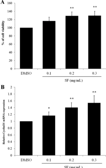
오미자의 신경줄기세포 증식에 미치는 영향 − 오미자가 신경줄기세포의 증식 혹은 사멸에 미치는 영향을 확인하기 위하여, growth 배지에 오미자의 70%에탄올 추출물을 0, 0.1, 0.2, 0.3 mg/mL농도로 24시간 처리한 후 MTT assay를 실시하였다. 그 결과 오미자 추출물을 처리한 신경줄기세포 는 세포 생존율이 농도의존적으로 증가하였다(Fig. 2A). 이
러한 세포 생존율의 증가가 증식에 의한 것인지 알아보기 위해, 세포 주기 단백질인 Cyclin D1의 mRNA 발현을 real- time PCR을 이용하여 확인한 결과, Cyclin D1의 발현이 농 도의존적으로 증가함을 확인하였다(Fig. 2B). 이 결과로부 터 오미자 추출물이 신경줄기세포의 증식을 유도함을 확인 하였다.
오미자의 신경줄기세포 분화에 미치는 영향 − 오미자가 신경줄기세포의 신경세포로의 분화를 유도했는지를 알아보
Fig. 1. Nestin was expressed in NSCs derived from embryonic stem cells. Cells were fixed and Nestin expression was detected by confocal microscopy as described in Material &
Method. Nestin was stained using anti-Nestin monoclonal anti- body and goat anti-mouse antibody coupled to Alexa Fluor 488.
Fig. 2. SF increases NSC viability and mRNA expression of Cyclin D1. NSCs were treated with 0.1, 0.2, or 0.3 mg/mL of SF for 24 h in growth media. Control cells were treated with vehicle (DMSO) only. (A) MTT assay. After the reduced pur- ple-blue MTT formazan crystals were solubilized by DMSO, cell viability was measured using the absorbance at 540 nm with ELISA reader. Each bar represents the percentage com- pared with control (DMSO) (±S.D.). (B) relative mRNA expression of Cyclin D1. Cyclin D1 expression was detected by real-time RT-PCR analysis and normalized to that of GAPDH. Each bar represents the mean fold increase with respect to control (±S.D.). Differences were statistically signif- icant at *P<0.05 and **P<0.01.
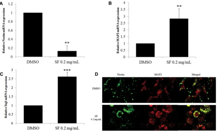
기 위하여, 신경줄기세포를 오미자 추출물 0.2 mg/mL를 첨 가한 differentiation 배지에서 4일간 배양한 후, real-time PCR를 시행하여, 신경줄기세포 마커 분자와 신경세포 마커 분자의 발현을 비교하였다. 0.2 mg/mL 오미자 추출물을 처 리한 실험군에서 신경줄기세포의 마커인 Nestin의 발현은 감소(Fig. 3A)한 반면, 신경세포 특이적인 단백질인 MAP2(Fig. 3B)와 Tuj1(Fig. 3C)의 mRNA발현은 증가함을 확인할 수 있었다. 이를 면역형광염색법으로 Nestin과 MAP2 항체를 사용하여 염색한 결과, 대조군에 비하여 0.2 mg/mL 오미자 추출물을 처리한 실험군에서는 Nestin을 발현하는 세포는 줄어들고 MAP2를 발현하는 세포는 늘어났음을 확 인하였다(Fig. 3D).
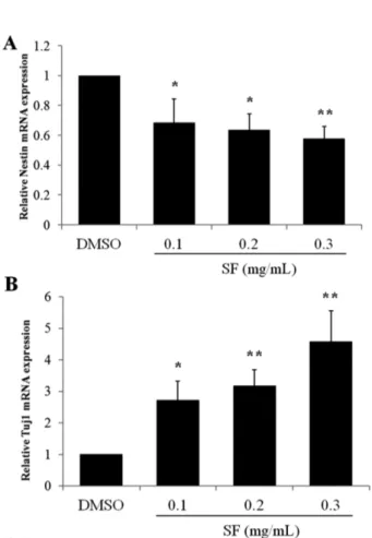
오미자가 growth 배지에서도 단시간에 신경세포로의 분 화 유도가 가능한지 확인하기 위하여, growth 배지에서 0, 0.1, 0.2, 0.3 mg/mL의 오미자 추출물을 72시간 처리한 후, 신경줄기세포 및 신경세포의 마커 단백질의 mRNA 발현 정 도를 real-time PCR을 이용하여 비교하였다. 그 결과, Nestin 의 발현은 감소(Fig. 4A)한 반면, 신경세포 특이적인 단백
질인 Tuj1(Fig. 4B)의 mRNA 발현은 농도의존적으로 증가 함을 확인할 수 있었다.
오미자가 BDNF 발현에 미치는 영향 − 오미자가 신경줄 기세포의 증식과 분화에 중요한 역할을 하는 성장 인자인 BDNF의 발현에 관여하는지 확인하기 위하여 real-time PCR 를 시행하였다. 신경줄기세포를 오미자 추출물 0.2 mg/mL 를 첨가한 differentiation 배지에서 4일간 배양한 후, real- time PCR를 시행한 결과, BDNF mRNA발현이 약 1.7배 증 가함을 확인하였다(Fig. 5A). growth 배지에서 0, 0.1, 0.2, 0.3 mg/mL의 오미자 추출물을 신경줄기세포에 처리하였을 때도BDNF mRNA의 발현이 농도의존적으로 증가하였다 (Fig. 5B).
신경줄기세포는 자기복제능과 신경세포로의 분화능을 가 진 세포로 퇴행성 뇌질환을 비롯하여 신경 손상에 의해 발 병하는 다양한 질환의 세포치료법으로 각광받고 있다. 신경 줄기세포를 이식하여 신경 분화를 촉진시키거나 손상된 신 경 세포를 대체하여 신경전달물질의 분비를 유도하는 세포 치료 방법이 활발히 연구 중이다.2,3)이러한 치료법의 가장
Fig. 3. SF decreases mRNA expression of Nestin and increases that of Tuj1 and MAP2 in differentiation process. NSCs were treated with 0.2 mg/mL of SF for 4 days in differentiation media. Control cells were treated with vehicle (DMSO) only. Total RNA was then extracted from the treated NSCs and used to synthesize cDNA. The mRNA expression of Nestin (A), MAP2 (B), and Tuj1 (C) was detected by real-time RT-PCR and normalized to that of GAPDH. Each bar represents the mean fold increase with respect to control (±S.D.). Differences were statistically significant at **P<0.01 and ***P<0.005. (D) Double-labeled immunocy- tochemistry was performed using antibodies against Nestin (green) and MAP2 (red). The images shown are representative of at least three experiments.
큰 어려움은 신경줄기세포를 원하는 신경세포로 정확하고 완벽하게 분화시키지 못한다는 점이다. 본 연구에서는 오미 자의 70% 에탄올 추출물이 배아줄기세포 유래의 신경줄기 세포의 증식과 신경세포로의 분화를 촉진시킴을 확인하였다.
선행 연구 결과에 의하면, 쥐 해마로부터 분리한 신경줄 기세포에 삼칠(Panax Notoginseng)의 saponin을 처리하면 신경줄기세포의 증식이 증가하며, 신경세포와 교세포로의 분화 역시 증가시킴을 확인하였다.23) 본 연구에서도 선행 결 과인 삼칠 saponin과 유사하게 오미자가 신경줄기세포의 증 식을 증가시키고 MAP2와 Tuj1의 mRNA 발현을 증가(Fig.
3)시켜 신경세포 분화를 촉진함을 확인할 수 있었다. 하지 만, 오미자는 GFAP의 발현을 증가시키지 않는 것으로 보아, 교세포로의 분화에는 영향을 미치지 않는 것으로 보인다.
특히 오미자는 신경줄기세포의 증식과 분화의 중요한 성장 조절 인자인 BDNF mRNA의 발현을 증가시켜 신경줄기세
포에 유익한 작용을 하는 것으로 보인다. 본 연구에서 얻어 진 오미자 추출물이나, 선행 문헌에서 얻어진 삼칠 saponin 을 신경줄기세포에 처리하여 신경줄기세포를 좀 더 효율적 으로 신경세포로 분화시킬 수 있다면, 신경줄기세포를 이용 한 세포 치료법의 성공률을 높이는데 기여할 수 있을 것이 라 사료된다.
또한, 오미자는 신경학적 증상의 치료에 널리 사용되어 온 한약제제 중의 하나이며 많은 선행 연구 결과들을 통하 여 그 신경보호 효과,16)기억력 회복 효과14)가 밝혀졌다. 특 히, 오미자의 성분 중 schisandrin B는 SVZ에서 세포의 증 식을 증가시키는 역할을 하는 반면, schisandrin A는 신경줄 기세포의 이동 경로인 rostral migratory stream에서 neuroblast의 형성을 유도한다는 보고가 있다.22) 따라서 본 연구에서 얻어진 오미자의 신경줄기세포 증식과 분화에 대 Fig. 4. SF decreases mRNA expression of Nestin and
increases that of Tuj1 in growth media. NSCs were treated with 0.1, 0.2, or 0.3 mg/mL of SF for 72 h in growth media.
Control cells were treated with vehicle (DMSO) only. The mRNA expression of Nestin (A) and Tuj1 (B) was detected by real-time RT-PCR and normalized to that of GAPDH. Each bar represents the mean fold increase with respect to control (±S.D.). Differences were statistically significant at *P<0.05 and **P<0.01.
Fig. 5. SF increased BDNF mRNA expression in both growth and differentiation media. NSCs were treated as described in Fig. 3 and 4. The mRNA expression of BDNF in differenti- ation media (A) and growth media (B) was detected by real- time RT-PCR and normalized to that of GAPDH. Each bar represents the mean fold increase with respect to control (±S.D.). Differences were statistically significant at *P<0.05 and **P<0.01.
한 효과는 schisandrin A와 schisandrin B 둘 다의 효과가 합 쳐진 결과인 것으로 판단된다. 오미자 추출물의 효과는 다 양한 성분들의 효과가 더해진 것으로 부작용을 줄이고 순 수한 효과를 얻기 위해서는 앞으로 성분을 특정하는 연구 가 필요할 것으로 판단된다.
오미자가 배아줄기세포 유래의 신경줄기세포의 증식, 분 화를 유도하므로, 실제 신경 손상 환자가 복용 시 성체 신 경줄기세포에도 유사한 효과를 가져올 수 있을지도 모른다.
실제로 in-vitro 실험을 통해 신경줄기세포의 증식과 분화 효 과가 밝혀진 삼칠의 경우, in-vivo에서 허혈 손상된 쥐의 해 마의 SVZ, SGZ 영역에서 신경줄기세포의 증식, 이동, 분화 를 증진시킨다는 연구 결과가 밝혀졌다.24) 오미자 또한 기 억력 감퇴 쥐 모델에 투여 시, CA1, CA3 영역의 신경세포 밀도가 증가한다는 보고25)가 있어, in-vivo에서도 성체신경 줄기세포의 증식과 분화를 유도할 가능성이 있다. 앞으로 오미자를 투여한 신경퇴행성 질환 동물 모델이나 뇌졸중 동 물 모델을 이용하여 실제로 뇌의 신경줄기세포에서 증식과 분화가 유도되는지에 대한 in-vivo 연구가 필요할 것으로 판 단된다.
결 론
본 연구에서는 오미자의 70% 에탄올 추출물을 이용하여 신경줄기세포의 증식과 분화에 오미자가 미치는 영향에 관 하여 분석하고자 하였다. 그 결과, 오미자는 growth 배지에 서 신경줄기세포의 증식을 증가시켰으며, differentiation 배 지에서는 신경줄기세포의 신경세포로의 분화를 촉진하였다.
오미자 추출물은 신경줄기세포의 분화 시 Nestin의 mRNA 발현은 낮추고, MAP2와 Tuj1의 발현은 증가시켰다. 특히, 오미자는 주요 성장인자인 BDNF mRNA의 발현을 증가시 켰다. 본 연구 결과와 선행 연구들을 토대로 오미자 추출물 은 신경줄기세포를 이용한 세포 치료 시의 신경세포 분화 촉진제로 개발 가능하며, 지속적 연구를 통하여 퇴행성 뇌 질환이나 뇌졸중 환자 뇌에서 직접 신경줄기세포의 증식과 분화를 촉진하여 질환을 개선하는 치료제로의 개발도 가능 할 것으로 사료된다.
사 사
본 연구는 보건복지부 한의약선도기술개발사업(HI13C0516) 으로 수행된 연구결과입니다.
인용문헌
1. Reynolds, B. A., Tetzlaff, W. and Weiss, S. (1992) A mul- tipotent EGF-responsive striatal embryonic progenitor cell
produces neurons and astrocytes. J. Neurosci. 12: 4565-4574.
2. Kim, S. U., Lee, H. J. and Kim, Y. B. (1999) Neural stem cell-based treatment for neurodegenerative diseases. Neuro- pathology 33: 491-504.
3. Hao, L., Zou, Z., Tian, H., Zhang, Y., Zhou, H. and Liu, L.
(2014) Stem cell-based therapies for ischemic stroke.
Biomed. Res. Int. 2014: 468748.
4. Wernig, M., Zhao, J. P., Pruszak, J., Hedlund, E., Fu, D., Soldner, F., Broccoli, V., Constantine-Paton, M., Isacson, O.
and Jaenisch, R. (2008) Neurons derived from reprogrammed fibroblasts functionally integrate into the fetal brain and improve symptoms of rats with Parkinson's disease. Proc.
Natl. Acad. Sci. U. S. A. 105: 5856-5861.
5. Kriks, S., Shim, J. W., Piao, J., Ganat, Y. M., Wakeman, D.
R., Xie, Z., Carrillo-Reid, L., Auyeung, G., Antonacci, C., Buch, A., Yang, L., Beal, M. F., Surmeier, D. J., Kordower, J. H., Tabar, V. and Studer, L. (2011) Dopamine neurons derived from human ES cells efficiently engraft in animal models of Parkinson's disease. Nature 480: 547-551.
6. Moghadam, F. H., Alaie, H., Karbalaie, K., Tanhaei, S., Nasr Esfahani, M. H. and Baharvand, H. (2009) Transplantation of primed or unprimed mouse embryonic stem cell-derived neu- ral precursor cells improves cognitive function in Alzhe- imerian rats. Differentiation 78: 59-68.
7. Morshead, C. M., Reynolds, B. A., Craig, C. G., McBurney, M. W., Staines, W. A., Morassutti, D., Weiss, S. and van der Kooy, D. (1994) Neural stem cells in the adult mammalian forebrain: a relatively quiescent subpopulation of subependy- mal cells. Neuron 13: 1071-1082.
8. Palmer, T. D., Takahashi, J. and Gage, F. H. (1997) The adult rat hippocampus contains primordial neural stem cells. Mol.
Cell. Neurosci. 8: 389-404.
9. An, L., Wang, Y., Wang, C., Fan, M., Han, X., Xu, G., Yuan, G., Li, H., Sheng, Y., Wang, M., Sun, J., Zhan, J., Sun, H., Li, N., Ding, F. and Du, P. (2014) Protective effect of Schisan- drae chinensis oil on pancreatic beta-cells in diabetic rats.
Endocrine: doi: 10.1007/s12020-014-0375-y.
10. Qu, Y., Chan, J. Y., Wong, C. W., Cheng, L., Xu, C., Leung, A. W. and Lau, C. B. (2014) Antidiabetic Effect of Schisan- drae Chinensis Fructus Involves Inhibition of the Sodium Glucose Cotransporter. Drug Dev Res: doi: 10.1002/ddr.
21233.
11. Wang, S. Y., Fu, L. L., Zhang, S. Y., Tian, M., Zhang, L., Zheng, Y. X., Wang, J. H., Huang, J. and Ouyang, L. (2015) In silico analysis and experimental validation of active com- pounds from fructus Schisandrae chinensis in protection from hepatic injury. Cell Prolif. 48: 86-94.
12. Li, P. C., Poon, K. T. and Ko, K. M. (1996) Schisandra chin- ensis-dependent myocardial protective action of sheng-mai- san in rats. Am. J. Chin. Med. 24: 255-262.
13. Hsieh, M.-T., Tsai, M.-L., Peng, W.-H. and Wu, C.-R. (1999) Effects of Fructus schizandrae on cycloheximide-induced
amnesia in rats. Phytothera Res 13: 256-257.
14. Jeong, E. J., Lee, H. K., Lee, K. Y., Jeon, B. J., Kim, D. H., Park, J. H., Song, J. H., Huh, J., Lee, J. H. and Sung, S. H.
(2013) The effects of lignan-riched extract of Shisandra chin- ensis on amyloid-beta-induced cognitive impairment and neurotoxicity in the cortex and hippocampus of mouse. J.
Ethnopharmacol. 146: 347-354.
15. Ma, C. J., Kim, S. H., Lee, K. Y., Oh, T., Kim, S. Y., Sung, S. H. and Kim, Y. C. (2009) ESP-102, a combined extract of Angelica gigas, Saururus chinensis and Schizandra chinensis, protects against glutamate-induced toxicity in primary cul- tures of rat cortical cells. Phytother. Res. 23: 1587-1591.
16. Kim, J. H., Jeong, C. H., Choi, G. N., Kwak, J. H., Choi, S.
G. and Heo, H. J. (2009) Antioxidant and neuronal cell pro- tective effects of methanol extract from Schizandra chinensis using an in vitro system. Korean J. Food Sci. Technol. 41:
712-716.
17. Wang, C. P., Li, G. C., Shi, Y. W., Zhang, X. C., Li, J. L., Wang, Z. W., Ding, F. and Liang, X. M. (2014) Neuro- protective effect of schizandrin A on oxygen and glucose deprivation/reperfusion-induced cell injury in primary culture of rat cortical neurons. J. Physiol. Biochem. 70: 735-747.
18. Lee, M. S., Chao, J., Yen, J. C., Lin, L. W., Tsai, F. S., Hsieh, M. T., Peng, W. H. and Cheng, H. Y. (2012) Schizandrin pro- tects primary rat cortical cell cultures from glutamate-induced apoptosis by inhibiting activation of the MAPK family and the mitochondria dependent pathway. Molecules 18: 354-372.
19. Zeng, K.-W., Zhang, T., Fu, H., Liu, G.-X. and Wang, X.-M.
(2012) Schisandrin B exerts anti-neuroinflammatory activity by inhibiting the Toll-like receptor 4-dependent MyD88/IKK/
NF-κB signaling pathway in lipopolysaccharide-induced microglia. EurJ Pharmacol 692: 29-37.
20. Giridharan, V. V., Thandavarayan, R. A., Sato, S., Ko, K. M.
and Konishi, T. (2011) Prevention of scopolamine-induced memory deficits by schisandrin B, an antioxidant lignan from Schisandra chinensis in mice. Free Radic. Res. 45: 950-958.
21. Xu, X., Zhou, X., Zhou, X.-W., Zhang, Z., Liao, M.-J., Gao, Q. and Luo, H.-M. (2012) Schizandrin prevents dexam- ethasone-induced cognitive deficits. Neuroscience bulletin 28: 532-540.
22. Sun, Y. X., Cong, Y. L., Liu, Y., Jin, B., Si, L., Wang, A. B., Cai, H., Che, G. Y., Tang, B., Wang, C. F., Li, Z. Y. and Zhang, X. M. (2014) Schisandrin A and B affect subven- tricular zone neurogenesis in mouse. Eur. J. Pharmacol. 740:
552-559.
23. Si, Y.-C., Zhang, J.-P., Xie, C.-E., Zhang, L.-J. and Jiang, X.- N. (2011) Effects of Panax Notoginseng Saponins on Pro- liferation and Differentiation of Rat Hippocampal Neural Stem Cells. Am. J. Chin. Med. 39: 999-1013.
24. Si Yin-chu, Li Jin-wei, Zhang Li-juan, Wu Hai-xia, Xu Hong and Pei-chun, Z. (2008) Panax notoginseng saponins promote the proliferation and differentiation of neural stem cells in subependymal zone of the lateral ventricle in rat brain after intracerebral hemorrhage J. Clin. Rehabil. Tissue Eng. Res.
12: 1414-1417.
25. Jin-Ho, K., Han-na, C., Eun-hye, P., Jong-Kil, J., Kyeong-Ok, K. and Jeong-Sang, K. (2013) Effects of Gastrodia elata Extracts on Scopolamine-induced Memory Impairment in Rats. J. Korean Soc. Food Sci. Nutr. 42: 595-599.
(2015. 2. 9 접수; 2015. 3. 2 심사; 2015. 3. 10 게재확정)