Existence of a Neuropathic Pain Component in Patients with Osteoarthritis of the Knee
Seiji Ohtori, Sumihisa Orita, Masaomi Yamashita, Tetsuhiro Ishikawa, Toshinori Ito, Tomonori Shigemura, Hideki Nishiyama, Shin Konno, Hideyuki Ohta, Masashi Takaso, Gen Inoue,
Yawara Eguchi, Nobuyasu Ochiai, Shunji Kishida, Kazuki Kuniyoshi, Yasuchika Aoki, Gen Arai, Masayuki Miyagi, Hiroto Kamoda, Miyako Suzkuki, Junichi Nakamura, Takeo Furuya,
Gou Kubota, Yoshihiro Sakuma, Yasuhiro Oikawa, Masahiko Suzuki, Takahisa Sasho, Koichi Nakagawa, Tomoaki Toyone, and Kazuhisa Takahashi
Department of Orthopaedic Surgery, Graduate School of Medicine, Chiba University, Chiba, Japan.
Received: August 24, 2011 Revised: October 10, 2011 Accepted: October 11, 2011 Corresponding author: Dr. Seiji Ohtori, Department of Orthopaedic Surgery, Graduate School of Medicine, Chiba University, 1-8-1 Inohana, Chuo-ku, Chiba 260-8670, Japan.
Tel: 81-43-226-2117, Fax: 81-43-226-2116 E-mail: [email protected]
∙ The authors have no financial conflicts of interest.
© Copyright:
Yonsei University College of Medicine 2012 This is an Open Access article distributed under the terms of the Creative Commons Attribution Non- Commercial License (http://creativecommons.org/
licenses/by-nc/3.0) which permits unrestricted non- commercial use, distribution, and reproduction in any medium, provided the original work is properly cited.
Purpose: Pain from osteoarthritis (OA) is generally classified as nociceptive (in- flammatory). Animal models of knee OA have shown that sensory nerve fibers in- nervating the knee are significantly damaged with destruction of subchondral bone junction, and induce neuropathic pain (NP). Our objective was to examine NP in the knees of OA patients using painDETECT (an NP questionnaire) and to evaluate the relationship between NP, pain intensity, and stage of OA. Materials and Meth- ods: Ninety-two knee OA patients were evaluated in this study. Pain scores using Visual Analogue Scales (VAS), Western Ontario and McMaster Universities Osteo- arthritis Index (WOMAC), painDETECT, duration of symptoms, severity of OA using the Kellgren-Lawrence (KL) system, and amount of joint fluid were evaluat- ed and compared using a Spearman’s correlation coefficient by rank test. Results:
Our study identified at least 5.4% of our knee OA patients as likely to have NP and 15.2% as possibly having NP. The painDETECT score was significantly correlated with the VAS and WOMAC pain severity. Compared with the painDETECT score, there was a tendency for positive correlation with the KL grade, and tendency for negative correlation with the existence and amount of joint fluid, but these correla- tions were not significant. Conclusion: PainDETECT scores classified 5.4% of pain from knee OA as NP. NP tended to be seen in patients with less joint fluid and increased KL grade, both of which corresponded to late stages of OA. It is impor- tant to consider the existence of NP in the treatment of knee OA pain.
Key Words: Knee, osteoarthritis, neuropathic pain, nociceptive pain
INTRODUCTION
Knee osteoarthritis (OA) is a common and progressive joint disease. With an esti- mated incidence rate of 240 per 100000 person-years, it is a major public health problem in the US and often results in early retirement and joint replacement.1
of NP in the knees of OA patients using painDETECT, and to evaluate the relationship between NP and stage of OA.
MATERIALS AND METHODS
The ethics committee of our institution approved the protocol for the human procedures used in this study. Furthermore, the protocol and publication of the study were approved by our institutional review board and informed consent was ob- tained from each of the participants.
The patients who participated in this study were selected from outpatients who attended our hospital for knee pain.
These 92 patients were selected from 112 knee pain pa- tients who matched the following criteria. All patients had knee pain for more than 1 month. Inclusion criteria were the observation of the existence of OA of the knee joint on examination of an anterior-posterior X-ray image. Patients who had history of knee surgery, infection, and rheumatoid arthritis were excluded from this study. Ultimately, 20 pa- tients were excluded, and 92 patients having OA of the knee joint were evaluated. The patients completed a self- administered questionnaire on sociodemographic factors (age and gender) and duration of knee symptoms. The background of the subjects is summarized in Table 1.
Pain scores
Various screening tools have been developed to identify NP, including LANSS, DN4, NPQ, painDETECT, and ID Pain.9-13 These tools have been developed for evaluation of NP such as nerve injury, radicular pain, and diabetic neurop- athy. Hochman, et al.15 selected a list of NP descriptors from these 5 validated questionnaires and used them for knee OA pain. Five items were commonly identified to distinguish nociceptive pain from NP in people with other chronic pain conditions. They reported that 34% of knee OA patients de- scribed their pain using quality descriptors suggestive of NP, and concluded that the 5 items are reliable indicators.15 Bennett, et al.14 reviewed the strengths and weakness of these 5 validated questionnaires and reported that painDE- TECT did not require a clinical examination, and showed a slightly higher sensitivity and specificity in comparison with the other tools. Thus, in the current study, we selected painDETECT for this advantage.
All patients completed the Visual Analogue Scale (VAS), Western Ontario and McMaster Universities Osteoarthritis In- dex (WOMAC), and painDETECT screening.9,16 WOMAC Chronic pain is classified into nociceptive (inflammatory)
pain and neuropathic pain (NP). Nociceptive pain, such as that in rheumatoid arthritis, results from continuously stim- ulating nociceptors through chronic inflammation. NP is generated from damage or dysfunction to the nervous sys- tem. Major causes of NP include diabetes, lumbar or cervi- cal radiculopathies, and spinal cord injury.2,3 Knee OA is generally classified as nociceptive (inflammatory) pain.
It has been reported that intra-articular injection of mono- iodoacetate into the femorotibial joint space in mice and rats induces a linear pathology with similarities to OA, and this model is now widely used for investigating the patho- genesis of knee pain.4 In the early phase of rat OA, the level of inflammatory neuropeptides in sensory nerves innervat- ing the knee increases.5 On the other hand, as subchondral bone is densely innervated, subchondral bone pathology may cause neuropathy after destruction of chondral struc- ture in the late phases of OA. In the late phase of OA in rat models, markers of nerve injury in sensory nerves innervat- ing the knee are significantly increased compared with con- trols.6 Understanding whether nerve damage or neuropathy underpins the lack of pharmacological sensitivity to cele- coxib or diclofenac, but not morphine observed at later time points would clarify the origin of pain.7,8 These observa- tions suggest that the pain of knee OA is both nociceptive pain and NP. However, the origin of pain is generally un- known in clinical cases.
For measurement of NP in patients with other chronic pain conditions the PainDETECT score, the Leeds Assess- ment of Neuropathic Symptoms and Signs pain scale with a self-report version (LANSS), the Douleur Neuropathique with 4 questions (DN4), ID Pain, and the Neuropathic Pain Questionnaire (NPQ) have been developed.9-13 Compared with expert physician diagnosis of NP versus non-NP, the sensitivity, specificity, and positive predictive values of these measures ranges from 66.6% to 85%, 74% to 90%, and 71.4% to 86%, respectively.14 Only one report has described the relationship between OA pain and NP in patients with OA of the knee. Hochman, et al.15 previously reported that 34% of patients with OA of the knee had pain with an NP component; however, the relationships among NP, pain in- tensity, and stage of OA have not been examined.
Our hypothesis is that there is an NP component in knee OA pain, and that the NP is particularly seen in late phases of OA related to the severity of pain, severity of OA using radiographic evaluation, and reduced amounts of joint fluid.
The purpose of our current study was to examine the extent
73 (79.3%) as unlikely NP.
Table 3 evaluates KL grade, BOP, and amount of joint fluid of knee. There was no patient with KL0 and all pa- tients were distributed from KL1 to KL4. The proportion of patients with BOP was 33% of all patients and average amount of joint fluid was 9.3±2.5 mL.
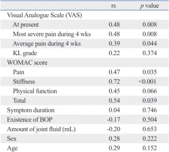
Table 4 shows the correlation between the painDETECT consists of subsections for pain, stiffness, and physical func-
tion. PainDETECT is a patient-based questionnaire consist- ing of seven weighted sensory descriptor items and two items relating to the spatial (radiating) and temporal charac- teristics of the patient’s pain pattern. The painDETECT score was distributed from 0 to 38. Patients were divided into three groups: neuropathic pain is likely (score ≥19), possible (score ≥13 to ≤18), and unlikely (score ≤12). VAS was evaluated at 3 time points. VAS at entry point, of se- verest pain during 4 weeks, and of average pain during 4 weeks were evaluated for all patients.
Radiographic evaluation
Anterior-posterior view of X-ray examination was per- formed in all patients. We used the Kellgren-Lawrence (KL) severity classification system, which is a validated method of classifying individual joints into one of five grades, with 0 representing normal and 4 being the most severe radio- graphic disease.17 Evaluation was blinded and performed by 5 observers. If at least 3 of the observers concurred, the grading score was used to define KL grade.
Evaluation of knee joint fluid
Ballottment of the patella (BOP) was examined for all pa- tients. If the BOP sign was positive, needle puncture into the knee was performed in all positive patients and the amount of joint fluid was evaluated.
Statistical Analysis
Data were compared using a Spearman’s correlation coeffi- cient by rank test. p<0.05 was considered statistically sig- nificant.
RESULTS
Table 1 shows demographic characteristics of patients.
Ninety-two patients (71 female, 21 male) with a mean age of 70.3 years were included in the study during the entire period. Average duration of symptoms was 37.5 months.
The patients suffered from knee pain originating from OA for at least 1 month. Pain at entry point using VAS was 5.5 and total WOMAC score was 57.1.
Table 2 shows the painDETECT score and indicates like- ly NP (score ≥19), possible NP (score ≥13 to ≤18), and un- likely NP (score ≤12). Within this study population, 5 (5.4%) were classified as likely NP, 14 (15.2%) as possible NP, and
Table 1. Demographic Characteristics
Number of patients 92
Sex Male: 21
Female: 71
Age (mean±SD yrs) (range) 70.3±8.0
(53-81) Symptom duration (mean±SD months)
(range) 37.5±8.0
(1-180) Pain scores
Visual Analogue Scale (VAS)
At entry point (mean±SD) 5.5±2.0 Severest pain during 4 wks
(mean±SD) 6.1±2.6
Average pain during 4 wks
(mean±SD) 5.1±2.1
WOMAC scores
Pain (mean±SD) 12.3±3.6
Stiffness (mean±SD) 4.7±2.3
Physical function (mean±SD) 40.3±11.4
Total (mean±SD) 57.1±15.3
WOMAC, western ontario and McMaster universities osteoarthritis index.
Table 2. PainDETECT Score
Score Number of patients (%)
(total; 92 patients)
0-12 73 (79.3)
13-18 14 (15.2)
19-38 5 (5.4)
Neuropathic pain is likely (score ≥19), possible (score ≥13 to ≤18), and unlikely (score ≤12).
Table 3. X-ray Evaluation, BOP, and Amount of Joint Fluid
KL grade Number of patients (%)
(total; 92 patients)
0 0
1 24 (26)
2 25 (27)
3 33 (36)
4 10 (11)
BOP Number of patients (%)
+ 30 (33)
- 62 (67)
Amount of joint fluid (mean ± SD mL) (range: mL) 9.3±2.5 (1-40) BOP, ballottment of the patella; KL, Kellgren-Lawrence.
Some authors have reported that joint pain arises mainly from free nerve endings that exist in the capsule or in the synovium,19-21 whereas others have reported innervation of the osteochondral junction in human knee OA samples and indicated a possible contribution of the subchondral area to OA knee pain.22,23 Regarding the mechanism of NP in knee OA, it is highly possible that NP occurs in association with damage to nerves innervating subchondral bone because of its weight-bearing surface in late stage OA. In early phases, synovitis produces joint fluid and stimulates free nerve end- ings in synovium. On the other hand, a decrease in the amount of joint fluid and destruction of the osteochondral junction may induce injury to the nerve endings in this area in late phases of OA. The current study showed the possi- bility of NP pathogenesis in OA pain; however, this was not clear because the correlations were not significant. There was a tendency for positive correlation between painDE- TECT score and the KL grade, and tendency for negative correlation between the painDETECT score and the amount of joint fluid. Further study using a larger population is probably needed to clarify this.
Another mechanism has been postulated for NP in OA.
Approximately 60-80% of OA patients achieve pain relief after local anesthetic treatment or surgical replacement of the affected joint, indicating peripheral mechanisms driving the pain, although central mechanisms are also thought to play a role in some patients and dysfunction of diffuse nox- ious inhibitory controls has been described.24-26 These cen- tral mechanisms may overlap with those in other pain states such as neuropathy.
There are several limitations of this study. The reliability of painDETECT for NP in knee OA has not been fully eval- uated, therefore, further study is needed to clarify this.
In conclusion, the PainDETECT scores for pain from knee OA revealed that 5.4% are likely NP, and 15.2% are possible NP. NP tended to be seen in patients with less joint fluid and a KL grade that correspond to late stages of OA. It is important to be aware of consider the existence of NP in the treatment of knee OA pain.
REFERENCES
1. Centers for Disease Control and Prevention. National Center for Chronic Disease Prevention and Health Promotion. Arthritis types:
overview. 2008. http://www.cdc.gov/arthritis/osteoarthritis.htm.
2. Daousi C, MacFarlane IA, Woodward A, Nurmikko TJ, Bundred PE, Benbow SJ. Chronic painful peripheral neuropathy in an ur-
score and each factor. A strongly significant correlation was seen with the WOMAC stiffness score (rs=0.72, p<0.001), and a moderately significant correlation was seen with the VAS scores (rs=0.39, p=0.044), WOMAC pain (rs=0.47, p=0.035) and total scores (rs=0.54, p=0.039). There was a tendency for positive correlation between the painDETECT score and KL grade (rs=0.22, p=0.374), and tendency for negative correlation between the painDETECT score, exis- tence of BOP (rs=-0.17, p=0.504), and amount of joint fluid (rs=-0.20, p=0.653). However, there was no significant posi- tive or negative correlation between painDETECT score, KL grade (p=0.374), existence of BOP (p=0.504), or amount of joint fluid (p=0.653).
There was no significant correlation between the pain- DETECT score and duration of symptoms (p=0.746), sex (p=0.22), or age (p=0.15).
DISCUSSION
OA has a high prevalence in the elderly population and is a major cause of disability within this group.18 Generally, the origin of OA pain has been considered to be nociceptive, but our study identified at least 5.4% of our knee OA patients as likely to have NP and 15.2% as possibly having NP. pain- DETECT scores were correlated with VAS and WOMAC assessments of pain severity. Therefore, some patients with severe pain and disability from the pain may have a NP com- ponent in their knee OA pain.
Table 4. PainDETECT and Correlation Coefficients
rs p value
Visual Analogue Scale (VAS)
At present 0.48 0.008
Most severe pain during 4 wks 0.48 0.008 Average pain during 4 wks 0.39 0.044
KL grade 0.22 0.374
WOMAC score
Pain 0.47 0.035
Stiffness 0.72 <0.001
Physical function 0.45 0.066
Total 0.54 0.039
Symptom duration 0.04 0.746
Existence of BOP -0.17 0.504
Amount of joint fluid (mL) -0.20 0.653
Sex 0.28 0.222
Age 0.29 0.152
WOMAC, western ontario and McMaster universities osteoarthritis index;
BOP, ballottment of the patella.
pain. Pain 2007;127:199-203.
15. Hochman JR, French MR, Bermingham SL, Hawker GA. The nerve of osteoarthritis pain. Arthritis Care Res (Hoboken) 2010;
62:1019-23.
16. Bellamy N, Buchanan WW, Goldsmith CH, Campbell J, Stitt LW.
Validation study of WOMAC: a health status instrument for mea- suring clinically important patient relevant outcomes to antirheu- matic drug therapy in patients with osteoarthritis of the hip or knee. J Rheumatol 1988;15:1833-40.
17. Kellgren JH, Lawrence JS. Radiological assessment of osteo-ar- throsis. Ann Rheum Dis 1957;16:494-502.
18. Mäntyselkä P, Kumpusalo E, Ahonen R, Kumpusalo A, Kauhanen J, Viinamäki H, et al. Pain as a reason to visit the doctor: a study in Finnish primary health care. Pain 2001;89:175-80.
19. Grönblad M, Korkala O, Liesi P, Karaharju E. Innervation of syno- vial membrane and meniscus. Acta Orthop Scand 1985;56:484-6.
20. Fortier LA, Nixon AJ. Distributional changes in substance P noci- ceptive fiber patterns in naturally osteoarthritic articulations. J Rheumatol 1997;24:524-30.
21. Saito T, Koshino T. Distribution of neuropeptides in synovium of the knee with osteoarthritis. Clin Orthop Relat Res 2000:172-82.
22. Suri S, Gill SE, Massena de Camin S, Wilson D, McWilliams DF, Walsh DA. Neurovascular invasion at the osteochondral junction and in osteophytes in osteoarthritis. Ann Rheum Dis 2007;66:
1423-8.
23. Ogino S, Sasho T, Nakagawa K, Suzuki M, Yamaguchi S, Higashi M, et al. Detection of pain-related molecules in the subchondral bone of osteoarthritic knees. Clin Rheumatol 2009;28:1395-402.
24. Creamer P, Hunt M, Dieppe P. Pain mechanisms in osteoarthritis of the knee: effect of intraarticular anesthetic. J Rheumatol 1996;23:1031-6.
25. Ethgen O, Bruyère O, Richy F, Dardennes C, Reginster JY.
Health-related quality of life in total hip and total knee arthroplas- ty. A qualitative and systematic review of the literature. J Bone Joint Surg Am 2004;86-A:963-74.
26. Kosek E, Ordeberg G. Lack of pressure pain modulation by het- erotopic noxious conditioning stimulation in patients with painful osteoarthritis before, but not following, surgical pain relief. Pain 2000;88:69-78.
ban community: a controlled comparison of people with and with- out diabetes. Diabet Med 2004;21:976-82.
3. Siddall PJ, McClelland JM, Rutkowski SB, Cousins MJ. A longi- tudinal study of the prevalence and characteristics of pain in the first 5 years following spinal cord injury. Pain 2003;103:249-57.
4. van der Kraan PM, Vitters EL, van de Putte LB, van den Berg WB. Development of osteoarthritic lesions in mice by “metabolic”
and “mechanical” alterations in the knee joints. Am J Pathol 1989;135:1001-14.
5. Ochiai N, Ohtori S, Sasho T, Nakagawa K, Takahashi K, Taka- hashi N, et al. Extracorporeal shock wave therapy improves motor dysfunction and pain originating from knee osteoarthritis in rats.
Osteoarthritis Cartilage 2007;15:1093-6.
6. Ivanavicius SP, Ball AD, Heapy CG, Westwood FR, Murray F, Read SJ. Structural pathology in a rodent model of osteoarthritis is associated with neuropathic pain: increased expression of ATF-3 and pharmacological characterisation. Pain 2007;128:272-82.
7. Fernihough J, Gentry C, Malcangio M, Fox A, Rediske J, Pellas T, et al. Pain related behaviour in two models of osteoarthritis in the rat knee. Pain 2004;112:83-93.
8. Pomonis JD, Boulet JM, Gottshall SL, Phillips S, Sellers R, Bun- ton T, et al. Development and pharmacological characterization of a rat model of osteoarthritis pain. Pain 2005;114:339-46.
9. Freynhagen R, Baron R, Gockel U, Tölle TR. painDETECT: a new screening questionnaire to identify neuropathic components in patients with back pain. Curr Med Res Opin 2006;22:1911-20.
10. Bennett M. The LANSS Pain Scale: the Leeds assessment of neu- ropathic symptoms and signs. Pain 2001;92:147-57.
11. Bouhassira D, Attal N, Alchaar H, Boureau F, Brochet B, Bruxelle J, et al. Comparison of pain syndromes associated with nervous or somatic lesions and development of a new neuropathic pain diag- nostic questionnaire (DN4). Pain 2005;114:29-36.
12. Portenoy R. Development and testing of a neuropathic pain screening questionnaire: ID Pain. Curr Med Res Opin 2006;22:
1555-65.
13. Krause SJ, Backonja MM. Development of a neuropathic pain questionnaire. Clin J Pain 2003;19:306-14.
14. Bennett MI, Attal N, Backonja MM, Baron R, Bouhassira D, Freynhagen R, et al. Using screening tools to identify neuropathic