Dracocephalum palmatum
Stephan 잎 헥산 분획 추출물의 인간 유래 전립선 암세포 사멸에 대한 작용이민지1#, 이세은1#, 최나리2, 조성현2, 조수인2*
1 : 부산대학교 한의학전문대학원 한의과학과, 2 : 부산대학교 한의학전문대학원 한의학과
Effects of hexane fraction of
Dracocephalum palmatum
Stephan leaf on human-derived prostate cancer cell deathMin Ji Lee
1#, Se-Eun Lee
1#, Na Ri Choi
2, Sung Hyeon Jo
2, Suin Cho
2*1 : Department of Korean Medical Science, School of Korean Medicine, Pusan National University, Yangsan, Gyeongnam, Republic of Korea
2 : Department of Korean Medicine, School of Korean Medicine, Pusan National University, Yangsan, Gyeongnam, Republic of Korea
ABSTRACT
Objectives :
Dracocephalum palmatumStephan (Lamiaceae) is a medicinal plant used by the East-Russian nomads but there were few studies on this plant. This study was to evaluate anti-cancer effects of
D. palmatumStephan leaf hexane fraction on human derived prostate cancer cell death.
Methods : The dried leaves of
D. palmatumwere dissolved in methanol, and hexane fraction (DpLH) was again obtained from lyophilized methanol extract (DpLM). DpLH was investigated by measuring by MTT assay and annexin V/PI staining to evaluate its effects on the cell viability and apoptosis of PC-3 cells. The ROS generations were detected by DCF-DA dye. The protein expressions were confirmed by p-AKT, Bcl-2, Bax, procaspase-3 activities.
Results : After treatment of DpLH to PC-3 cells, the cell proliferation was significantly inhibited, and in addition, DpLH treatment also accelerated apoptosis of PC-3 cells. When DpLH was treated to the PC-3 cells, its ROS production significantly decreased. The proportion of all proteins (p-AKT/actin, Bcl-2/Bax and procaspase-3/actin ratios) showed decreasing tendency of expression compared with the control group.
Conclusions : As shown in the above results, the extract from
D. palmatuminhibits ROS production and promotes cell death, which is considered to be a relatively safe induction of cell death when administered to a living body. In conclusion, these results suggested that DpLH may have anti-cancer effect in human prostate cancer cell.
1)
Key words :
Dracocephalum palmatumStephan, anti-cancer, prostate cancer cell, apoptosis.
Ⅰ. 서 론
2017년 자료에 의하면 미국에서 전립선 암세포는 암 관련
사망의 3번째 주요한 원인이 되고 있다
1). 전립선 암 치료에는 수술, 호르몬 요법 및 화학요법 등 다양한 방법이 전 세계적 으로 사용되고 있지만 심각한 부작용 때문에 한계가 있으며
2-5),
*Corresponding author : Suin Cho, Department of Korean Medicine, School of Korean Medicine, Pusan National University.
·Tel : +82-51-510-8457 ·E-mail : [email protected]
#First author: Min Ji Lee, Department of Korean Medical Science, School of Korean Medicine, Pusan National University.
·Tel : +82-10-7429-1052 ·E-mail : [email protected]
#Co-first author: Se-Eun Lee, Department of Korean Medical Science, School of Korean Medicine, Pusan National University.
·Tel : +82-10-2462-7299 ·E-mail : [email protected]
·Received : 01 June 2018 ·Revised : 25 June 2018 ·Accepted : 25 July 2018
이러한 이유로 인해 효과적이고 비교적 안전한 화학 치료제 또는 천연물에서 유래한 치료용 의약품들의 개발이 필요하다.
Dracocephalum palmatum
Stephan은 꿀풀과 용머리속에 속하는 식물로 우리나라에서는 자생하지 않으나 중국 북동부와 러시아 등지에서 볼 수 있는데, 우리나라에 자생하는 식물 중 분류학상 가까운 식물로 용머리가 있으며 같은 용머리속에 속 하고 Fisch. ex Link라는 학명을 가지고 있다. 용머리는 생 약명으로는 광악청란 (光萼靑蘭)이라고 불리며, 성미는 苦, 寒 하며 消炎, 鎭痛 등의 효과가 있고 한국, 일본, 중국 동북부 또는 시베리아 동부에 분포하며 산 숲에서 자란다
6).
산소 (oxygen)는 몸의 대사 과정에서 필요하지만 체내에서 발생하는 산화적 스트레스 (oxidative stress)와 활성산소 (reactive oxygen species; ROS)는 병의 진행을 가속화시키 기도 한다
7,8). 활성산소는 몸 속에 남아 세포의 산화를 촉진시 켜 암 세포 성장과 노화 및 각종 질병에 관여한다
9-12).
본 연구에서는 시베리아 지역에서 직접 채취하고 기원을 현지 학자로부터 동정 받은 위 식물 잎의 메탄올 추출물을 얻 었으며, 이의 용매 분획 중 헥산 분획이 인간 유래 전립선 암 세포의 사멸에 어떠한 작용을 미치는지 확인하여 그 결과를 보고하는 바이다.
Ⅱ. 재료 및 방법
1. 재료
1) 용매 및 시약메탄올은 (Korea)에서 Dimethyl sulfoxide (DMSO)은 Bio Basic InC. (Canada)에서 Tris는 Duchefa Biochemie (Netherlands)에서 구입하였다. Thiazolyl blue tetrazolium bromide (MTT) 와 2',7'-dichlorodihydro-fluorescein diacetate (DCFH-DA)는 Sigma-Aldrich Co. (USA)에서 구입하였다.
실험에 사용한 Dulbecco’ s Modified Eagle’ s Medium (DMEM)과 fetal bovine serum (FBS)은 Corning Inc, (USA)에서 구입하였고, Penicillin-streptomycin (P/S)은 Gibco (USA)에서 구입하였다. 단백질 정량에 사용된 BCA reagent와 albumin standard는 Thermo scientific (USA) 에서 구입하였다.
Primary antibodies (일차 항체)로 Bcl-2, Bax와 procaspase-3는 Cell Signaling 제품을 사용하였고, p-AKT와 β -actin는 Santa Cruz 제품을 사용하였다.
Secondary antibody (이차 항체)로 goat anti-rabbit IgG, pAb와 goat anti-mouse IgG, pAb는 Enzo life sciences Inc. (USA) 제품을 사용하였다. West-Q chemiluminescent substrate 은 GenDEPOT (USA)을 사용하였다.
2) PC-3 암세포
인간 유래 전립선 암인 PC-3는 ATCC (American Type Culture Collection, Tockville, MD)에서 분양 받아 본 실험에 사용하였다.
2. 방법
1) 약물의 제조본 연구에 사용된 약물인
D. palmatumS.은 러시아 북동 부에 소재한 북동연방대학교의 식물생태학 전공 교수가 채집 및 기원을 동정한 후 멸균건조한 시료의 상태로 제공 받았다.
이 중
D. palmatumS.의 건조된 잎 (시료 보관 번호; 16- DPL-42) 을 따로 선별하여 무수메탄올에 넣어 2일 동안 잘 섞어 추출한 다음 2번 메탄올 추출물을 여과시켰다. 이 후, EYELA 1L rotary evaporator (TOKYO RIKAKIKAI CO.
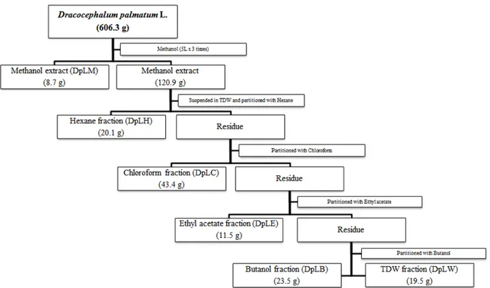
LTD)를 통하여 메탄올 추출물 (추출물 번호; 16EX-DPL- 03)만을 얻어, freeze dryer (Ilshin Bio Base Co., Ltd)를 사용하여 동결건조 분말을 얻었다. 앞으로의 후속 연구에서의 메탄올 추출물과 분획물의 비교를 위해 메탄올 추출물을 소량 보관 후, 나머지를 증류수에 현탁하여 같은 양의 헥산, 클로 로포름, 에틸 아세테이트, 부탄올 순으로 5가지 분획물을 만들 었으며 (Fig. 1), 본 연구에서는 헥산 분획 (DpLH)만을 실험에 사용하였다.
2) PC-3 암세포 배양
PC-3세포는 10% heat-inactivated fetal bovine serum and 1% penicillin-streptomycin이 포함된 DMEM media 에서 배양되었으며, 세포들은 배양기 온도가 37℃ 이며, 5%
이산화탄소의 조건하에서 배양되었다.
3) HPLC 분석
메탄올 추출물과 DpLH 구성 성분 패턴의확인은 HPLC (YL 9100 HPLC system)을 사용하였다. 사용한 분석기기는 HPLC vacuum degasser, quaternary pump, UV/VIS detector 과 manual injection로 구성되어 있으며, data는 Autochro-3000 프로그램을 사용하여 분석하였다 (Table 1.)
4) MTT assay
세포 생존율은 MTT assay로 측정되었다. 48-well plate에 5×10
3cells/well으로 하루 동안 배양 시킨 후, 다음날 약물을 농도 별로 처리하였다. 24시간 후 MTT solution (0.5 ㎎/㎖)을 2 시간 처리하여 formazan crystal을 확인하였다. 배지를 제거 한 후, formazan crystal을 DMSO에 용해시켜 570 ㎚의 파장 에서 확인하였다.
5) 세포자멸 확인
세포자멸 확인은 Annexin V-PI double staining을 통하여
확인하였다. 세포를 1×10
6cells/well으로 하루 동안 배양시
킨 후, 다음날 약물을 농도 별로 처리하였다. 24시간 후 세포를
0.05% trypsin으로 모아 1x PBS로 3번 washing 한 뒤, 1x
binding buffer (500 ㎕)으로 세포를 풀어준 뒤 염색을 암실
에서 15분간 염색 후 상온에서 반응시킨 다. 이 후 세포자멸
확인을 위해 10,000개의 세포를 flow cytometry (BD
FACSCanto Ⅱ, USA)로 분석을 진행하였다.
6) 세포 내 활성산소 측정
세포 내 활성산소의 발생량은 DCF-DA fluorescent dye를 사용하여 확인하였다. DCF-DA fluorescent dye는 H
2DCF- DA의 형태로 세포에 처리되며, 세포막을 통과한 이후 H
2DCF 의 형태까지는 형광을 띠지 않으나 이후 H
2O
2등과 반응하여 DCF의 구조를 가지면서 형광을 띠는데, 이 정도가 ROS 발생 량으로 간주된다
9-12). 따라서 본 실험에서는 세포를 1×10
6cells/well으로 하루 동안 배양시킨 후, 다음날 약물을 농도 별로 처리하였다. 24시간 후 DCF-DA를 최종농도 5 μM로 맞춰 30분 반응 시킨 뒤, 세포를 1x PBS로 3번 washing하여 flow cytometry (BD FACS Canto Ⅱ, USA)로 분석하였다.
7) 단백질 분석
세포 자멸의 단백질 분석은 western blot을 통하여 분석되 었다. 세포를 1×10
6cells/well으로 하루 동안 배양시킨 후, 다음날 약물을 농도 별로 처리하였다. 세포를 모아 Pro-prep (iNtRON, Gyeonggi-do, Korea)으로 단백질을 추출해냈으며, BCA 방법으로 단백질을 정량 하였다. 각각의 단백질 (30 ㎍)은 sodium dodecyl sulfate polyacrylamide gel (SDS gel) electrophoresis (SDS-PAGE)를 통해 분리한 후 PVDF (Millipore, Darmstadt, Germany) membrane으로 transfer 하였다. 이 membrane은 1시간 동안 5% skim milk에서 blocking 하였다. 그 후, 하루 동안 4℃ 저온실에서 1차 항체인 Bcl-2 (1:2000), Bax (1:2000), and β-actin (1:1000)을 반응시켰다. 다음날 membrane을 1x TBST에서 3번 씻어낸 뒤, 2차 항체인 horse radish peroxidase (HRP) conjugated
goat anti-rabbit IgG, pAb (1:5000)와 HRP conjugated goat anti-mouse IgG pAb (1:3000)를 두 시간 동안 상온 에서 반응시켰다. 다시 1x TBST에서 3번 씻어낸 뒤, membrane을 ECL solution (GenDEPOT, Hous-ton, TX,.
USA)에 반응시켜 LAS (photosensitive luminescent analyzer system; Amersham
™Imager 600, UK)를 이용 하여 결과를 확인하였다. 또한 image J software program (NIH)를 이용하여 분리된 단백질을 정량하였다.
8) 통계처리
본 연구에서 수행되어 얻어진 모든 데이터는 3회 반복한 실험을 평균 ± 표준편차로 (means ± S.D.) 나타내었다. 각 그룹 사이의 통계적 유의성은 Kruskall-Wallis 분석을 통해 확인한 후 Dunnette 의 분석 방법을 사용해 사후 검정하였다.
이러한 분석에 사용된 프로그램은 SIGMAPLOT 12.0 version 이며,
P값이 5% 미만일 경우 유의성이 있는 것으로 판정하 였다.
Ⅲ. 결 과
1. 추출물 및 분획물의 용량
건조 생약 형태 시료의 최초 건조 중량은 총 606.3 g이었고, 메탄올 추출물 (DpLM)은 총 129.6 g으로 수율은 21.38 %였다.
그 중 8.7 g은 따로 보관하였고, 나머지 120.9 g은 분획 획득 용도로 사용하였다. DpLH는 20.1 g이었다 (Fig. 1).
Fig. 1. Fractionation scheme of Dracocephalum palmatum Stephan leaf extracts.
2. DpLM과 DpLH 추출 패턴 비교
DpLH가 DpLM에서 분리가 잘 되었는지의 여부는 HPLC를 이용하여 확인하였다. 메탄올 추출물의 조건 (Table 1)은 용 머리 속에 속한 다른 종을 사용한 논문을 참고하였다
13). 결과는 약 14분과 18분 사이에서 DpLM과 DpLH의 성분이 비슷한 조건에서 나온 것을 확인할 수 있었다 (Fig. 2).
Description Condition
Mobile phase Acetonitrile (A) : 0.1% acetic acid (B)
Gradient elution 0 min, A : 10%; B : 90%
20 min, A : 100%; B : 0%
Flow rate 1.0 ㎖/min
Inject vol. 10 ㎕
Guard column 5㎛ Eclipse XDB-C18 column, 4.6×250 ㎜
Column temp. RT
UV wavelength 254 ㎚
Table 1. HPLC condition for analysis of Dracocephalum palmatum Stephan leaf methanol extract and hexane fraction
Fig. 2. HPLC/UV chromatograms from the methanol extract and hexane fraction of D. palmatum leaf. A, DpLM; B, DpLH.
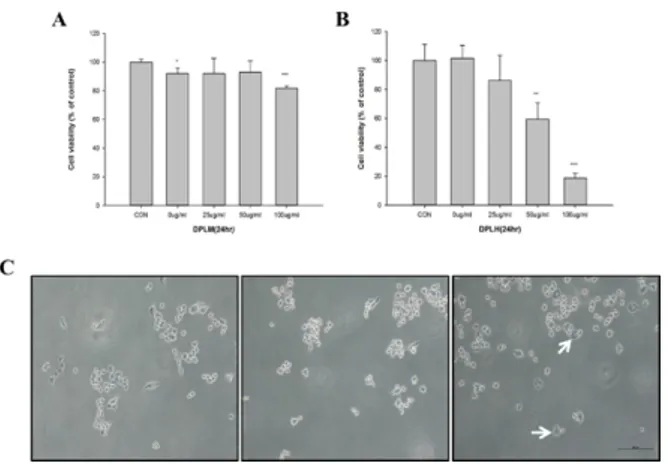
3. DpLH의 암세포 증식 억제 효과
PC-3 세포에서 DpLM과 DpLH 처리에 의한 암세포 증식 억제를 조사하기 위하여 각각의 시료들을 25 ㎍/㎖, 50 ㎍/㎖, 100 ㎍/㎖의 농도로 24 시간 처리하였다. DpLM은 100 ㎍/㎖
의 농도에서 암세포 증식 억제가 나타나기 시작하였고 20 ± 1.52%의 암세포 억제를 확인하였다. 한편, DpLH은 25 ㎍/㎖
의 농도에서 암세포 억제가 나타나기 시작하여 DpLH는 50
㎍/㎖에서 40 ± 11.25 %, 100 ㎍/㎖에서 80 ± 3.27 %의 결과를 얻었으며 유의적으로 암세포 증식 억제를 확인되었다.
DpLM과 DpLH를 같은 농도로 24시간 처리 후 비교 하였을 때 DpLH를 처리한 세포의 증식이 더욱 억제되었다 (**
p< 0.01,
***
p< 0.001; Fig. 3A-B).
또한 DpLH를 처리한 후 세포의 형태 변화에서는 비교군과 0 ㎍/㎖군은 세포 모양의 차이가 거의 나타나지 않았으며, 100
㎍/㎖에서는 세포 자멸에서 관찰되는 세포 소기관과 세포질의 분적과 같은 특징적인 형태의 변화를 관찰할 수 있었다 (Fig. 3C).
Fig. 3. Effects of DpLM and DpLH on cell viability of human prostate cancer cells in vitro. PC-3 cells were attached 48-well plate, and added DpLM (A) and DpLH (B) as indicated various concentrations (0, 50, and 100 ㎍/㎖, respectively) of DpLH (C). After 24 hr incubation, cell viability was measured using MTT methods. White arrows indicate cellular changes to cytoplasmic apoptotic body.
The data presented as means ± S.D. of three independent experiments. **p < 0.01 vs. Control; ***p < 00.001 vs. Control group.
4. DpLH의 암세포 자멸 효과
DpLH를 처리로 인한 PC-3 세포사의 형태를 알아보기 위 하여 annexin V/PI double stain을 시행하였다. DpLH의 농 도가 높아질수록 apoptosis가 유도됨을 확인 할 수 있었다 (Fig. 4A). 암세포는 25 ㎍/㎖에서 13.80 ± 1.15, 50 ㎍/㎖
에서 39.60 ± 0.36 그리고 100 μg/ml에서 52.23 ± 0.45%로 나타나 유의한 변화가 관찰되었다 (***
p< 0..001;
Fig. 4B).
Fig. 4. Effects of DpLH on apoptosis in PC-3 cells. PC-3 cells were treated with various concentrations of DpLH (0-100 ㎍/㎖) for 24 hr. A, the proportions of cell death by DpLH were determined using Annexin V and PI (Propidium Iodide) double staining analysis. (A), Control; (B), 0; (C), 25; (D), 50; (E), 100 ㎍/㎖ of DpLH treated. B, the percentage of the cell located areas were shown. The data presented as means ± S.D. of three independent experiments. ***p < 0.001 vs. Control group.
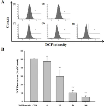
5. DpLH 처리에 의한 활성산소의 발생
DpLH가 PC-3 암세포의 ROS에 미치는 영향을 조사하기 위하여 DpLH를 처리하였다. 그 결과 DpLH를 처리한 암세포를 비교군과 비교하였을 때 그래프가 왼쪽으로 이동하는 것을 확인 할 수 있었고 (Fig. 5A), 25 ㎍/㎖에서 30 ± 7.77 %, 50 ㎍ /㎖에서 10 ± 2.84 % 그리고 100 ㎍/㎖에서 5 ± 1.44 %로 ROS가 현저히 감소하는 것을 확인할 수 있었다 (**
p< 0.01,
***
p< 0.001; Fig. 5B).
Fig. 5. Effects of DpLH on intracellular ROS generation in PC-3 cells. PC-3 cells were treated with various concentrations of DpLH (0-100 ㎍/㎖). A, the proportions of intracellular ROS generation by DpLH were determined using DCF-DA dye. (A), Control; (B), 0; (C), 25; (D), 50; (E), 100 ㎍/㎖ of DpLH treated. B, the percentage of the cell located areas were shown. The data presented as means ± S.D. of three independent experiments.
**p < 0.01 vs. Control group. ***p < 0.001 vs. Control group.
6. DpLH 처리에 의한 단백질 발현 변화
DpLH 처리에 의한 세포 사멸에서 단백질의 발현의 변화를 확인하기 위하여 western blot을 실시하였다. AKT는 암세포의 생존에 관여하며, Fig. 6A와 같이 p-AKT 의 발현은 감소하 였다. 또한 p-AKT/actin 비는 CON 대비 100 ㎍/㎖에서 0.3 ± 0.2 배로 유의하게 감소하는 것을 알 수 있었다 (**
p<
0.01; Fig. 6B).
Procaspase-3/actin의 발현을 살펴보았으며, Fig. 8A에 나타난 것과 같이 procaspase-3는 감소하였다. 또한 procaspase-3/actin 비는 CON 대비 100 ㎍/㎖에서 0.47
± 0.02 배로 유의하게 감소하는 것을 확인 할 수 있었다 (*
p<
0.05; Fig. 8B).
세포 사멸의 내인성 경로에 작동하는 단백질인 Bcl-2와 Bax의 발현을 살펴보았으며, Fig. 7A에 나타난 것과 같이 Bcl-2는 감소하였고 Bax의 발현은 증가하였다. 또한 Bcl-2/
Bax 비는 CON 대비 100 ㎍/㎖에서 유의하게 0.61 ± 0.21 배로 감소되었다 (*
p< 0.05; Fig. 7B).
Fig. 6. Expression of DpLH on p-AKT/actin ratio in PC-3 cells.
PC-3 cells were treated with various concentrations of DpLH (0-100 ㎍/㎖). A, the p-AKT/actin proteins in PC-3 cells cultured in 0, 25, 50 and 100 ㎍/㎖ of DpLH were detected by western blot analysis. (B) Quantification of p-AKT/actin protein expression ratio. The data presented as means ± S.D. of three independent experiments. **p < 0.01 vs. control.
Fig. 7. Expression of DpLH on Bcl-2/Bax ratio in PC-3 cells.
PC-3 cells were treated with various concentrations of DpLH (0-100 ㎍/㎖). A, the Bcl-2, Bax proteins in PC-3 cells cultured in 0, 25, 50 and 100 ㎍/㎖ of DpLH were detected by western blot analysis. (B) Quantification of Bcl-2/Bax protein expression ratio. The data presented as means ± S.D. of three independent experiments. *p < 0.05 vs. control.
Fig. 8. Expression of DpLH on Pro caspase-3/actin ratio in PC-3 cells. PC-3 cells were treated with various concentrations of DpLH (0-100 ㎍/㎖). A, the Pro caspase-3/actin proteins in PC-3 cells cultured in 0, 25, 50 and 100 ㎍/㎖ of DpLH were detected by western blot analysis. (B) Quantification of Pro caspase-3/actin protein expression ratio. The data presented as means ± S.D. of three independent experiments. *p < 0.05 vs.
control.
Ⅳ. 고 찰
암은 단일 세포의 유전자 돌연변이로부터 발생한다. 암세 포가 증식함에 따라 유전자 변형과 세포 사를 통하여 암세포 성장 억제 및 종양 억제를 유도한다
14). 세포의 발달, 성장, 생 존에 있어 필수적인 세포사는 괴사 (necrosis), 자가포식 (autophagy)과 세포자멸 (apoptosis)으로 크게 3가지로 나눌 수 있다
15). 첫 번째로 괴사 (necrosis)는 세포질 팽창에 의한 세포 소기관 및 원형질 막 파괴를 특징을 가지고 있으며 세포 내 성분을 방출시켜 염증을 일으킨다
16). 두 번째로 자가포식 (autophagy)은 손상된 세포 소기관 또는 고분자 구성 요소들 을 재활용하는 과정을 통하여 종양 성장 억제, 세포 내 미생 물의 제거 및 항원 제시 역할을 한다
17). 마지막으로 세포자멸 (apoptosis)은 프로그램화된 세포 죽음 (programed cell death)이라고 한다. 이것이 세포 내에 발생 하였을 때 나타나는 현상으로는 핵이 응축되고, 세포 부피가 감소하며, DNA분절 이 일어난다
18). 결론적으로 세포자멸은 외부적인 자극에 의한 손상이나 노화된 세포들이 세포자살을 결정하는 것을 의미한
다
15,19). 여러 연구자들에 의한 선행연구로 용머리속에 기원한
식물 자원들에 대한 연구는 계속되어 왔으며 주요 암세포들로 는 간, 혈액, 유방, 선암 등으로부터 유래된 암세포주들을 사용 하였다
20-22).
그러나 본 연구에 사용된
D. palmatumStephan은 그 자 생지가 중국의 동북부와 러시아 툰드라 지역에 제한적인 이유 로 인해 충분한 연구가 수행되지 못하였다.
따라서 본 연구팀은 위 시료 일부를 획득하여 DpLM이 항암 활성이 있음을 예비연구를 통해 확인하였으며, 본 연구에서는
전립선 암 세포주에 대해 세포사멸 및 자멸에 어떠한 영향을 미치는지를 관찰한 결과 DpLH를 24시간 처리하였을 때 apoptosis 가 유도됨을 알 수 있었다 (Fig. 3-4).
ROS는 세포의 산화, 대사 과정에서 생기는 유해한 산소로, 세포 손상과 생리적 기능 장애를 일으켜 DNA로부터 조직까 지 파괴 할 수도 있다
23-25). 따라서 DpLH의 처리가 세포 내 ROS의 양을 감소시킴으로써 암세포가 산화적인 스트레스를 받는 것을 확인하였다 (Fig. 5).
AKT는 PI3K의 주요 하위 신호로 세포 주기, 분화 및 생존에 관여하고, PDK1과 PDK2를 통화여 인산화되어 암세포의 진행 과정에 중심적인 역할을 담당하며, AKT1, AKT2, AKT3로 나뉜다. AKT1은 세포자멸 과정을 억제하여 세포의 성장을 일으키고, AKT2는 인슐린 신호 전달에 관여하며 AKT3는 불 분명하지만 뇌와 같은 신경 조직에서 많이 발견되는 것으로 보인다
26). 활성화 된 AKT는 미토콘드리아의 cytochrome C의 방출을 막아 apoptosis를 억제한다
27).
Apoptosis를 유발하는 경로는 크게 외인성 경로 (extrinsic pathway)와 내인성 경로 (intrinsic pathway)로 나뉜다. 그 중 내인성 경로에 Bcl-2, Bax 단백질이 속해있으며, 미토콘드 리아 경로 또는 스트레스 경로라고도 불린다
28).
세포 자멸이 일어날 때 pro-apoptotic protein인 Bax는 증가하고 anti-apoptotic protein인 Bcl-2의 발현이 감소한다.
이 때 단백질들이 활성화되어 cytochrome C가 세포 기질로 방출되며 이어서 procaspase-9과 procaspase-3를 활성화 시키기도 한다. 여기서 caspase는 세포 사멸의 과정에 관여 할 뿐만 아니라 DNA를 절단하는 역할을 한다
29).
본 실험에서는 DpLH의 처리가 p-AKT를 억제하여 세포 사에 관여함을 알 수 있었고 (Fig. 6), Bcl-2가 감소하고 Bax가 증가하며 (Fig. 7), procaspase-3 가 감소하는 것을 확인 할 수 있었다 (Fig. 8).
앞으로 저자들은 국내에 자생하는 용머리 추출물도 획득하여 DpLM 및 DpLH 등의 항암 활성을 비교 연구할 계획이며 동 일한 속에 속한 유사 식물들 간의 활성의 차이 유무 탐색을 통해 대체 한약 자원의 개발 가능성도 함께 탐구해보고자 한다.
Ⅴ. 결 론
여러 논문에서 이미 용머리속에 속한 식물들에 대한 항암 효과가 보고되어 있지만, 본 연구에서는 전립선암 세포주와
D. palmatumStephan의 건조된 잎의 메탄올 추출물을 사용 하였는데, 이러한 연구는 최초로 시도되었다. DpLH은 뚜렷한 세포자멸 효과를 보였으며 향후 유기 용매인 헥산 분획물이라는 단점을 최소화 시키고 임상에 적용 가능한 추출 방법들의 개발 등 해결점을 찾는다면 앞으로 항암 연구에 기대해볼 만한 자원 식물이 될 것으로 생각된다.
감사의 글
이 논문은 부산대학교 기본연구지원사업(2년)에 의하여 연
구되었음.
References