이 논문은 2012년 6월 22일 접수하여 2012년 8월 23일 채택되었음.
책임저자:김영범, 고대구로병원 방사선종양학과 Tel: 02)2626-1387, Fax: 02)2626-1399 E-mail: [email protected]
Purpose: The respiration is one of the most important factors in respiratory gating radiation therapy (RGRT). We have developed an unique respiratory guidance system using an audio-visual system in order to support and stabilize individual patient’s respiration and evaluated the usefulness of this system.
Materials and Methods: Seven patients received the RGRT at our clinic from June 2011 to April 2012. After breathing exercise with the audio-visual system, we measured their spontaneous respiration and their respira- tion with the audio-visual system respectively. With the measured data, we yielded standard deviations by the superficial contents of respiratory cycles and functions, and analyzed them to examine changes in their breathing before and after the therapy.
Results: The PTP (peak to peak) of the standard deviations of the free breathing, the audio guidance system, and the respiratory guidance system were 0.343, 0.148, and 0.078 respectively. The respiratory cycles were 0.645, 0.345, and 0.171 respectively and the superficial contents of the respiratory functions were 2.591, 1.008, and 0.877 respectively. The average values of the differences in the standard deviations among the whole patients at the CT room and therapy room were 0.425 for the PTP, 1.566 for the respiratory cycles, and 3.671 for the respiratory superficial contents. As for the standard deviations before and after the application of the PTP respiratory guidance system, that of the PTP was 0.265, that of the respiratory cycles was 0.474, and that of the respiratory superficial contents. The results of t-test of the values before and after free breathing and the audio-visual guidance system showed that the P-value of the PTP was 0.035, that of the cycles 0.009, and that of the respiratory superficial contents 0.010.
Conclusion: The respiratory control could be one of the most important factors in the RGRT which determines the success or failure of a treatment. We were able to get more stable breathing with the audio-visual respi- ratory guidance system than free breathing or breathing with auditory guidance alone. In particular, the above system was excellent at the reproduction of respiratory cycles in care units. Such a system enables to reduce time due to unstable breathing and to perform more precise and detailed treatment.
Key words: free breathing, respiratory guidance, reproducibility
INTRODUCTION
The movement of tumors and surrounding organs due to patients’ breathing and the involuntary movement of organs delivers an unintended dose of radiation to the target ab- sorbed dose and surrounding normal organs as it has influence on the distribution of radiation dose in radiation treatment.1) This can bring results other than the effects of radiation
treatment intended in the process of planning.2) Such a phe- nomenon mainly occurs with tumors in the chest and abdo- men, and the movement is prominent with breathing.3-5) Thus, much research has been devoted to the movement of chest and abdomen tumors with respect to breathing.6-8)
The respiratory gating radiation therapy1) in consideration of patients' breathing in radiation treatment is done in meth- ods in which patients’ breathing is adjusted to the purpose,9,10) in which irradiation is done in a certain respiratory cycle,11) or in which an established target is tracked for treatment.12) In order to apply such a series of methods, we should be able to directly observe the movement of tumors or surrounding
Fig. 1. Flow of RGRT in Korea University Guro Hospital.
organs or indirectly obtain related signals.11,13) This is directly related with the movement of tumors or surrounding organs, and methods for attaining respiratory signals with prominent relevance are mainly used. We can measure respiratory signals in methods in which a spirometer is used,14) in which a belt system is used,15) or in which infrared cameras and reflectors are used (Real-time Position Management System).16)
In case that the respiratory gating radiation therapy based on respiratory signals is performed, we can get results con- sistent with a radiation treatment plan when respiratory cy- cles, respiratory volumes, and respiratory strengths are regu- lar, and unexpected treatment results can be brought when regular breathing does not occur.17) Thus, it is important in the respiratory gating radiation therapy to maintain regular and stable breathing. Our institution applies the respiratory gating radiation therapy to those patients who are determined to be appropriate to that therapy after evaluating the possi- bility of respiratory control via respiratory training before treatment and find out respiratory cycles appropriate to them (Fig. 1). This study took as its subjects 7 patents who under- went the respiratory gating radiation therapy from June 2011 to April 2012.
Developing our own respiratory guidance system for main- taining stable and regular breathing among patients in the course from the CT scan to radiation treatment, this study has compared free breathing and audio-based breathing with voice files, and measured and quantitatively analyzed respiratory signals via the comparison with our own developed voice and image based respiratory system. In doing this, we would like to evaluate the usefulness of our institution’s own developed respiratory guidance system in the course of the respiratory gating radiation therapy.
MATERIALS AND METHODS
1. Respiratory Signal Obtaining System
The respiratory signal obtaining system used in this study consists of the Ream-Time Position Management System (RPM, Varian) using infrared cameras and reflectors and the inter- face (RPM Computer, Varian) showing signals real time. As seen in Fig. 2, the system puts the infrared reflectors on a certain abdomen site and then visualizes time and position information. The CT simulator used in the experiment is BrightSpeed Elite (GE) and the linear accelerator is Trilogy
Fig. 3. Visual and auditory guidance system. (A) Used LCD monitor, (B) Goggle.
Fig. 2. CT simulation of RGRT patient.
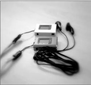
Fig. 4. MP3 player for auditory guidance system.
nute), and those respiratory files appropriate to patients are selected via preliminary respiratory training for them. The visual system consists of three parts. The first part is a 17”
LCD monitor (Multisync LCD 1770NX, NEC) put in a cas- sette holder for those who have bad vision or are abhorrent
one can apply one of them in accordance with patients’
characteristics.
3. Analysis of Results from the Application of the Respiratory Guidance System
This study took as its subjects 7 patients who received the respiratory gating radiation therapy from June 2011 to April 2012 in order to examine the inspiratory with the use of our own developed respiratory guidance equipment. Of the sub- jects, 4 had lung cancer and 3 liver cancer.
In order to compare respiratory data from each system, we respectively obtained data from free breathing without any application of the systems, from the application of the audi- tory guidance system, and from the simultaneous visual and auditory system of respiratory stability developed by our in- stitution. For the purpose of the evaluation of the usefulness of the respiratory guidance system in the course of treatment, we obtained data by measuring respiratory signals with or without the application of the respiratory guidance system for 5 minutes a day over a 5 day period 3days after the attain-
Fig. 5. Three kinds of visual guid- nace system. (A) LCD monitor, (B) Goggle, (C) LED monitor mounted monitor arm.
Table 1. Respiratory rate per minute during free breathing Patient Diagnosis Sex/age Breath per min.
pt.1 Lung ca. M/59 12/min
pt.2 Liver mets F/66 15/min
pt.3 HCC M/65 18/min
pt.4 Lung ca. M/30 19/min
pt.5 Lung ca. M/69 16/min
pt.6 HCC M/51 10/min
pt.7 Lung ca. M/53 16/min
Fig. 6. The standard deviation of PTP during free breathing.
ment of the signals.
By means of the subjects’ respiratory data obtained in this way, we yielded the PTP (peak to peak) of each respiratory cycle, and the superficial contents of the respiratory cycles and functions. Then, we yielded the standard deviations of these items and examined the changes in respiratory cycles and the similarities among respiratory volumes. Also, we eval- uated the usefulness of the respiratory guidance system via t-test of the standard deviations.
RESULTS
The respiration rates per minute with which the 7 patients undergoing the respiratory gating radiation therapy from June 2011 to April 2012 felt comfortable diversely ranged from 10 to 19 times per minute (Table 1). In the measurement of the free breathing, and of the respiratory cycles using the audi- tory and audio-visual guidance system with audio files, we measured the free breathing without the guidance of auditory or visual senses when the patients felt most comfortable. The standard deviations of the PTP values of the respiratory cy-
cles measured in free breathing turned out to be 0.070 mini- mally and 0.827 maximally (Fig. 6), while those of the respi- ratory cycles ranged from 0.242 to 1.164 (Fig. 7). Also, the standard deviations of the free breathing based on the changes in the superficial contents of the respiratory functions turned out to be 0.497 minimally and 7.204 maximally.
The standard deviation of the PTP in the whole patients was 0.343 in the free breathing and 0.148 in the application of the auditory guidance system, but it reduced to 0.078 in the application of the our own developed audio-visual guid- ance system. Similar to the case of the PTP, the standard de- viations of the respiratory cycles gradually reduced from 0.645 in the free breathing, 0.345 in the auditory guidance system, and 0.171 in the audio-visual guidance system. As the stand- ard deviations due to the changes in the superficial contents of the respiratory functions were 2.591, 1.008, 0.877 respec- tively, we can see that the values reduced by 66% compared
Fig. 8. The standard deviation of patients with irregular breathing.
Fig. 7. The standard deviation of breathing cycle.
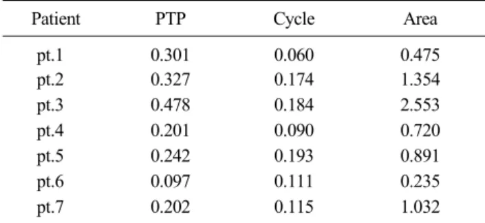
Table 4. The difference of standard deviation of the system before and after during free breathing
Patient PTP Cycle Area
pt.1 0.301 0.060 0.475
pt.2 0.327 0.174 1.354
pt.3 0.478 0.184 2.553
pt.4 0.201 0.090 0.720
pt.5 0.242 0.193 0.891
pt.6 0.097 0.111 0.235
pt.7 0.202 0.115 1.032
Table 3. The standard deviation of the difference between CT simulation ronn and treatment room
Patient PTP Cycle Area
pt.1 0.531 5.052 9.579
pt.2 0.538 2.494 8.152
pt.3 0.997 5.958 9.452
pt.4 0.632 3.504 4.458
pt.5 0.889 3.641 10.204
pt.6 0.224 0.165 0.612
pt.7 0.286 0.179 1.681
Table 2. The standard deviation according to the type of brea- thing system
Free breathing Audio-guidance Audio-visual feedback
PTP 0.343 0.148 0.078
Cycle 0.645 0.345 0.171
Area 2.590 1.001 0.877
the PTP, cycles, and superficial contents reduced from 0.827, 0.974, and 1.608 to 0.072, 0.179, and 0.429 in patient 1 while they changed from 0.210, 1.164, and 1.925 to 0.080, 0.187, and 0.507.
Also, in order to investigate the reproducibility of breathing in actual care units before the application of the audio-visual guidance system, we measured the averages of the PTP of free breathing, respiratory cycles, and the changes in the su- perficial contents of respiratory functions in the CT room and
contents of the respiratory functions (Table 4).
The results of t-test of the values of before and after the free breathing and the application of the audio-visual guid- ance system showed that the p-values were 0.035, 0.009, and 0.010 for the PTP, cycles, and respiratory function superficial contents respectively.
DISCUSSION AND CONCLUSION
As patients’ respiration causes the movement of tumor tis- sues and surrounding tissues, and changes the positions of tu- mors and target organs in radiation treatment, we cannot get the planned radiation distribution. Due to such a problem, there are gradually increasing treatment methods that mini- mize the influence of respiration by intentionally adjusting patients’ respiration or applying methods like the one in which irradiation is done only in a certain cycle consistently with the respiratory cycle. With the respiratory gating radiation ther- apy performed in our institution based on respiratory signals, we can get results consistent with the radiation treatment plan when the respiratory cycles, respiratory volumes, and respira- tory strengths are regular, and it tends to increase. However, an unexpected result can be caused when regular respiration is not achieved in the course of the respiratory gating radia- tion therapy. Thus, it is important to maintain regular and stable respiration in the respiratory gating radiation therapy.
In the existing respiratory training system, the concept of res- piration cycle is educated before the plan of treatment, and free breathing is performed on the basis of this education in actual treatment. It is not easy with such a system to monitor whether the actually educated respiration is identically repro- duced in care units and the reproducibility can be insufficient.
Thus, our institution’s own developed audio-visual respiratory guidance system is designed to find out a respiratory cycle appropriate to a patient via patient education before a treat- ment plan, and on the basis of this, identically reproduce the respiratory cycle that the patient felt most comfortable by means of the audio-visual respiratory guidance system in ac- tual treatment.
From the values of 0.343 in the PTP, 0.148 in the applica- tion of the auditory guidance system, and 0.078 in the appli- cation of the audio-visual respiratory guidance system with respec to the free breathing, we can learn that the respiratory cycles became regular when the cycles were notified by voice files, and that this means that patients breathed more stably in the application of the audio-visual guidance system. As the patients regularly breathed with the standard deviations of the respiratory cycles from 0.645 to 0.345 and 0.171 in the appli- cation of the systems as is the case with the PTP, we believe that treatment effects can be increased by more precise treat-
ment along with the reduction in treatment time due to stable breathing. Also, from the standard deviations of the changes in the superficial contents of the respiratory functions such as 2.590 in the free breathing, 1.001 in the application of the auditory guidance system, and 0.877 in the application of the audio-visual respiratory guidance system, we can tell that the changes in respiratory volumes are also regular in the applica- tion of the audio-visual respiratory guidance system. More precise treatment is possible as disproportionate dose distribu- tions of target tumors due to respiratory irregularity are im- proved and their influence on surrounding organs is mini- mized if changes in respiratory volumes are regular.
Our institution’s own developed audio-visual respiratory guidance system has the merit of applying customized systems to conditions of various patients by simultaneously using audi- tory and visual senses. The visual system also is applicable to all patients as it can be flexibly applied on the bases of pa- tients’ ages, whether or not patients wear glasses, and abhor- rence. As can be seen from the experiment, we can increase the preciseness of radiation treatment in accordance with sta- ble respiratory cycles and the regular maintenance of respira- tory volumes by applying this system, and enhance the tem- poral efficiency with the reduction of the whole treatment time.
We believe that the induction of people with the thoracic respiration into the abdominal respiration in the respiratory gating radiation therapy, the interrelationship between the movement of infrared reflectors due to breathing (skin move- ment) and the movement of the target, and the direction of the movement of surrounding organs.
REFERENCES
1. Keall PJ, Mageras GS, Balter JM, et al.: The management of respiratory motion in radiation oncology report of AAPM TG 76. Med Phys 2006;33:3874-3900
2. Pan T, Lee TY, Rietzel E, Chen GTY: 4D-CT Imaging of a volume influenced by respiratory motion on multi-slice CT.
Med Phys 2004;31:333-340
3. Ross CS, Hussey DH, Pennington EC, et al.: Analysis of move- ment of intrathoracic neoplasms using ultrafast computerized tomography. Int J Radiat Oncol Biol Phys 1990;18:671-677 4. Shimizu S, Shirato H, Kagei K, et al.: Impact of respiratory movement on the computed tomographic images of small lung tumors in three-dimensional (3D) radiotherapy. Int J Radiat
7. Kim MS, Back GM, Kim DS, et al.: How to detemine the moving target exactly condidering target size and respiratory motion. Korean J Radiothera Techno 2010;2:145-153 8. Seppenwoolde Y, Shirato H, Kitamura K, et al.: Precise and
real-time measurement of 3D tumor motion in lung due to breathing and heartbeat, measured during radiotherapy. Int J Radiat Oncol Biol Phys 2002;4:822-834
9. Wong JW, Sharpe MB, Jaffray DA, et al.: The use of active breathing control (ABC) to reduce margin for breathing motion. Int J Radiat Oncol Biol Physics 1999;44:911-919 10. Hanley J, Debois NM, Mah D, et al.: Deep inspiration
breathing technique for lung tumors: the potential value of target immobilization and reduced lung density in dose esca- lation. Int J Radiat Oncol Biol Physics 1999;45:603-611
2000;48:435-442
14. Zhang T, Keller H, O'Brien MJ, et al.: Application of the spirometer in respiratory gated radiotherapy. Med Phys 2003;
30:3165-3172
15. Ali I, Lovelock D, Kang H, et al.: Extraction of internal and external marker 3D-motion in liver patients with compression belt using kv cone-beam radiographic projections. Med Phys 2007;34:2392
16. Keall P, Vedam S, George R, et al.: The clinical implemen- tation of respiratory-gated intensity-modulated radiotherapy.
Med Dosim 2006;31:15262
17. Song JY, Nah BS, Jung WK, et al.: Development of error analysis program for phase-based respiratory gating radiation therapy. Korean J Med Physics 2006;17:136-143
Abstract
호흡동조 방사선치료 시 호흡유도시스템의 유용성 평가
고려대학교 구로병원 방사선종양학과
이영철ㆍ김선명ㆍ도경민ㆍ박근용ㆍ김건오ㆍ김영범
목 적: 호흡동조방사선치료(Respiratory Gating Radiation Therapy, RGRT)에서 호흡의 안정성은 매우 중요한 인자이다. 이러 한 호흡의 안정을 위해 본인의 호흡주기를 직접 확인할 수 있도록 시청각시스템을 이용한 호흡유도시스템을 개발하였고 이 의 유용성을 평가하고자 하였다.
대상 및 방법: 2011년 6월부터 2012년 4월까지 본원에서 호흡동조방사선치료를 받은 7명의 환자를 대상으로 시청각시스템 을 이용하지 않는 자유호흡을 먼저 측정하고 자체개발한 호흡유도시스템을 이용한 호흡을 측정하였다. 시청각시스템을 이 용한 호흡연습 후에는 치료실내에서의 자가호흡과 시청각시스템을 이용한 호흡을 각각 측정하였다. 측정된 데이터는 호흡 주기, 호흡함수의 면적을 구하여 표준편차를 구하였으며, 이를 분석하여 치료전후의 호흡변화를 알아 보았다.
결 과: 자유호흡과 오디오 유도시스템, 시청각 유도시스템의 표준편차는 PTP (peak to peak)가 각각 0.343, 0.148, 0.078이다.
호흡주기는 각각 0.645, 0.345, 0.171이며, 호흡함수의 면적은 각각 2.591, 1.008, 0.877로 나타났다. 전체 환자의 CT실과 치료 실에서의 차이를 평균한 값은 PTP가 0.425, 호흡주기가 1.566, 호흡면적이 3.671로 측정되었다. 호흡유도시스템 적용전후의 표준편자는 PTP가 0.265, 호흡주기가 0.474, 호흡면적이 1.714의 차이를 나타내었다. 자유호흡과 시청각유도시스템 적용전후 의 값을 T-검정한 결과에서는 PTP, 주기, 호흡함수면적에서 각각 P-value 0.035, 0.009, 0.010의 값을 나타냈다.
결 론: 호흡동조방사선치료에서 호흡조절은 치료의 성패를 좌우할 만큼 중요한 인자이다. 자유호흡이나 청각에 의존한 호 흡주기 획득에 비해 시청각 호흡유도 시스템을 이용한 경우에 보다 안정적인 호흡을 얻을 수 있었다. 특히, 치료실에서도 같은 시스템을 이용하여 호흡을 조절함으로써 호흡주기의 재현성이 뛰어났다. 이러한 시스템은 호흡불안정에 의한 치료시 간의 지연을 줄이고 좀 더 정확하고 정밀한 치료가 가능하게 되었다.
핵심용어: 자유호흡, 호흡유도, 재현성