• 검색 결과가 없습니다.

Progression-Free Survival: An Important Prognostic Marker for Long-Term Survival of Small Cell Lung Cancer

N/A
N/A
Protected

Academic year: 2021

Share "Progression-Free Survival: An Important Prognostic Marker for Long-Term Survival of Small Cell Lung Cancer"

Copied!
8
0
0

로드 중.... (전체 텍스트 보기)

전체 글

(1)

Progression-Free Survival: An Important Prognostic Marker for Long-Term Survival of Small Cell Lung Cancer

Myoung-Rin Park, M.D.

1

, Yeon-Hee Park, M.D.

2

, Jae-Woo Choi, M.D.

2

, Dong-Il Park, M.D.

2

, Chae- Uk Chung, M.D., Ph.D.

2

, Jae-Young Moon, M.D., Ph.D.

2

, Hee-Sun Park, M.D., Ph.D.

2

, Sung-Soo Jung, M.D., Ph.D.

2

, Ju-Ock Kim, M.D., Ph.D.

2

, Sun-Young Kim, M.D., Ph.D.

2

and Jeong-Eun Lee, M.D., Ph.D.

2

1

Department of Public Health Administration, The Province of Chungcheongnam-do, Hongseong,

2

Department of Internal Medicine, Chungnam National University Hospital, Chungnam National University School of Medicine, Daejeon, Korea

Background: Small cell lung cancer (SCLC) is an extremely aggressive tumor with a poor clinical course. Although many efforts have been made to improve patients' survival rates, patients who survive longer than 2 years after chemotherapy are still very rare. We examined the baseline characteristics of patients with long-term survival rates in order to identify the prognostic factors for overall survivals.

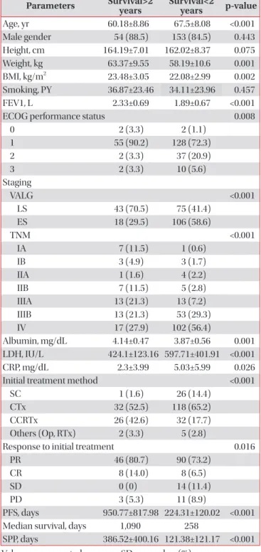
Methods: A total of 242 patients with cytologically or histologically diagnosed SCLC were enrolled into this study. The patients were categorized into long- and short-term survival groups by using a survival cut-off of 2 years after diagnosis.

Cox’s analyses were performed to identify the independent factors.

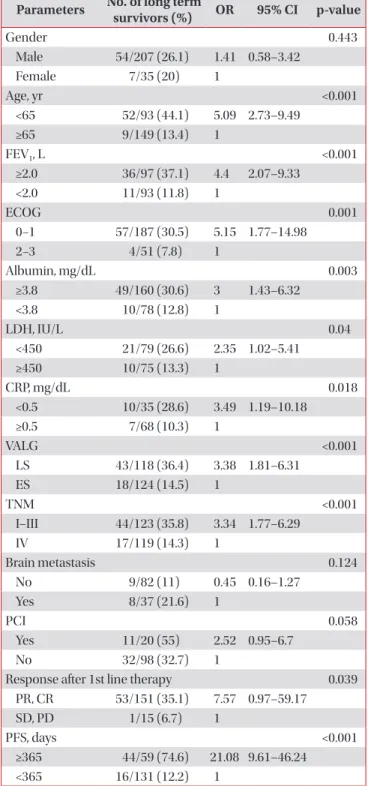
Results: The mean patient age was 65.66 years, and 85.5% were males; among the patients, 61 of them (25.2%) survived longer than 2 years. In the multivariate analyses, CRP (hazard ratio [HR], 2.75; 95% confidence interval [CI], 1.25−6.06;

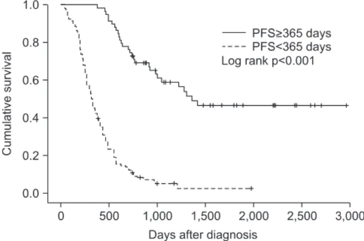
p=0.012), TNM staging (HR, 3.29; 95% CI, 1.59−6.80; p=0.001), and progression-free survival (PFS) (HR, 11.14; 95% CI, 2.98−41.73; p<0.001) were independent prognostic markers for poor survival rates.

Conclusion: In addition to other well-known prognostic factors, this study discovered relationships between the long- term survival rates and serum CRP levels, TNM staging, and PFS. In situations with unfavorable conditions, the PFS would be particularly helpful for managing SCLC patients.

Keywords: Small Cell Lung Carcinoma; Prognosis; Disease-Free Survival

Introduction

Small cell lung cancer (SCLC) is an extremely aggressive tumor with a poor clinical course. Without treatment, the me- dian length of survival after diagnosis is 1−3 months. Despite the initially high response rate to chemotherapy, the overall survival (OS) rate is low, with only 10−15% of patients with limited stage (LS)- or extensive stage (ES)-SCLC alive 2 years after diagnosis

1

. The median survival time for patients with ES-SCLC is 12−20 months, depending on the disease stage

1

.

Causes of the poor outcome of SCLC have been described, including more rapid tumor doubling times, a higher growth fraction, and earlier development of widespread metastases compared to non-small cell lung cancer (NSCLC). The tumor Copyright © 2014

The Korean Academy of Tuberculosis and Respiratory Diseases.

All rights reserved.

Address for correspondence: Jeong-Eun Lee, M.D., Ph.D.

Department of Internal Medicine, Chungnam National University Hospital, Chungnam National University School of Medicine, 282 Munhwa-ro, Jung-gu, Daejeon 301-721, Korea

Phone: 82-42-280-7147, Fax: 82-42-257-5753 E-mail: [email protected]

Received: Jan. 24, 2014 Revised: Feb. 24, 2014 Accepted: Mar. 14, 2014

cc It is identical to the Creative Commons Attribution Non-Commercial License (http://creativecommons.org/licenses/by-nc/3.0/).

(2)

doubling time of primary lung cancer is known to be a signifi- cant prognostic factor for lung cancer patients

2

. A short dou- bling time is the most reliable risk factor for multiple metastases, early recurrence, and a poor prognosis

3

. SCLC is considered the prototype of rapid-growing malignancies, with doubling times in the range of 25−217 days according to several studies

4-6

.

Although the results have been inconsistent, prior studies have identified a number of prognostic factors for SCLC

7-9

. Those factors, including age, sex, smoking history, syndrome of inappropriate secretion of antidiuretic hormone (SIADH), platelet count, and levels of albumin, lactate dehydrogenase (LDH), neuron-specific enolase (NSE), carcinoembryonic antigen, and cytokeratin-19 fragment, were determined to be potential prognostic factors for survival

8-10

. The International Association for the Study of Lung Cancer (IASLC) staging sys- tem has been used as a prognostic factor because it was found to be better than the Veterans Administration Lung Study Group (VALG) staging system for estimating long-term sur- vival in patients with SCLC

11

. National Comprehensive Can- cer Network (NCCN) 2014 guidelines (version 2) suggested that performance status, VALG staging, weight loss, and serum LDH level were prognostic factors for SCLC. Additionally, depending on the VALG stage, sex, age, serum creatinine level, and the number of metastatic organs at the time of diagnosis could be prognostic factors

12

.

Despite increased effort to find prognostic factors for SCLC, we could not find consistent and meaningful markers. In this study, we investigated prognostic factors that are related with treatment strategy, new drug targets, and clinical trials.

Materials and Methods

1. Data collection

This retrospective study included patients who were cyto- logically or histologically diagnosed with SCLC at Chungnam National University Hospital (Daejeon, Korea) between Janu- ary 2003 and July 2009. Medical records, as well as radiologic and nuclear medicine reports, were reviewed. Additionally, data were collected regarding the patients’ age, physical measurements, smoking history, pulmonary function, per- formance status, stage, and levels of serum albumin, LDH, and C-reactive protein (CRP) at the time of diagnosis. Data regarding the patients’ sex, existence of prophylactic cranial ir- radiation (PCI), progression-free survival (PFS), survival post- progression (SPP), response to initial treatment, and total sur- vival length were also collected. Patients with lost to follow-up within 2 years or active concurrent infection were excluded.

2. Clinical variables

The final follow-up date was 31 July 2011. The patients

were categorized into two groups according to the duration of survival after diagnosis: >2 years or <2 years. Patients who sur- vived >2 years after they were first diagnosed with SCLC were defined as long-term survivors. Physical measurement data included height, body weight, and body mass index (BMI). We used the pack/year unit for patients with a history of smoking before the diagnosis of SCLC, regardless of whether they had stopped smoking. Pulmonary function was evaluated spiro- metrically using the forced expiratory volume in 1 second (FEV

1

), which was performed within 2 months of the initial diagnosis. Performance status was defined using the Eastern Cooperative Oncology Group (ECOG) scale (stages 0−3) at the time of diagnosis. Two staging systems, the VALG system, which has been used only in SCLC, and the IASLC system (American Joint Committee on Cancer [AJCC] 7th edition), which has been used both in SCLC and NSCLC, were used to estimate stage based on chest computed tomography (CT), positron emission tomography-CT (PET-CT), brain CT, and brain magnetic resonance imaging. We used the highest levels of serum albumin, LDH, and CRP obtained between the date of SCLC diagnosis and that of initial treatment. After initial diagnosis, patients received treatment for SCLC with one of the 5 types of treatment arm which consist of chemotherapy, concurrent chemoradiotherapy, operation, radiotherapy, sup- portive care. Response evaluation using chest CT scan was performed after every 2−3 cycles of chemotherapy. Patients who received radiotherapy or operation, chest CT scan was conducted 4 weeks after the initial treatment. After the treat- ment, chest CT scan was recommended every 2 months for the first year, 3 months for the second year, every 6 months for the next several years, at least yearly after 5 years. Data for the treatment response, which was based on Response Evaluation Criteria in Solid Tumors from chest CT or PET-CT findings after initial treatment, were collected, excluding data of sup- portive care. In the case of disease progression, subsequent chemotherapy with single agent (i.e., IV topotecan) was con- ducted in favorable performance status. PFS was defined as the period from the time of initial treatment until an increased size of the primary mass or new lesion was found on radiolog- ic or nuclear medicine examination. PFS was not measured in patients who underwent supportive care. SPP was calculated by subtracting OS from PFS.

3. Statistical analyses

All analyses were performed using PASW Statistics (version

18.0; SPSS Inc., Chicago, IL, USA). Independent t-tests were

used to assess relationships between the existence of long-

term survival and continuous variables such as age, physi-

cal measurements, smoking history, FEV

1

, PFS, and serum

albumin, LDH, and CRP levels. Chi-squared tests were used

to assess the relationship between long-term survival and

categorical variables such as sex, performance status, stage,

(3)

conduction of PCI, and treatment response, as well as some continuous variables classified as categorical, including age, FEV

1

, and serum creatinine, albumin, LDH, and CRP levels. In addition, for variables identified as having a significant effect on long-term survival, the Kaplan-Meier method was used to determine survival curves. Significant differences between survival curves were estimated using log-rank tests. To identify prognostic factors, multivariate analyses using Cox’s regres- sion analysis were conducted.

4. Ethical considerations

The Institutional Review Board at Chungnam National Uni- versity Hospital reviewed and approved the study protocol (project approval number: CNUH 2012-07-006-001).

Results

1. Patient information

Two hundred and forty-two patients participated in the present study. The mean patient age was 65.66 years, and 85.5% of the patients were male. The median survival length was 349.5 days; the maximum was 2,961 days. Sixty-one (25.2%) patients were long-term (>2 years) survivors. Many variables had missing data except for age, sex, stage, initial treatment method, and survival length factors, with CRP level demonstrating the most missing patient data (n=139). The patients’ baseline characteristics are described in Table 1.

2. Relationship between long-term survival and other factors

In an analysis of the relationship between long-term sur- vival and factors for which means could be calculated, height (p=0.075) and smoking history (p=0.457) were not significant.

However, factors such as mean age, weight, BMI, FEV

1

, PFS, albumin, LDH, and CRP levels differed significantly according to long-term survival (Table 1).

More men (26.1%) than women (20%) survived >2 years, but sex was not significantly related to long-term survival with SCLC (odds ratio [OR], 1.41; 95% confidence interval [CI], 0.58−3.42; p=0.443). Age <65 years was related to long-term survival (OR, 5.09; 95% CI, 2.73−9.49; p<0.001), as were ECOG performance statuses 0 and 1 (OR, 5.52; 95% CI, 1.77−14.98;

p=0.001). Concerning VALG stage, LS was related to long-term survival (OR, 3.376; 95% CI, 1.81−6.31; p<0.001), and metasta- sis group based on TNM staging showed a similar relationship (OR, 3.34; 95% CI, 1.77−6.29; p<0.001). However, the absence of brain metastasis also showed no relationship to long-term survival (OR, 0.447; 95% CI, 0.16−1.27; p=0.124). An FEV

1

≥2.0 L was related to long-term survival (OR, 4.40; 95% CI, 2.07−9.33;

Table 1. Baseline characteristics Parameters Survival>2

years Survival<2

years p-value

Age, yr 60.18±8.86 67.5±8.08 <0.001

Male gender 54 (88.5) 153 (84.5) 0.443

Height, cm 164.19±7.01 162.02±8.37 0.075

Weight, kg 63.37±9.55 58.19±10.6 0.001

BMI, kg/m

2

23.48±3.05 22.08±2.99 0.002

Smoking, PY 36.87±23.46 34.11±23.96 0.457

FEV1, L 2.33±0.69 1.89±0.67 <0.001

ECOG performance status 0.008

0 2 (3.3) 2 (1.1)

1 55 (90.2) 128 (72.3)

2 2 (3.3) 37 (20.9)

3 2 (3.3) 10 (5.6)

Staging

VALG <0.001

LS 43 (70.5) 75 (41.4)

ES 18 (29.5) 106 (58.6)

TNM <0.001

IA 7 (11.5) 1 (0.6)

IB 3 (4.9) 3 (1.7)

IIA 1 (1.6) 4 (2.2)

IIB 7 (11.5) 5 (2.8)

IIIA 13 (21.3) 13 (7.2)

IIIB 13 (21.3) 53 (29.3)

IV 17 (27.9) 102 (56.4)

Albumin, mg/dL 4.14±0.47 3.87±0.56 0.001

LDH, IU/L 424.1±123.16 597.71±401.91 <0.001

CRP, mg/dL 2.3±3.99 5.03±5.99 0.026

Initial treatment method <0.001

SC 1 (1.6) 26 (14.4)

CTx 32 (52.5) 118 (65.2)

CCRTx 26 (42.6) 32 (17.7)

Others (Op, RTx) 2 (3.3) 5 (2.8)

Response to initial treatment 0.016

PR 46 (80.7) 90 (73.2)

CR 8 (14.0) 8 (6.5)

SD 0 (0) 14 (11.4)

PD 3 (5.3) 11 (8.9)

PFS, days 950.77±817.98 224.31±120.02 <0.001

Median survival, days 1,090 258

SPP, days 386.52±400.16 121.38±121.17 <0.001 Values are presented as mean±SD or number (%).

BMI: body mass index; PY: pack years; FEV

1

: forced expiratory volume in one second; ECOG: Eastern Cooperative Oncology Group; VALG: Veteran’s Administration Lung Group; LS: limited stage; ES: extensive stage; LDH: lactate dehydrogenase; CRP:

c-reactive protein; SC: supportive care; CTx: chemotherapy;

CCRTx: concurrent chemoradiotherapy; Op: operation; RTx:

radiotherapy; PR: partial remission; CR: complete remission; SD:

stable disease; PD: progressive disease; PFS: progression free

survival; SPP: survival post-progression.

(4)

p<0.001), as were an albumin level≥3.8 mg/dL, LDH level<450 IU/L, and CRP level<0.5 mg/dL (ORs, 3.00, 2.35, and 3.49, respectively; 95% CIs, 1.43−6.32, 1.02−5.41, and 1.19−5.41, respectively; p=0.003, p=0.04, and p=0.018, respectively). Ad- ditionally, in the LS patients, performing PCI showed no sig- nificant relationship to long-term survival (OR, 2.52; 95% CI, 0.95−6.70; p=0.058). To determine the relationship between treatment response and long-term survival, we classified the initial treatment response into four categories: partial remis- sion, complete remission, stable disease, and progressive dis- ease. We then analyzed the relationship between the remis- sion groups and long-term survival, which showed statistical significance (OR, 7.57; 95% CI, 0.97−59.17; p=0.039). However, when the partial remission, complete remission, and stable disease groups were designated as the control group and the progressive disease group as the non-control group, the con- Table 2. Categorical variables and long-term survival rates

Parameters No. of long term

survivors (%) OR 95% CI p-value

Gender 0.443

Male 54/207 (26.1) 1.41 0.58–3.42

Female 7/35 (20) 1

Age, yr <0.001

<65 52/93 (44.1) 5.09 2.73–9.49

≥65 9/149 (13.4) 1

FEV

1

, L <0.001

≥2.0 36/97 (37.1) 4.4 2.07–9.33 <2.0 11/93 (11.8) 1

ECOG 0.001

0−1 57/187 (30.5) 5.15 1.77–14.98

2−3 4/51 (7.8) 1

Albumin, mg/dL 0.003

≥3.8 49/160 (30.6) 3 1.43–6.32 <3.8 10/78 (12.8) 1

LDH, IU/L 0.04

<450 21/79 (26.6) 2.35 1.02–5.41

≥450 10/75 (13.3) 1

CRP, mg/dL 0.018

<0.5 10/35 (28.6) 3.49 1.19–10.18

≥0.5 7/68 (10.3) 1

VALG <0.001

LS 43/118 (36.4) 3.38 1.81–6.31

ES 18/124 (14.5) 1

TNM <0.001

I–III 44/123 (35.8) 3.34 1.77–6.29

IV 17/119 (14.3) 1

Brain metastasis 0.124

No 9/82 (11) 0.45 0.16–1.27

Yes 8/37 (21.6) 1

PCI 0.058

Yes 11/20 (55) 2.52 0.95–6.7

No 32/98 (32.7) 1

Response after 1st line therapy 0.039

PR, CR 53/151 (35.1) 7.57 0.97–59.17

SD, PD 1/15 (6.7) 1

PFS, days <0.001

≥365 44/59 (74.6) 21.08 9.61–46.24 <365 16/131 (12.2) 1

Statics were analyzed by chi-square test.

OR: odds ratio; CI: confidence interval; FEV

1

: forced expiratory volume in one second; ECOG: Eastern Cooperative Oncology Group; LDH: lactate dehydrogenase; CRP: C-reactive protein;

VALG: Veteran’s Administration Lung Group Stage; LS: limited stage; ES: extensive stage; PCI: prophylactic cranial irradiation; PR:

partial remission; CR: complete remission; SD: stable disease; PD:

progressive disease; PFS: progression free survival.

Figure 1. Kaplan-Meier estimates of overall survival, according to serum C-reactive protein (CRP) level.

Figure 2. Kaplan-Meier estimates of overall survival, according to

TNM stage.

(5)

trol group had no significant relationship to long-term survival (OR, 1.77; 95% CI, 0.47−6.60; p=0.391). PFS≥365 days was related to long-term survival (OR, 21.08; 95% CI, 9.61−46.24;

p<0.001). Table 2 shows the relationships between long-term survival and nominal variables.

Significant differences in median OS were found for se- rum CRP levels, TNM staging, and PFS. The median OS for CRP<0.5 (n=35) was 408 days and for CRP≥0.5 (n=68) was 196 days (p=0.004) (Figure 1). The median OS for stage I (n=14) could not be calculated because over 50% of candidates sur- vived, stage II (n=17) was 740 days, stage III (n=92) was 470 days, and stage IV (n=119) was 230 (p<0.001) (Figure 2). The median OS for PFS≥365 (n=59) and for PFS<365 (n=131) was 324 days (p<0.001) (Figure 3). In multivariate analyses using Cox’s regression, CRP (hazard ratio [HR], 2.75; 95% CI, 1.25−6.06; p=0.012), TNM staging (HR, 3.29; 95% CI, 1.59−6.80;

p=0.001), and PFS (HR, 11.14; 95% CI, 2.98−41.73; p<0.001) (Table 3) were independent prognostic markers for poor sur- vival.

Discussion

In this study, previously known prognostic factors, including age, weight, BMI, serum albumin levels, LDH levels, perfor- mance status, and staging, were related to survival for >2 years.

In particular, serum CRP levels, TNM staging and PFS were prognostic factors in SCLC patients by multivariate analyses.

The CRP test is sensitive and widely used in the diagnosis of acute and chronic inflammation. In malignant tumors, inflam- matory cells produce various cytokines; particularly, interleu- kin (IL)-6 induces necrosis and inflammation of tumor tissue.

CRP is produced by hepatocytes, the level of which is affected by IL-6

13

. CRP is known to be a prognostic factor in many ma- lignant tumors such as hepatocellular carcinoma, esophageal

cancer, and colon cancer

14-16

. A few studies of NSCLC have also used CRP as an independent risk factor

17,18

. Some stud- ies have shown that dynamic changes in the serum CRP level can be used as a predicting factor for treatment response in SCLC

19,20

. One recent study examined the use of pretreatment CRP level as a prognostic factor

21

. In the current study, the long-term survival group had a significantly lower CRP level at the time of diagnosis, which seemed valuable as a prognostic factor for SCLC; however, data were missing for 139 patients, which is the largest amount among all variables. To gain statis- tical reliability, a study with more subjects is needed.

The VALG system has been used for staging SCLC since 1957

22

. Radiation therapy plays an important role in managing SCLC patients with cytotoxic chemotherapy. Thus, although the staging system is old, many treatment guidelines for SCLC are based on the VALG system, which is categorized by whether all known lesions are included in a single radiation field. However, the revised TNM staging system that was em- braced in AJCC (7th edition) and was approved for prognostic significance in NSCLC as well as SCLC, is used in selecting treatment modalities for SCLC by NCCN 2014 guidelines (version 2)

12,23

. The fact that TNM staging system was a prog- nostic factor for survival in this study is appropriate based on recent trends.

The US Food and Drug Administration defines PFS as “the time from randomization until objective tumor progression or death”

24

. Generally, PFS is not commonly considered a prog- nostic factor in malignant tumors because its correlation with OS is rare. Additionally, no report to date has shown that PFS is a prognostic factor in SCLC. In this study, however, longer PFS was related to long-term survival. Recently, PFS has come to the forefront as a primary endpoint, replacing OS in clinical trials developing anticancer agents, particularly in phase II or Figure 3. Kaplan-Meier estimates of overall survival, according to

length of progression free survival (PFS).

Table 3. Independent factors concerning overall survivals

Parameters HR 95% CI p-value

Gender 0.77 0.28−2.14 0.617

Age 0.73 0.30−1.80 0.499

FEV

1

1.79 0.59−5.40 0.301

ECOG 0.65 0.17−2.49 0.532

Albumin 0.71 0.24−2.13 0.542

LDH 2.21 0.88−5.56 0.091

CRP 2.75 1.25−6.06 0.012

TNM staging 3.29 1.59−6.80 0.001

PFS 11.14 2.98−41.73 <0.001

Statistics were analyzed by Cox’s regression test.

HR: hazard ratio; CI: confidence interval; FEV

1

: forced expiratory volume in one second; ECOG: Eastern Cooperative Oncology Group; LDH: lactate dehydrogenase; CRP: c-reactive protein; PFS:

progression free survival.

(6)

III trials. PFS has many advantages in clinical trials compared with OS. Disease progression can be observed earlier than the expiration of candidates, leading to lighter burdens with fewer candidates and shorter trial durations. In addition, the effects of subsequent therapy (after the study period) do not interfere with PFS. Longer PFS that delays tumor-associated morbidity, including mental and physical aspects, may improve quality of life (QOL) because PFS has an intimate connection with tumor progression. Most anticancer agents that have been de- veloped recently target angiogenesis or growth factor recep- tors (i.e., they are focused on cytostatic features). In the past, there was a different perspective because drugs focused on the death of tumor cells or decreased numbers of tumor cells.

Therefore, in phase II or III trials, PFS that covers stable dis- ease is preferable to an objective tumor response, which was previously used as an endpoint

25

. PFS, however, has limita- tions in replacing OS in clinical trials. Judging disease progres- sion precisely by radiologic examination would depend on subjective interpretation. Moreover, the definitions of disease progression and time of response evaluation may be different between studies

25,26

.

It thus appears that PFS cannot replace OS perfectly, but there is a correlation between PFS and OS under specific circumstances. Although the exact mechanism is unknown, PFS can represent OS in specific malignant tumors such as colorectal cancer and recurrent high-grade glioma

27,28

. Fur- thermore, one study emphasized that tumors with a shorter SPP (<12 months) had a relationship between PFS and OS compared to tumors with a longer SPP (≥12 months) (r=0.64 and r=0.38, respectively)

29

. In this study, the median SPP of the SCLC patients was 120 days (<12 months); therefore, our results support the association between PFS and OS.

In this study, PFS was not short in all patients with SCLC.

Therefore, we propose that PFS reflects such tumor character- istics as growth rate or metastatic ability. For example, SCLC could be divided into rapid- and slow-growing types by PFS;

this distinction may be related to molecular genetic factors that were not analyzed in this study. Currently, primary muta- tions in oncogenes and tumor suppressor genes are known to be molecular genetic factors in SCLC

30

. Primary oncogenes direct transcription, cell growth, and cell cycle progression.

MYC oncogenes (CMYC, NMYC, and LMYC) are related to SCLC. The overactivation of MYC results in the rapid growth of cancer cells and differentiation failure, which are found in 40% of patients with disease progression after chemothera- py

31,32

. The tumor suppressor genes FHIT, RASSF1, retinoic acid receptor β, and FUS1 are particularly associated with SCLC, and mutation of these genes is directly or indirectly re- lated to tumor growth and differentiation

30

. However, the type of genetic factor that is related to long-term survival in SCLC remains unknown; thus, additional studies are needed.

A potential conclusion drawn from this study is that PFS

reflects tumor characteristics or survival and is useful for man- aging SCLC patients by clinicians. According to one study, the tumor volume doubling time in SCLC was shorter than that in NSCLC (39−64 days vs. 61−269 days)

33

. A poor SCLC progno- sis is due to its rapid progression rate and vulnerability of me- tastasis to other organs. In addition, there are few anticancer agents against SCLC compared with NSCLC, and drug resis- tance occurs more easily in SCLC patients. Accordingly, after first-line therapy, sequential treatment should be focused on tumors in life-threatening organs or that concern QOL, includ- ing pain or nutritional intake. Sequential therapy begun just after the detection of slight increases in tumor size can result in the absence of the treatment arm at a critical period due to drug resistance, which occurs from previously used drugs. To overcome these limitations in managing SCLC patients, mak- ing treatment decisions while considering the tumor charac- teristics before progressing to second-line treatment would be helpful. Prognostic factors related to survival will contribute to the prevention of QOL deterioration due to cytotoxic chemo- therapy and drug resistance due to unnecessary treatment.

This study has three major limitations. First, 21.7% of pa- tients were lost to follow-up. Although we lost track of these patients, those who survived >2 years were counted as long- term survivors. However, in patients with <2 years of follow- up, we could not estimate the actual survival length and they were not classified as long-term survivors. The latter group was comprised of 39 patients (14.5% of all patients). Second, differences in staging might exist regarding whether PET-CT was performed. In patients diagnosed after 2007, PET-CT was used for initial staging because the equipment was available at Chungnam National University Hospital. However, before 2007, some patients had PET-CT performed at another hos- pital, and PET-CT was not performed at our hospital; thus, tumor staging might be over- or underestimated. Third, some parameters such as SIADH, NSE which are known as prog- nostic factors were not analyzed in the present study. Because urine sodium level which is essential for diagnosing SIADH and positivity of NSE were not measured in most of patients.

In addition to the many prognostic factors discussed in the Introduction, this study found a relationship between long- term survival and serum CRP levels, TNM staging, and PFS.

Among these factors, PFS in particular reflects the character of malignant tumors and may be useful for determining whether sequential therapy is necessary. Studies of the potential re- lationship between molecular factors involved in SCLC and long-term survival are needed.

Conflicts of Interest

No potential conflict of interest relevant to this article was

reported.

(7)

References

1. Chua YJ, Steer C, Yip D. Recent advances in management of small-cell lung cancer. Cancer Treat Rev 2004;30:521-43.

2. Usuda K, Saito Y, Sagawa M, Sato M, Kanma K, Takahashi S, et al. Tumor doubling time and prognostic assessment of pa- tients with primary lung cancer. Cancer 1994;74:2239-44.

3. Tanaka K, Shimada H, Miura M, Fujii Y, Yamaguchi S, Endo I, et al. Metastatic tumor doubling time: most important pre- hepatectomy predictor of survival and nonrecurrence of he- patic colorectal cancer metastasis. World J Surg 2004;28:263- 70.

4. Kerr KM, Lamb D. Actual growth rate and tumour cell pro- liferation in human pulmonary neoplasms. Br J Cancer 1984;50:343-9.

5. Ost D, Fein AM, Feinsilver SH. Clinical practice. The solitary pulmonary nodule. N Engl J Med 2003;348:2535-42.

6. Souhami RL, Law K. Longevity in small cell lung cancer. A report to the Lung Cancer Subcommittee of the United King- dom Coordinating Committee for Cancer Research. Br J Can- cer 1990;61:584-9.

7. Paesmans M, Sculier JP, Lecomte J, Thiriaux J, Libert P, Sergy- sels R, et al. Prognostic factors for patients with small cell lung carcinoma: analysis of a series of 763 patients included in 4 consecutive prospective trials with a minimum follow-up of 5 years. Cancer 2000;89:523-33.

8. Ando S, Suzuki M, Yamamoto N, Iida T, Kimura H. The prognostic value of both neuron-specific enolase (NSE) and Cyfra21-1 in small cell lung cancer. Anticancer Res 2004;24:1941-6.

9. Ou SH, Ziogas A, Zell JA. Prognostic factors for survival in extensive stage small cell lung cancer (ED-SCLC): the im- portance of smoking history, socioeconomic and marital statuses, and ethnicity. J Thorac Oncol 2009;4:37-43.

10. Singh S, Parulekar W, Murray N, Feld R, Evans WK, Tu D, et al.

Influence of sex on toxicity and treatment outcome in small- cell lung cancer. J Clin Oncol 2005;23:850-6.

11. Micke P, Faldum A, Metz T, Beeh KM, Bittinger F, Hengstler JG, et al. Staging small cell lung cancer: Veterans Administra- tion Lung Study Group versus International Association for the Study of Lung Cancer--what limits limited disease? Lung Cancer 2002;37:271-6.

12. National Comprehensive Cancer Network. The NCCN clini- cal practice guidelines in oncology: small cell lung cancer (version V.II.2014) [Internet]. Fort Washington: National Comprehensive Cancer Network; 2013 [cited 2013 Oct 3].

Available from: http://www.nccn.org/professionals/physi- cian_gls/pdf/sclc.pdf.

13. Wang CS, Sun CF. C-reactive protein and malignancy: clinico- pathological association and therapeutic implication. Chang Gung Med J 2009;32:471-82.

14. McMillan DC, Canna K, McArdle CS. Systemic inflammatory response predicts survival following curative resection of

colorectal cancer. Br J Surg 2003;90:215-9.

15. Nozoe T, Saeki H, Sugimachi K. Significance of preoperative elevation of serum C-reactive protein as an indicator of prog- nosis in esophageal carcinoma. Am J Surg 2001;182:197-201.

16. Nagaoka S, Yoshida T, Akiyoshi J, Akiba J, Torimura T, Adachi H, et al. Serum C-reactive protein levels predict survival in hepatocellular carcinoma. Liver Int 2007;27:1091-7.

17. Hara M, Matsuzaki Y, Shimuzu T, Tomita M, Ayabe T, Eno- moto Y, et al. Preoperative serum C-reactive protein level in non-small cell lung cancer. Anticancer Res 2007;27:3001-4.

18. Scott HR, McMillan DC, Forrest LM, Brown DJ, McArdle CS, Milroy R. The systemic inflammatory response, weight loss, performance status and survival in patients with inoperable non-small cell lung cancer. Br J Cancer 2002;87:264-7.

19. Milroy R, Shapiro D, Shenkin A, Banham SW. Acute phase reaction during chemotherapy in small cell lung cancer. Br J Cancer 1989;59:933-5.

20. Arpin D, Trillet-Lenoir V, Lasset C, Souquet PJ, Riou R, Yoani- dis I, et al. [Value of C-reactive protein determination in small cell lung cancer]. Bull Cancer 1993;80:1063-8.

21. Hong S, Kang YA, Cho BC, Kim DJ. Elevated serum C-reactive protein as a prognostic marker in small cell lung cancer. Yon- sei Med J 2012;53:111-7.

22. Zelen M. Keynote address on biostatistics and data retrieval.

Cancer Chemother Rep 3 1973;4:31-42.

23. Shepherd FA, Crowley J, Van Houtte P, Postmus PE, Carney D, Chansky K, et al. The International Association for the Study of Lung Cancer lung cancer staging project: proposals regarding the clinical staging of small cell lung cancer in the forthcoming (seventh) edition of the tumor, node, metastasis classification for lung cancer. J Thorac Oncol 2007;2:1067-77.

24. Guidance for industry: clinical trial endpoints for the ap- proval of cancer drugs and biologics [Internet]. Rockville: U.S.

Department of Health and Human Services, Food and Drug Administration, Center for Drug Evaluation and Research, Center for Biologics evaluation and Research; 2007 [cited 2013 Oct 2]. Available from: http://www.fda.gov/downloads/

Drugs/GuidanceComplianceRegulatoryInformation/Guid- ances/ucm071590.pdf.

25. Bergmann L, Hirschfeld S, Morris C, Palmeri S, Stone A.

Progression-free survival as an end-point in clinical trials of biotherapeutic agents. Eur J Cancer Suppl 2007;5:23-8.

26. Dancey JE, Dodd LE, Ford R, Kaplan R, Mooney M, Rubin- stein L, et al. Recommendations for the assessment of pro- gression in randomised cancer treatment trials. Eur J Cancer 2009;45:281-9.

27. Buyse M, Burzykowski T, Carroll K, Michiels S, Sargent DJ, Miller LL, et al. Progression-free survival is a surrogate for survival in advanced colorectal cancer. J Clin Oncol 2007;25:5218-24.

28. Lamborn KR, Yung WK, Chang SM, Wen PY, Cloughesy TF,

DeAngelis LM, et al. Progression-free survival: an important

end point in evaluating therapy for recurrent high-grade glio-

(8)

mas. Neuro Oncol 2008;10:162-70.

29. Amir E, Seruga B, Kwong R, Tannock IF, Ocana A. Poor corre- lation between progression-free and overall survival in mod- ern clinical trials: are composite endpoints the answer? Eur J Cancer 2012;48:385-8.

30. Jackman DM, Johnson BE. Small-cell lung cancer. Lancet 2005;366:1385-96.

31. Johnson BE, Makuch RW, Simmons AD, Gazdar AF, Burch D, Cashell AW. myc family DNA amplification in small cell lung cancer patients’ tumors and corresponding cell lines. Cancer

Res 1988;48:5163-6.

32. Brennan J, O’Connor T, Makuch RW, Simmons AM, Russell E, Linnoila RI, et al. myc family DNA amplification in 107 tu- mors and tumor cell lines from patients with small cell lung cancer treated with different combination chemotherapy regimens. Cancer Res 1991;51:1708-12.

33. Friberg S, Mattson S. On the growth rates of human malig-

nant tumors: implications for medical decision making. J Surg

Oncol 1997;65:284-97.

수치

Table 1. Baseline characteristics Parameters Survival&gt;2  years Survival&lt;2 years p-value Age, yr 60.18±8.86 67.5±8.08 &lt;0.001 Male gender 54 (88.5) 153 (84.5) 0.443 Height, cm 164.19±7.01 162.02±8.37 0.075 Weight, kg 63.37±9.55 58.19±10.6 0.001 BMI,
Figure 1. Kaplan-Meier estimates of overall survival, according to  serum C-reactive protein (CRP) level.
Table 3. Independent factors concerning overall survivals

참조

관련 문서

Prognostic Significance of Circulating Tumor Cells in Small- Cell Lung Cancer Patients: a Meta-analysis.. Jiao Zhang 1 , Hai-Tao Wang 1 *,

Background: The aim of this study was to determine the survival rate of patients with non-small cell lung cancer (NSCLC) who were preoperatively diagnosed with a negative N2

In this study, we evaluated the expression of fascin and its prognostic significance in stage I non-small cell lung cancer (NSCLC).. Methods: Immunohistochemical

Mutation of epidermal growth factor receptor (EGFR) is a major driver oncogene of non-small cell lung cancer (NSCLC), especially adenocarcinoma and it is more

In this study, we evaluated the expression of fascin and its prognostic significance in stage I non-small cell lung cancer (NSCLC).. Methods:

Prognostic Significance of Cyclooxygenase-2(COX-2) Expression in Primary, Resected Non-Small Cell Lung Cancer.. Hak Ryul Kim, M.D., Sei Hoon Yang, M.D., Eun

Abbreviations: ADC = adenocarcinoma, EGFR = epidermal growth factor receptor, HPV = human papillomavirus, MCPyV = Merkel cell polyomavirus, NSCLC = nonsmall cell lung cancer,

Transglutaminase 2 Expression Predicts Progression Free Survival in Non-Small Cell Lung Cancer Patients Treated with Epidermal Growth Factor Receptor Tyrosine Kinase