http://dx.doi.org/10.5090/kjtcs.2013.46.1.49 ISSN: 2233-601X (Print) ISSN: 2093-6516 (Online)
Departments of
1Thoracic and Cardiovascular Surgery and
2Pathology, Keimyung University Dongsan Medical Center, Keimyung University School of Medicine
Received: May 18, 2012, Revised: August 27, 2012, Accepted: August 29, 2012
Corresponding author: Chang Kwon Park, Department of Thoracic and Cardiovascular Surgery, Keimyung University Dongsan Medical Center, Keimyung University School of Medicine, 56 Dalseong-ro, Jung-gu, Daegu 700-712, Korea
(Tel) 82-53-250-7342 (Fax) 82-53-250-7307 (E-mail) [email protected]
C
The Korean Society for Thoracic and Cardiovascular Surgery. 2013. All right reserved.
CC
This is an open access article distributed under the terms of the Creative Commons Attribution Non-Commercial License (http://creative- commons.org/licenses/by-nc/3.0) which permits unrestricted non-commercial use, distribution, and reproduction in any medium, provided the original work is properly cited.
Long Term Survival of Patients with Unsuspected N2 Disease in Non-Small Cell Lung Cancer
Deok Heon Lee, M.D.
1, Jae Bum Kim, M.D.
1, Dong Yoon Keum, M.D.
1, Ilseon Hwang, M.D.
2, Chang Kwon Park, M.D.
1Background: The aim of this study was to determine the survival rate of patients with non-small cell lung cancer (NSCLC) who were preoperatively diagnosed with a negative N2 lymph node, but postoperatively confirmed as a positive N2 node based on a pathological evaluation. Materials and Methods: The hospital records of 248 pa- tients from 1994 to 2009 with resected primary NSCLC who were preoperatively diagnosed with negative N2 lymph node, were retrospectively reviewed. Of these, after surgery, there were 148 (59.7%) patients with pathological N0, 54 (21.8%) with pathological N1 and 46 (18.5%) with pathological N2. Results: The median follow-up period was 24 months (range, 1 to 132 months). The 5-year disease free survival rates were 60% in pN0, 44% in pN1, and 29% in pN2. The 5-year overall survival rates were 63.1% in pN0, 51.9% in pN1, and 33.5% in pN2. There were no statistically significant differences between pN1 and pN2 (p=0.326 and p=0.106, respectively). Thirty-three (71.7%) of the 46 pN2 patients had single-zone metastasis, and 13 patients (28.3%) had multiple-zone metastases over the two nodal zone metastasis. There were no statistical differences in the 5-year disease free survival rate and the 5-year overall survival rates between the two groups. Conclusion: The 5-year disease free survival and the overall survival rate of the patients with unsuspected N2 disease were statistically similar with that of the pa- tients with pathological N1 disease. There was no statistically significant difference between the patients with a sin- gle-zone metastasis and a multiple zone metastasis.
Key words: 1. Carcinoma, non-small-cell lung 2. Lymph nodes
3. Neoplasm staging
INTRODUCTION
Mediastinal nodal involvement is generally considered to be one of the most important prognostic factors in patients with non-small cell lung cancer (NSCLC). In patients with N2 disease, identified preoperatively, pulmonary resection is not recommended by the American College of Chest Physicians Evidence-Based Clinical Practice Guidelines [1].
However, the optimal treatment approach for clinical stage
IIIA has been controversial, because N2 disease is defined
just as the presence of metastasis in the ipsilateral mediastinal
lymph node by the 7th American Joint Committee on Cancer
tumor-node-metastasis (AJCC/TNM) staging for lung cancer
[2]. Stage IIIA NSCLC, especially N2 disease, shows a broad
spectrum of the disease. Several published reports have sug-
gested that there were significant variabilities of the long
term outcomes, according to the heterogeneity of N2 disease, such as a single N2 disease versus a multiple N2 disease, skip metastasis (negative N1 and positive N2 disease), the number of positive N2 disease, and unsuspected, occult or in- cidental N2 disease [3-11].
Recently, the nodal zone concept was proposed as a new descriptor of the intrathoracic lymph node by International Association for the Study of Lung Cancer (IASLC) [12]. The lymph node maps, which have been widely used for several decades, were classified by Mountain and Dressler [13], Naruke [14], and Naruke et al. [15]. However, there are sev- eral discrepancies in the definition of the nomenclature be- tween the two classifications. The nodal zone concept was proposed as able to achieve uniformity, an accurate assess- ment of lymph node involvement, and to promote future anal- yses of a planned prospective international database.
The purpose of this study is to investigate the long term outcomes for patients with unsuspected N2 (negative N2 dis- ease on the preoperative diagnosis, but positive N2 disease on the postoperative pathologic diagnosis) disease, and to identi- fy the effects of the ‘nodal zone’ relationship to the long-term outcomes of unsuspected N2 disease.
MATERIALS AND METHODS
The hospital records of 485 patients with primary NSCLC who underwent pulmonary resection and mediastinal lympha- denectomy in Keimyung University Dongsan Medical Center from May 1994 to December 2009, were retrospectively reviewed. The preoperative work-up included the following:
standard chest posteroanterior radiography, chest computed to- mography (CT), fiberoptic bronchoscopy, whole body bone scan, and brain magnetic resonance imaging (MRI). Positron emission tomography-CT (PET-CT) scans have been part of the preoperative work-up at Keimyung University Dongsan Medical Center since March 2007. Preoperative mediastino- scopic biopsy was also performed in selected patients. In this study, we included any patients 1) who had primary NSCLC, 2) who underwent anatomical resection, and 3) who were negative for malignancy of a mediastinal lymph node by me- diastinoscopic biopsy among selected patients. We excluded any patients 1) who had mediastinal lymph nodes, which
were over 1 cm of short axis diameter on CT, 2) who had mediastinal lymph nodes, which showed a maximum stand- ardized uptake value of over 2.5 on PET-CT scan, 3) who had metastasis of mediastinal lymph node confirmed by pre- operative mediastinoscopic biopsy, 4) who had received neo- adjuvant chemotherapy or radiotherapy, 5) who had multiple primary lung cancer, 6) who were diagnosed with T4 staging by postoperative pathologically evaluation, or 7) who under- went minor resection or incomplete pulmonary resection, or 8) who had died within 30 days due to postoperative morbidity. We retrospectively reviewed the hospital records of 248 patients who met the inclusion and exclusion criteria.
Surgery was performed using posterolateral thoracotomy.
Anatomical resection and complete mediastinal lymph node dis- section or mediastinal lymph node sampling were performed in the enrolled patients. The lymph node description used in this study was ‘The Lymph Node Classification of Mountain and Dressler’ [13]. Involved mediastinal lymph nodes (N2) were re- classified with ‘four nodal zone,’ which was recently proposed by the IASLC [12]. The 1 to 4 station of the mediastinal lymph node, according to ‘The Lymph Node Classification of Mountain and Dressler,’ were united with the upper zone, the 5 and 6 station with the aortopulmonary zone, the 7 station with the subcarinal zone, and the 8 and 9 station with the lower zone. All of the patients were surgically staged, according to the 7th AJCC/TNM staging classification of lung cancer [2].
The patients were followed up every 3 months for the first 2 years after surgery, and every 6 months thereafter. The chest radiographs were obtained during every follow-up, and CT was obtained every 6 months for the first two years, and annually thereafter. Other specific studies, such as a PET-CT scan, brain MRI, a whole body bone scan, or bronchoscopy were performed for selected patients during the follow-up period.
All statistical analyses were carried out with the statistical software SPSS ver. 18.0 (SPSS Inc., Chicago, IL, USA).
Continuous data were presented as the mean and standard de-
viation, and the discrete data were presented as a number and
percentage. The association between the data was analyzed
using the independent t-test or the Mann-Whitney U-test for
continuous variables, and the Pearson’s χ
2test or Fisher’s
exact test was used for the discrete variables. The disease
free survival was defined as the period of staying free from
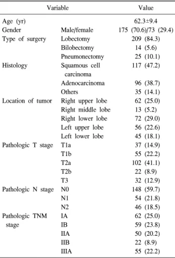
Table 1. Characteristics of the study population
Variable Value
Age (yr) Gender
Type of surgery
Histology
Location of tumor
Pathologic T stage
Pathologic N stage
Pathologic TNM stage
Male/female Lobectomy Bilobectomy Pneumonectomy Squamous cell
carcinoma Adenocarcinoma Others
Right upper lobe Right middle lobe Right lower lobe Left upper lobe Left lower lobe T1a
T1b T2a T2b T3 N0 N1 N2 IA IB IIA IIB IIIA
62.3±9.4 175 (70.6)/73 (29.4)
209 (84.3) 14 (5.6) 25 (10.1) 117 (47.2)
96 (38.7) 35 (14.1) 62 (25.0) 13 (5.2) 72 (29.0) 56 (22.6) 45 (18.1) 37 (14.9) 55 (22.2) 102 (41.1) 22 (8.9) 32 (12.9) 148 (59.7) 54 (21.8) 46 (18.5) 62 (25.0) 59 (23.8) 50 (20.2) 22 (8.9) 55 (22.2) Values are presented as mean±standard deviation or number (%).
TNM, tumor-node-metastasis.
Table 2. Location of the nodal zone according to the tumor loca- tion in the patients with a single-zone metastasis
Tumor location (patients) No. of patients
U AP Sub L
Right upper lobe (9) Right middle lobe (3) Right lower lobe (6) Left upper lobe (8) Left lower lobe (7)
7 1 1 1 0
0 0 0 6 0
2 2 5 0 4
0 0 0 1 3 U, upper zone; AP, aortopulmonary zone; Sub, subcarinal zone;
L, lower zone.
Table 3. Location of the nodal zone according to the tumor loca- tion in the patients with a multiple-zone metastasis
Tumor location (patients)
Location of nodal zone
No. of patients Right upper lobe (1)
Right middle lobe (2)
Right lower lobe (4)
Left upper lobe (4)
Left lower lobe (2)
U and Sub U and Sub Sub and L U and Sub U and L Sub and L U and AP AP and L AP, Sub, and L Sub and L Sub and L
1 1 1 2 1 1 1 1 1 1 2 U, upper zone; Sub, subcarinal zone; AP, aortopulmonary zone;
L, lower zone.
disease after surgery. The confirmed loco-regional recur- rences, distant metastases, and deaths were used as the specif- ic events for disease free survival. The overall survival was defined as the duration from the operation to death from any cause. The survival rates were derived using the Kaplan-Meier method, and the differences between the subgroups were as- sessed using the log-rank test. The multivariate analysis of the survival influence was performed using a Cox propor- tional hazards model. A statistically significant difference was defined as a p-value of less than two-sided 0.05. The institu- tional review board of the University of Keimyung in South Korea approved this study.
RESULTS
There were 248 patients who met the inclusion and exclusion
criteria of this study. There were 175 males (70.6%) and 73 fe- males (29.4%), with a mean age of 62.3±9.4 years. The median follow-up period was 24 months (range, 1 to 132 months).
Lobectomy was performed in 209 patients (84.3%), bilobectomy in 14 (5.6%), and pneumonectomy in 25 (10.1%). The patients’
characteristics are described in Table 1. The most common his- tology was squamous cell carcinoma (117 patients, 47.2%).
There were 96 patients (38.7%) with adenocarcinoma, and 35 other patients (14.1%) presented with different histologies.
A total of 148 (59.7%) patients presented with pathological N0 (pN0) disease, 54 (21.8%) with pathological N1 (pN1), and 46 (18.5%) with pathological N2 (pN2), after surgery.
Thirty-three (71.7%) of the 46 pN2 patients had single-zone
metastasis, and 13 patients (28.3%) had multiple-zone meta-
stases over two nodal zone metastasis. The patterns of meta-
Fig. 1. (A) The disease free survival rate and (B) overall survival rate among the patients with N0, N1, and N2 disease. pN, pathologic node.
stasis of N2 disease are shown in Tables 2, 3. There were 109 patients (43.9%) that received adjuvant therapy, accord- ing to the local protocol for NSCLC. Adjuvant chemotherapy was administered to 66 patients (26.6%), radiotherapy to 16 (6.4%), and concurrent chemo-radiotherapy to 27 patients (10.8%).
The 5-year disease free survival rates were 60.4% in the pN0 group, 44.1% in the pN1 group, and 29.4% in the pN2 group. The disease free survival rate was significantly better in the pN0 group than in that of the pN1 or pN2 group (p=0.030 and p=0.000, respectively). However, there was no statistically significant differences between the pN1 and pN2 groups (p=0.326). The 5-year overall survival rates were 63.1% in the pN0 group, 51.9% in the pN1 group, and 33.5% in the pN2 group. The overall survival was sig- nificantly better in the pN0 group than in that of the pN2 group (p<0.000). However, there was no statistically sig- nificant differences between the pN0 and pN1 group (p=0.088), and between the pN1 and pN2 groups (p=0.106) (Fig. 1).
The 5-year disease free survival rates were 29.5% in the single-zone metastasis group and 28.0% in the multiple-zone metastasis group (p=0.635). The 5-year overall survival rates were 35.1% in the single-zone metastasis group and 30.8% in the multiple-zone metastasis group (p=0.857). There were no statistical differences between the two groups.
There were recurrences in 91 patients (36.7%). Of these, 46 patients (18.5%) had loco-regional recurrence and 45 pa- tients (18.1%) had distant recurrence. Loco-regional re- currence was most common in the postoperative pN1 group (20 [37.0%] of the 54 patients), and distant recurrence was most common in postoperative pN2 group (22 [47.8%] of the 46 patients).
The significant risk factors associated with disease free sur- vival and overall survival based on the univariate analysis were pathologic N staging (p=0.001) in disease free survival and pathologic T staging (p=0.001) and pathologic N staging (p=0.001) in the overall survival. Table 4 shows that the pathologic N stage was the independent predictor in disease free survival, and pathologic T stage and pathologic N stage were the independent predictors in the overall survival on multivariate analyses.
DISCUSSION
Surgery is recommended for patients with early stage
NSCLC (stage I and II) or surgery with adjuvant chemo-
therapy or radiotherapy; it is widely considered to result in a
better chance for a patient’s long-term survival. However, the
role of surgery for N2 disease is controversial. Most physi-
cians agree that patients with mediastinal lymph node meta-
stasis have a poor long-term outcome, and surgery should not
Table 4. Multivariate analyses for disease free survival and overall survival with a Cox proportional hazards model
Variable Disease free survival Overall survival
p-value HR (95% CI) p-value HR (95% CI)
Age (<60 vs. ≥60) Gender (male/female) Operation (L/P) Location (U/ML) Side (left/right) Pathologic pT stage pT1 vs. pT2 pT2 vs. pT3 Pathologic pN stage pN0 vs. pN1 pN1 vs. pN2
0.447 0.709 0.840 0.120 0.689 0.112 0.532 0.468 0.003
a)0.001
a)0.251
1.185 (0.765–1.836) 1.093 (0.685–1.745) 0.966 (0.691–1.351) 1.410 (0.915–2.172) 1.094 (0.705–1.695)
0.789 (0.375–1.660) 1.296 (0.643–2.615)
0.407 (0.241–0.687) 0.717 (0.406–1.266)
0.153 0.594 0.119 0.058 0.077 0.003
a)0.002
a)0.251 0.031
a)0.008
a)0.148
1.367 (0.890–2.100) 1.133 (0.717–1.790) 1.259 (0.942–1.684) 1.514 (0.987–2.322) 0.684 (0.449–1.042)
0.356 (0.186–0.682) 0.714 (0.401–1.270)
0.500 (0.299–0.838) 0.665 (0.383–1.156) HR, hazard ratio; CI, confidence interval; L, lobectomy; P, pneumonectomy; U, upper lobe; ML, middle and lower lobe; pT, patho- logical tumor; pN, pathological node.
a)