절제된 IIIA N2 병기 비소세포형 폐암에 있어서 Nodal Station의 의의
Nodal Station as a Prognostic Factor in Resected Stage IIIA N2 Non-Small Cell Lung Cancer
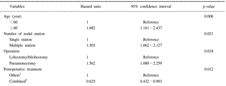
Background: To clarify the prognostic implication of the location and number of the metastatic mediastinal nodes in resected stage IIIA N2 non-small cell lung cancer. Material and Method: One hundred and seventy-four patients with resected non-small cell lung cancer who eventually proved to have pathologic stage IIIA N2 disease were studied. Patients who received preoperative induction therapy, non-curative operation or defined as operative mortality were excluded from this study. Result: In upper lobe tumors, there was no difference in 5-year survival according to the involvement of lower mediastinal nodes (32.3% vs 25.6%, p=0.86). In lower lobe tumors, no differencee was found in 5-year survival according to the involvement of upper mediastinal nodes (25.1% vs 14.1%, p=0.33). There was no significant difference in 5-year survival between patients with or without metastatic subcarinal node (20.9% vs 25.6%, p=0.364). In terms of the number of metastatic mediastinal nodes, 5-year survival was better in single station group (26.3%) than multiple station group (18.3%) (p=0.048). In multiple station N2 group, the patients who received postoperative chemotherapy and radiation therapy had better 5-year survival (34.2%) (p=0.01). Cox's proportional hazards model revealed that the age 60 (O.R: 1.682, p=.006), multiple station N2 (O.R: 1.503. p=0.021), pneumonectomy (O.R: 1.562, p=0.018), postoperative chemotherapy and radiation therapy (O.R: 0.625, p=0.012) were the factors affecting the postoperative survival. Conclusion: Multiple station N2 disease was the important prognostic factor for postoperative survival in resected stage IIIA N2 non-small cell lung cancer. Postoperative chemotherapy and radiotherapy were thought to improve the survival in case of multiple station N2 disease.
(Korean J Thorac Cardiovasc Surg 2003;36:489-496) Key words
:1. Neoplasm staging
2. Carcinoma, non-small cell, lung
3. Lymph node
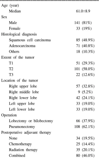
Table 1. Patient characteristics
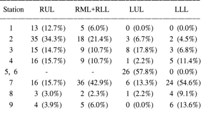
Table 2. Prevalent N2 stations according to the primary location of the tumor
Fig. 1. Actuarial survival according to the involvement of lower mediastinal node(s) in patients with upper lobe tumor. LMN+
(solid line): involvement of the lower mediastinal node(s), LMN- (dotted line): involvement of the upper mediastinal node(s) only.
Fig. 2. Actuarial survival according to the involvement of upper mediastinal node(s) in patients with lower lobe tumor. UMN+
(solid line): involvement of the upperr mediastinal node(s), LMN- (dotted line): involvement of the lower mediastinal node(s) only.
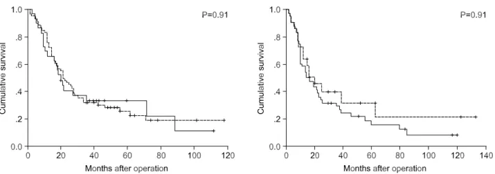
Fig. 3. Actuarial survival according to the involvement of subcarinal node in patients with upper lobe tumor (left) and lower lobe tumor (right). Solid line: involvement of subcarinal node, Dotted line: no involvement of subcarinal node.
Fig. 4. Actuarial survival according to the number of nodal station. Survival was better in single station group (dotted line) than multiple station group (solid line) (p=0.048).
Fig. 5. Actuarial survival according to the postoperative therapy.
Combined (bold solid line): chemotherapy and radiation therapy, CT (bold dashed line): chemotherapy alone, RT (dotted line):
radiation therapy alone, None (dashed line): no postoperative treatment.
Fig. 6. Actuarial survival according to the postoperative therapy in multiple station group. Combined (bold solid line): chemo- therapy and radiation therapy. CT (bold dashed line): chemo- therapy alone, RT (dotted line): radiation therapy alone, None (dashed line): no postoperative treatment.
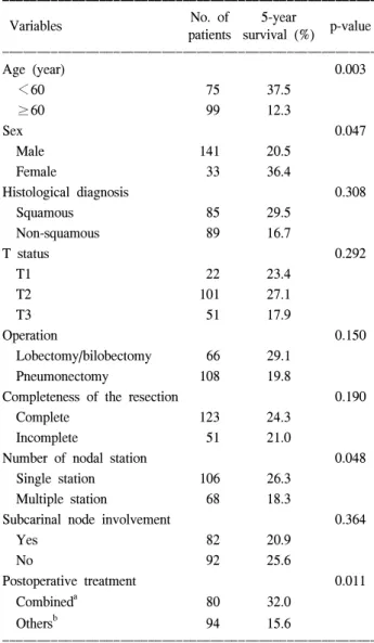
Table 3. Univariate analysis of the risk factor for postopera- tive survival
Table 4. Multivariate analysis
=국문 초록=
배경:
대상 및 방법:
결과:
결론:
중심 단어