□
증 례□
흉막삼출액의 관례적 천자 후 발생한 국소성 재팽창성 폐 부종 1예
경희대학교 의과대학 내과학교실
손성동, 유지홍, 최천웅, 박명재, 강홍모
=Abstract=
A Case of Focal Reexpansion Pulmonary Edema after Conventional Thoracentesis of Pleural Effusion
Sohn Seong Dong, M.D., Yoo Jee Hong, M.D., Choi Cheon Woong, M.D., Park Myung Jae, M.D., Kang Hong Mo, M.D.
Department of Internal Medicine, Kyung Hee University College of Medicine, Seoul, Korea
A 60-year old male patient admitted with complaints of dyspnea and pleuritic chest pain. The chest X-ray demonstrated right pleural effusion. We planed to do the conventional thoracentesis to evaluate the characteristics of pleural effusion and to relieve the symptom of the patient. Focal reexpansion pulmonary edema was seen on the follow-up chest X-ray. After the 5-day conservative management, the patient recovered without any complications. (Tuberculosis and Respiratory Diseases 2004, 56:297-301)
Key words : Focal reexpansion pulmonary edema, Conventional thoracentesis, Pleural effusion.
Address for correspondence : Yoo Jee Hong, M.D.
Department of Internal Medicine, Kyung Hee University College of Medicine, Seoul, Korea, 1 Hoegi-Dong, Dongdaemun-Gu, Seoul, 130-702, Korea
Phone : 02-958-8200 Fax : 02-968-1848 E-mail : [email protected] 서 론
재팽창성 폐 부종(reexpansion pulmonary edema) 은 다량의 기흉 또는 흉수를 단기간 혹은 강제적 흡인 시, 만성적으로 허탈 되었던 폐의 급격한 재팽 창으로 나타날 수 있는 드물지 않은 합병증이다1. 기전은 명확히 밝혀지지 않았으나 허탈 되었던 폐
의 저산소 손상에 의한 혈관 투과성 변화로 나타 나는 것으로 보고되어 있다. 재팽창성 폐 부종은 주로 재팽창된 폐의 전역에 걸쳐 미만성 폐 부종 이 나타나며, 국소성 폐 부종은 매우 드문 것으로 알려져 있으며, 일반적으로 관례적 천자 후에는 잘 발생하지 않는다. 저자들은 60세 남자에서 음압을 가하지 않은 비흡인성 경피적 천자 배액법으로 시
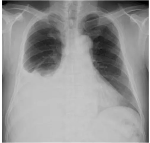
Fig. 2. Chest X-ray obtained 4hours after drai nage of 800 mL of effusion without nega tive pressure suction shows reexpansion pulmonary edema limited to right lower lung.
Fig. 1. Chest X-ray of 60-year-old man with right pleural effusion.
행한 800mL 정도의 관례적 흉수 천자 후 우하부 폐엽에 국한되어 나타난 재팽창성 폐 부종 1예를 경험하였기에 문헌고찰과 함께 보고하는 바이다.
증 례
환 자 : 이○○, 남자 60세 주 소 : 호흡곤란, 흉막성 흉통
현병력 : 평소 건강히 지내던 60세 남자 환자로 내 원 5일전부터 호흡곤란, 흉막성 흉통이 있어 개인 의원을 들러 촬영한 흉부 방사선 사진에서 우측 흉막 삼출액 소견 외 특이 소견 보이지 않아 자세 한 검사를 위해 전원 되었다.
과거력 : 특이사항 없음 가족력 : 특이사항 없음
진찰소견 : 혈압 120/80 mmHg, 맥박수 68회/분, 호흡수 24회/분, 체온 36.9 ℃ 였다. 흉부 청진에서 우하부 호흡음 및 성음 진탕이 감소되어 있었으며, 정상적인 심음을 보였다. 그 외 신체검사에서 특이 소견은 없었다.
검사실 소견 : 일반 혈액 검사상 혈색소 14.3 g/
dL, 적혈구 용적 42.4 %, 백혈구 5600/mm3, 혈소 판 299,000/mm3 이었다. 생화학 검사상 BUN 15
mg/dL, creatinine 0.6 mg/dL, sodium 135 mEq/L, potassium 4.2 mEq/L, AST 30 IU/L, ALT 32 IU/L, 총단백 5.9 g/dL, 알부민 3.4 g/dL 이었다.
소변검사 및 대변검사는 정상이었다. 심전도 소견 은 정상 동조율이었고, 흉부 방사선 사진(Fig. 1)에 서 우측 흉막 삼출액 소견이 관찰되었다. 음압을 가하지 않은 비흡인성 경피적 천자 배액으로 시행 한 흉수 검사에서 천자액은 약 800 mL 이고 황색 의 혼탁한 양상을 보였으며 백혈구 2040/mm3(호중 구 3 %, 임파구 84 %), LDH 551 IU/L, 단백질 5 g/dL, glucose 249 mg/dL, ADA 74 IU/L 으로 결 핵성 흉수에 합당한 소견이었다. 그러나 흉수 및 객담 도말검사에서 결핵균은 발견되지 않았다.
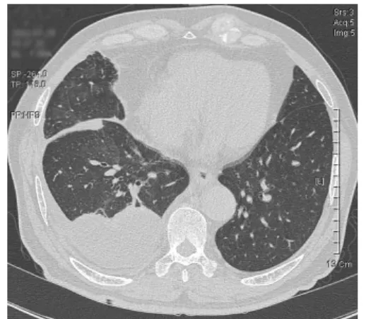
치료 및 경과 : 약 800 mL 정도의 흉수를 음압을 가하지 않은 비흡인성 배액법으로 배액하고 약 4 시간 경과 후 촬영한 흉부 방사선 사진(Fig. 2)에 서 우측 흉막 삼출액 소견은 감소되었으나, 우하부 폐야에 국한된 폐음영 증가 소견이 보였다. 배액 후 약 24시간 지나서 촬영한 흉부 전산화 단층 촬 영 사진(Fig. 3)에서 우하부 폐엽에 국한된 간유리 음영과 공기 기관지 음영이 관찰되었으며 소엽간 열이 두터워진 소견이 보였으나, 환자는 열이나 혈
Fig. 3. Computed tomography obtained through right lower lung shows reexpansion pul monary edema that had been collapsed and decreased pulmonary vascularity.
Fig. 4. Chest X-ray shows decreased pulmonary edema compared with previous films on 5days after thoracentesis with natural drainage.
Fig. 5. Computed tomography shows decreased pulmonary edema compared with previous films on 5days after thoracentesis with natural drainage.
액검사상 백혈구 증가 소견이 없고 화농성 객담이 동반되지 않아 폐렴보다는 국소성 폐 부종이 의심 되었다. 특별한 치료 없이 5일 후 추적 관찰한 흉 부 방사선 사진(Fig. 4) 및 전산화 단층 촬영 사진 (Fig. 5)에서 흉막 삼출액 소견은 더 이상 증가하 지 않았으며 우하부 폐야에 국한된 폐음영 증가 소견은 감소되었다. 따라서 환자는 결핵성 흉막염
삼출액의 관례적 천자 후 발생한 국소성 재팽창성 폐 부종으로 진단 되었고, 폐 부종은 별 치료 없이 자연적으로 소실 되었다. 현재는 결핵약을 복용하 며 외래 추적 관찰 중이다.
고 찰
흉막 삼출액의 천자 후 발생한 폐 부종은 1853년 에 Pinault가 처음으로 보고하였고2, 1875년에 Fou
cart과 1899년에 Ortner에 의해 처음으로 자세하게 보고된 것으로 알려져 있다3.
허탈되었던 폐의 재팽창성 폐 부종은 드물지 않 은 합병증으로 알려져 있다. 정확한 유병율은 현재 알려져 있지 않으나 일부 보고에 따르면 Matsuura 등은 14%, Takamura 등은 27%에서 기흉이나 흉 수를 제거한 후 재팽창성 폐부종이 발생한다고 보 고하였다4,5.
대부분의 예에서 흉막 삼출액이나 기흉을 짧은 시간 내에 많은 양을 제거하거나 과다한 음압을 이용하여 제거하는 경우와 같이 만성적으로 허탈 되었던 폐의 급작스런 재팽창이 일어나는 상황에 서 주로 발생하는 것으로 알려져 있다1,6.
보통 동측의 폐에 주로 발생하는 것으로 알려져 있으나 드물게 반대측의 폐에 발생하는 경우7뿐만 아니라 양측으로 발생하는 보고8도 있었다. 그리고 대부분 미만성 폐 부종의 형태로 발생한다. 또한 반복적인 재팽창성 폐 부종이 2개월의 기간을 두 고 반복 발생되었다는 보고도 있었다12.
나이에 따른 발생률의 차이도 보고되고 있는데 Yuichiro 등에 의하면 20~39세에서 재팽창성 폐 부종의 발생률이 40세 이상인 경우 보다 높으며, 이는 나이와 연관된 폐의 변화가 재팽창성 폐 부 종의 발생을 어느 정도 예방하는 것이라고 생각되 었다13.
발생 기전은 임상적으로 연구되는 경우가 흔치 않기 때문에 정확한 내용은 알려져 있지 않다. 그 러나 동물 실험을 바탕으로 알려진 대표적인 내용 을 살펴보면 재팽창된 폐의 흉강내 음압에 대한 혈관내 정수압의 증가로 인한 유출12, 음압을 가한 경우 미세 폐 혈관의 기계적 확장과 폐포벽의 긴 장으로 인한 폐 혈관의 투과성 변화10, 허탈되었던 폐 혈관의 저산소 손상으로 인한 모세 혈관 막의 투과성 변화11 등이 있다.
치료는 대증요법이 대부분을 차지한다. 단순히 산소 공급만으로도 호전되는 경우가 대부분이나 증세가 심할 경우 중환자실에서 집중 치료를 받기 도 한다.
예후는 재팽창성 폐 부종은 재팽창 후 몇 시간 이내에 생겨 5~7일 지난 후 사라지는 양성의 경 과를 밝는 것으로 알려져 있으나, 호흡 곤란, 저산 소증, 저혈압, 심부전, 그리고 많게는 20%정도가 사망에 이르는 등 심각한 합병증을 유발할 수도 있다14.
국소성 폐 부종은 현재 국내에는 문헌상 보고된 바가 없었고, 외국에도 대부분 미만성 폐 부종을 보고하고 있었고 국소성 폐 부종이 보고된 예는 매우 드물었다. 그 중 한 예15를 살펴보면 총 5명 의 환자 중 1명은 음압을 가하지 않았고 4명은 음
압을 가하여 다량(1100~4000 mL)의 흉수 천자 후 허탈되었던 폐부위에서 발생한 국소성 폐 부종을 보고하였는데, 이는 기계적인 손상보다도 저산소 손상으로 인한 모세 혈관 막의 투과성 변화를 주 요 원인으로 추정하였다.
본 증례와의 차이점은 음압을 가하지 않은 상태 에서 800 mL 정도의 흉수를 관례적 천자를 하였 으나 국소성 재팽창성 폐 부종이 발생한 것이 가 장 큰 차이점이다. 그리고 정확한 기전은 밝혀낼 수 없었으나 본 증례도 앞서 살펴본 다양한 발생 기전들이 복합적으로 작용하여 허탈되었던 폐의 손상이 유발되고 국소적인 재팽창성 폐부종이 발 생한 것으로 추정된다.
그리고 흉부 방사선이나 전산화 단층 촬영 사진 에서 흉막 유착의 객관적인 소견은 관찰되지 않았 으나, 내원 당시 흉부 방사선 사진에서 우측 흉막 삼출액의 air-fluid level 이 반원모양이 아닌 것으 로 보아 흉막 유착에 의한 부분적인 폐 허탈과 이 로 인한 국소성 재팽창성 폐 부종도 가능할 것으 로 생각된다.
요 약
저자들은 800 mL 정도의 흉막삼출액 관례적 천자 후 발생한 국소성 재팽창성 폐 부종 1예를 경험하 였기에 문헌고찰과 함께 보고하는 바이다.
참 고 문 헌
1. Robert DT, Lynn SB, Dewey JC. Reexpa
nsion pulmonary edema. Journal of Thoracic Imaging 1996;11:198-209.
2. Vuong TK, Dautheribes C, Robert J, and Laaban JP. Reexpansion pulmonary edema localized to a lobe. Chest 1989;95:1170.
3. Waqaruddin MB. A Re-expansion pulmonary
oedema. Thorax 1975;30:54-60.
4. Matsuura Y, Nominura T, Murakami H. Clini
cal anaysis of reexpansion pulmonary edema.
Chest 1991;100:1562-6.
5. Takamura K, Takamura M, Kobayashi H.
Current topics of re-expansion pulmonary edema. Respir Circ 1984;32:133-41.
6. Saade M, William RH, Benjamin LA, Peter B, Donald CW. Reexpansion pulmonary edema. Ann Thorac Surg 1988;45:340-5.
7. Heller BJ, Grathwohl MK. Contralateral re
expansion pulmonary edema. South Med J 2000;93(8):828-31.
8. 흉강천자 후 발생한 양측성 재팽창성 폐부종 1 례. 결핵 및 호흡기질환 2001;51(2):161-5.
9. Mahfood S, Hix WR, Aaron BL. Reexpansion pulmonary edema. Ann Thorac Surg 1988;45:
340-5.
10. Pavlin DJ, Nessly ML, Cheney FW. Increa
sed pulmonary vascular permeability as a
cause of re-repansion edema in rabbits. Am Rev Respir Dis 1981;124:422-7.
11. Jackson RM, Veal CF, Alexander CB. Re-ex
pansion pulmonary edema:a potential role for free radicals in its pathogenesis. Am Rev Respir Dis 1988;137:1165-71.
12. Shaw TJ, Caterine JM. Recurrent re-expa
nsion pulmonary edema. Chest 1984;86:784-6.
13. Yuichiro M, Takayuki N, Hironobu M, Ta
keshi M, Masayuki K, Hiroki K. Clinical analysis of reexpansion pulmonary edema.
Chest 1991;100:1562-6.
14. Douglas SK, Carol RD, William JF. Reexpa
nsion pulmonary edema after pneumothorax.
Southrn Medical Journal 1984;77:318-22.
15. John HW. Focal reexpansion pulmonary edema after drainage of large pleural ef
fusions. Southern Medical Journal 1997;90:
1176-82.