An anomalous origin of the right coronary artery from the main pulmonary artery (ARCAPA) is a rare congeni- tal cardiac malformation. As compared to the patients with a left coronary artery from the pulmonary artery (ALCAPA), most of the patients with ARCAPA remain asymptomatic. The premorbid diagnosis of ARCAPA has been made with conventional angiography for sev- eral decades. However, this imaging technique has limi- tations due to its invasive nature. The recent develop- ment of multidetector computed tomography (MDCT) allows accurate and non invasive depiction of the origin, course and termination of coronary artery anomalies.
We report here on the first case of ARCAPA that was noninvasively diagnosed and postoperatively followed up with 64-slice MDCT.
Case Report
A 63-year-old man was admitted to our hospital with complaints of exertional dyspnea and atypical chest pain. His heart rate was 75 beats/min with a regular pat- tern and his blood pressure was 130/90 mmHg.
Coronary angiography (Integris Allura 9; Phillips;
Hamburg; Germany) was performed. The aortogram showed the left coronary sinus without opacification of the right coronary sinus. The late phase left coronary an- giogram demonstrated retrograde filling of the right coronary artery (RCA) into the pulmonary trunk from multiple rich collaterals from the left coronary artery (LCA) (Fig. 1). He underwent 64-slice MDCT (Sensation Cardiac 64; Siemens; Forchheim, Germany) for further evaluation of the anomalous findings of the right coro- nary artery. The curved multiplanar reformatted (MPR) images of the coronary artery demonstrated the anom- alous origin of the RCA from the pulmonary trunk (AR- CAPA) (Fig. 2). There was no significant stenotic nar- rowing, wall thickening or intimal calcification in the coronary arteries. Tc-99m tetrofosmin (TF) rest/stress
The Noninvasive Diagnosis and Postoperative Evaluation of Anomalous Right Coronary Artery from the Pulmonary
Artery (ARCAPA) using Coronary MDCT: A Case Report1
Jae Hoon Lim, M.D., Song Choi, M.D.2, Yang Jun Kang, M.D.2, Hyun Ju Seon, M.D., Yun Hyeon Kim, M.D.
1Department of Radiology, Chonnam National University Hospital
2Department of Radiology, Chonnam National University Hwasun Hospital
Received July 29, 2009 ; Accepted September 29, 2009
Address reprint requests to : Song Choi, M.D., Department of Radiology, Chonnam National University Hwasun Hospital, 160, Ilsimri, Hwasun- eup, Hwasun-gun, Jeollanamdo 519-809, Korea.
Tel. 82-61-379-7109 Fax. 82-61-379-7133 E-mail: [email protected]
A 63-year-old man was admitted with complaints of exertional dyspnea and atypical chest pain. Coronary angiography and 64-slice multidetector computed tomography (MDCT) revealed an anomalous origin of the right coronary artery from the pul- monary artery (ARCAPA). He received a coronary artery bypass graft (CABG). The in- cidence of ARCAPA is extremely rare. We report here on the first case of ARCAPA that was noninvasively diagnosed and postoperatively followed up with 64-slice MDCT.
Index words :Coronary Artery Diseases Coronary Vessel Anomalies Pulmonary Artery
Tomography, X-ray Computed
myocardial perfusion SPECT (MSPECT) was performed due to the myocardial ischemic symptoms. MSPECT showed a fixed and partly reversible perfusion defect and hypokinesia in the inferior wall. Myocardial is- chemia and infarction were thought to have occurred in the RCA territory (Fig. 3). Thereafter, he underwent a coronary artery bypass graft (CABG) and postoperative follow up 64-slice MDCT. The postoperative curved MPR and the volume rendered images demonstrated ligation of the original os of the RCA from the pul- monary trunk and creation of an anastomosis between the right internal mammary artery (RIMA) and the RCA (Fig. 4).
Discussion
In 1885, Brooks was the first to show that coronary ar- teries may be anomalously originate from the pul- monary trunk (1). ARCAPA was first diagnosed angio- graphically in 1962 and it was diagnosed echocardio- graphically in 1985 (2). One-third of the patients with ARCAPA have other congenital cardiac malformations, and most of these patients have an aorto-pulmonary window (36%) or tetralogy of Fallot (23%) (2).
Soloff noted 4 possible types of anomalous origin of the coronary arteries from the pulmonary arteries (3).
The most common of these is the anomalous origin of the left coronary artery, and this occurs in approximate-
A B
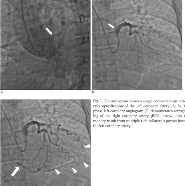
Fig. 1. The aortogram shows a single coronary sinus (arrow) and only opacification of the left coronary artery (A, B). The late phase left coronary angiogram (C) demonstrates retrograde fill- ing of the right coronary artery (RCA, arrow) into the pul- monary trunk from multiple rich collaterals (arrow heads) from the left coronary artery.
ly 1 in 300,000 children. The origin of the right coronary artery from the pulmonary artery is extremely rare.
Yamanaka and Hobbs detected 2 cases of ARCAPA out of 126,595 patients who underwent angiography and they concluded that ARCAPA accounts for 0.12% of all the coronary artery anomalies (4).
Comparisons between ARCAPA and ALCAPA were described by Williams (5). The most common presenta- tion of ARCAPA is a murmur. Unlike ALCAPA, there are no characteristic ECG findings associated with AR- CAPA, which often results in the classic syndrome of in- fant myocardial ischemia, infarction and when untreat- ed, death. Some authors have hypothesized that this is due to the lower oxygen demand of the right ventricle
compared to the left one and the smaller amount of my- ocardial territory fed by the RCA as compared to that of the LCA.
Although ARCAPA is uncommon, advanced diagnos- tic methods have led to an increase in diagnosing this malady during infancy and childhood. Patients with AR- CAPA are usually asymptomatic (5). Radke et al. re- viewed the literature and summarized 57 cases of this anomaly (6); it is typically revealed in children when they are examined for other congenital anomalies. Few of the cases of ARCAPA had myocardial ischemic symp- toms and they underwent re-implantation or CABG us- ing cardiopulmonary bypass. Therefore, more physi- cians will be faced with the dilemma of how to manage
C D
A B
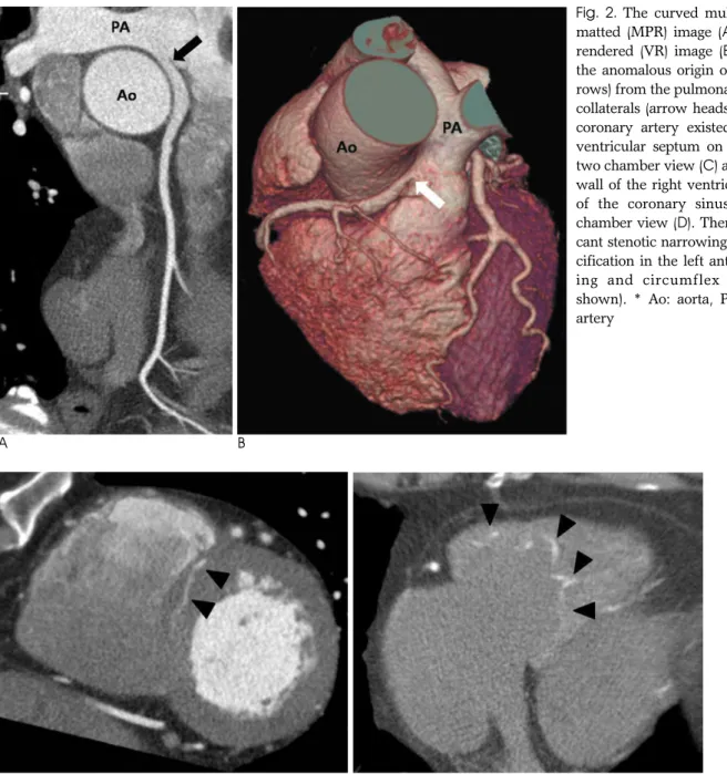
Fig. 2. The curved multiplanar refor- matted (MPR) image (A) and volume rendered (VR) image (B) demonstrate the anomalous origin of the RCA (ar- rows) from the pulmonary trunk. Rich collaterals (arrow heads) from the left coronary artery existed at the inter- ventricular septum on the short axis two chamber view (C) and the inferior wall of the right ventricle at the level of the coronary sinus on the four chamber view (D). There is no signifi- cant stenotic narrowing or intimal cal- cification in the left anterior descend- ing and circumflex arteries (not shown). * Ao: aorta, PA: pulmonary artery
Fig. 4. The postoperative curved MPR (A) and VR (B) images obtained after ligation of the original os of the RCA from the pulmonary trunk (arrow head) and creation of an anastomosis (arrow) between the right internal mammary artery (RIMA) and the RCA.
Fig. 3. Tc-99m tetrofosmin (TF) rest/stress myocardial perfusion SPECT (MSPECT). MSPECT shows the fixed and partly reversible perfusion defect and hypokinesia in the inferior wall. We thought that myocardial ischemia and infarction occurred in the RCA territory.
what has historically been considered a benign lesion.
Yet ARCAPA has been associated with cardiac symp- toms and sudden death (7, 8).
The accurate identification of coronary artery anom- alies is vital for patients with congenital heart disease, as the pattern and the course of the abnormality determine the need for treatment and this may affect the type of re- pair or the patients’ outcome. Coronary artery imaging with echocardiography may be difficult in some patients due to poor acoustic windows. Conventional coronary angiography is invasive and it may also be difficult be- cause of the lack of 3D information on the coronary artery as is related to its surrounding structures.
Recently, coronary angiography using MDCT has rapid- ly evolved as a promising, non-invasive method for as- sessing patients with coronary artery disease.
We report here on a first case of an elderly male pa- tient with isolated ARCAPA, which supplied rich collat- eral flows to the RCA and this was all detected by 64- slice MDCT. This is also the first report that describes the follow-up of ARCAPA by 64-slice MDCT after surgi- cal correction.
References
1. Radke PW, Messmer BJ, Haager PK, Klues HG. Anomalous origin of the right coronary artery: preoperative and postoperative hemo- dynamics. Am Thorac Surg 1998;66:1444-1449
2. Worsham C, Sanders SP, Burger BM. Origin of the right coronary artery from the pulmonary trunk: diagnosis by two-dimensional echocardiography. Am J Cardiol 1985;55:232-233
3. Soloff LA. Anomalous coronary arteries arising from the pul- monary artery. Am Heart J 1942;24:118-127
4. Yamanaka O, Hobbs RE. Coronary artery anomalies in 126, 595 patients undergoing coronary arteriography. Cathet Cardiovasc Diagn 1990;21:28-40
5. Williams IA, Gersony WM, Hellenbrand WE. Anomalous right coronary artery arising from the pulmonary artery: a report of 7 cases and a review of the literature. Am Heart J 2006;152:1004.e9- e17
6. Radke PW, Messmer BJ, Haager PK, Klues HG. Anomalous origin of the right coronary artery: preoperative and postoperative hemo- dynamics. Am Thorac Surg 1998;66:1444-1449
7. Kobayashi K, Tokunaga T, Isobe M. Images in cardiaology: a case of anomalous origin of right coronary artery from pulmonary artery complicated by acute myocardial infarction. Heart 2005;91:1130
8. Bossert T, Walther T, Doll N, Gummert JF, Kostelka M, Mohr FW.
Anomalous origin of the right coronary artery from the pulmonary artery combined with aortic valve stenosis. Ann Thoracic Sur 2005;79:347-348
대한영상의학회지 2010;62:113-117
폐동맥에서 기시한 우관상동맥:
MDCT를 이용한 진단과 수술 후 추적검사의 1예 보고11전남대학교병원 영상의학과
2화순전남대학교병원 영상의학과
임재훈∙최 송2∙강양준2∙선현주∙김윤현
63세 남자환자가 운동시 호흡곤란과 비정형적인 흉통을 주소로 내원하였다. 관상동맥조영술과 심전도 동조 64 채 널 컴퓨터 단층촬영에서 우관상동맥이 폐동맥에서 기시하는 관상동맥기형이 관찰되었다. 환자는 관상동맥우회술을 시행 받았다. 저자들은 심전도 동조 64 채널 컴퓨터 단층촬영을 이용하여 진단 및 수술 후 추적검사를 시행한 폐동 맥에서 기시하는 우관상동맥 기형의 증례를 보고하고자 한다.