59
DOI: 10.4046/trd.2009.67.1.59
ISSN: 1738-3536(Print)/2005-6184(Online) Tuberc Respir Dis 2009;67:59-62
CopyrightⒸ2009. The Korean Academy of Tuberculosis and Respiratory Diseases. All rights reserved.
흉관 삽관 후 발생한 국소성 재팽창성 폐부종 1예
순천향대학교 의과대학
1내과학교실,
2흉부외과학교실,
3영상의학교실
정혜경1, 장원호2, 김양기1, 이영목1, 황정화3, 김기업1, 어수택1A Case of Focal Reexpansion Pulmonary Edema after Chest Tube Insertion
Hye Kyoung Chung, M.D.
1, Won Ho Jang, M.D.
2, Yang Ki Kim, M.D.
1, Young Mok Lee, M.D.
1, Jung Hwa Hwang, M.D.
3, Ki-Up Kim, M.D.
1, Soo-Taek Uh, M.D.
1Departments of
1Internal Medicine,
2Radiology,
3Chest Surgery, School of Medicine, Soonchunhyang University, Seoul, Korea
Reexpansion pulmonary edema is not a common phenomenon after chest tube insertion but some reports from 0% to 14%. There are various resulting complications, including acute respiratory distress syndrome. We report a case of focal reexpansion pulmonary edema after chest tube insertion. A 49-year-old male came to the hospital due to ongoing dyspnea and left chest pain for 3 days. On chest X-ray, the patient had a left pneumothrax. We planned to insert a chest tube for symptom relief. To determine whether or not the chest had expanded as a result of the chest tube insertion, the patient underwent repeated chest X-rays the following day. The patient experienced brief respiratory symptoms upon initial suction; a chest PA showed patchy consolidated infiltration at the inserted site. After 5 days of conservative management, the recovered completely.Key Words: Reexpansion pulmonary edema, Pneumothorax, Chest tube insertion
Address for correspondence: Ki-Up Kim, M.D., Ph.D.
Department of Internal Medicine, School of Medicine, Soonchunhyang University, 22, Daesakwan-gil, Hannam- dong, Yongsan-gu, Seoul 143-740, Korea
Phone: 82-2-709-9027, Fax: 82-2-792-5812 E-mail: [email protected]
Received: May. 18, 2009 Accepted: Jul. 7, 2009
서 론
재팽창성 폐부종(reexpansion pulmonary edema)은 다량의 기흉 또는 흉수를 단기간에 과량 배출 시 만성적으 로 허탈 되었던 폐의 급격한 재팽창에 의하여 발생하는 폐부종으로, 흔하지 않지만, 심한 경우 급성 호흡부전증후 군으로 진행 될 수 있는 치명적 합병증이다1. 기전은 명확 하게 밝혀지지 않았으나, 허탈 되었던 폐의 계면활성 물질 의 감소와 저산소 손상에 의한 폐포의 혈관 투과성 변화 및 혈류 증가로 발생하는 것으로 알려져 있다2-4. 저자들은 국소적 기흉 환자에서 흉관 삽입 후 발생한 재팽창성 폐부 종이 발생된 환자를 경험하여 이달의 X-선에 증례를 보고
하는 바이다.
증 례
환 자: 백○○, 남자, 49세
주 소: 왼쪽 가슴의 통증과 호흡 곤란
현병력: 내원 3일 전 의자에 앉아 있던 도중 발생한 왼 쪽 가슴의 통증과 동반된 호흡 곤란으로 본원 외래 경유하 여 입원하였다.
과거력: 특이소견 없었다.
가족력: 특이소견 없었다.
진찰 소견: 내원 당시 혈압은 110/80 mmHg, 체온은 36.4oC, 맥박은 분당 76회, 호흡은 분당 16회이었다. 의식 은 명료하였으며, 흉부 청진에서 왼쪽 흉곽 하부의 호흡음 및 성음 진탕이 감소되었으나, 심음은 정상이었다. 그 외 신체검사에서 특이 소견은 없었다.
임상검사 소견: 내원 시 일반혈액검사는 백혈구 9,700/
mm3, 혈색소 15.4 g/dL, 혈소판 291,000/mm3, 임상화학
Image of the Month
HK Chung et al: Localized reexpansion pulmonary edema
60
Figure 1. Chest PA and left lateral from 49-year-old male, who had chest pain and dyspnea during three days, showed left pneu- mothorax. The patient had been shown total collap- sed at the area of lower lung (arrow) with small am- ount of pleural effusion but upper area had been spa- red due to adhesion by old tuberculous scar.
Figure 2. The next day af- ter chest tube insertion, chest PA showed infiltra- tion and consolidation at the left lower lung field.
Lateral film revealed loss of diaphragm shadow and prominent consolidation at the posterior basal area.
검사는 SGOT/SGPT 17/12 U/L, 알부민 4.1 g/dL, 혈청 Na+/K+/Cl- 142/3.8/102 mEq/L, BUN/Cr 21/1.0 mg/
dL이었다. 심전도 검사는 이상소견이 없었다.
영상의학 소견: 내원 당일 시행한 흉부 방사선 검사에 서 왼쪽 폐 하부에 기흉이 있고 적은 양의 흉수가 관찰되 었으며, 왼쪽 폐의 상부에는 병력에서는 확인되지 않았으 나 결핵에 의한 반흔을 발견할 수 있었고 기흉은 경한 정 도였다. 하부 폐는 총체적 허탈이 있었다(Figure 1).
치료 및 경과: 환자는 입원 당일 왼쪽 폐에 흉관을 삽입 후 10 cmH2O의 음압으로 흡입하였다. 삽관 후 다량의 기 포가 배출 된 후 기침과 호흡곤란을 호소하였으나 곧 증상
이 호전되었다. 고여 있었던 공기의 배출 후 더 이상의 공기의 누출은 없었다. 삽관 다음날 촬영한 흉부 방사선 사진에서 왼쪽 폐의 공기 음영은 소실되었으나 확장된 폐 에서 침윤성 경화를 보였고 측면 영상에서 왼쪽 후저부의 횡격막 경계의 소실과 경화를 보였다(Figure 2).
재팽창성 폐부종이 의심되어 흉부 전산화 단층 촬영을 시행하였고, 왼쪽 폐 첨부의 결핵의 자연치유 후 발생된 대기포가 다수 발견되어 기흉의 원인으로 판단되었고, 설 상엽 일부와 하엽의 의존부의 침윤 및 경화를 확인 할 수 있었다(Figure 3). 보존적 치료 후 증상의 호전과 침윤의 소실 그리고 더 이상의 공기의 누출이 없어 삽관을 제거하
Tuberculosis and Respiratory Diseases Vol. 67. No. 1, Jul. 2009
61
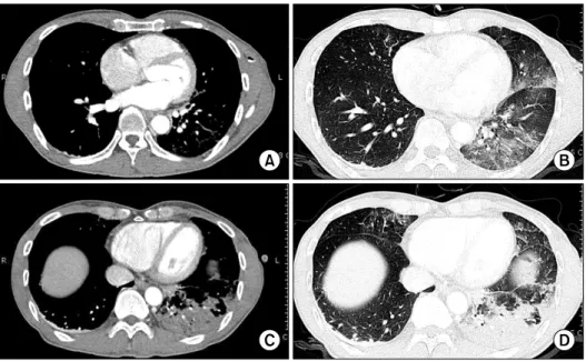
Figure 4. Chest PA and left lateral after removal of che- st tube after 5 day the le- sion, which had been occu- pied consolidation at post- erobasal area, were resolv- ed completely.Figure 3. Computerd tomography were checked immediately after detection of consolidation by chest radiography. At the upper level of totally collapsed area, parenchymal infiltrations were scattered from dependent area to lingular segment (A, B). The bottom level, patchy consolidation were detected almost all at the dependent area (C, D). Their differences of consolidation between upper and lower level were seems to reexpansion pressure from negative suction chamber.
고 입원 5일째 퇴원하였다(Figure 4).
고 찰
흉강 천자 후 발생한 재팽창성 폐부종은 1853년 Pinault 에 의해 처음으로 보고되었고, 대부분 보존적 치료 후 호 전을 보이나, 드물게 급성 호흡 증후군으로 인해 사망할
수 있다. 우리나라에서 보고는 일측성으로 발생되는 것이 대부분이나5,6, 긴장성 기흉의 감압 후 양측성 재팽창성 폐 부종의 발생도 보고 되었다7. 발병률은 아직 알려지지 않 았으며 일부의 보고에서는 0.9%, 그리고 다른 보고에서는 14%로 보고되었으며 40세 이하 그리고 기흉이 큰 경우에 발생 빈도가 높다8,9. 기전은 저산소 손상2,3 및 계면 활성 제 생산 감소로 인한 모세 혈관 막의 투과성 변화4, 재팽창
HK Chung et al: Localized reexpansion pulmonary edema
62
된 흉강 내 음압에 대한 혈관 내 정수압의 증가로 인한 유출 및 폐혈관의 투과성 변화로 추정되고 있다. 재팽창 성 폐부종은 환자가 최근 기흉이나 흉수로 인해 흉강 천자 를 시행한 병력, 호흡곤란 등의 증상과 방사선 소견의 변 화로 진단이 가능하다3. 무증상도 있으나, 발생 후 15분에 서 2시간 사이에 빈맥, 빈호흡, 호흡곤란과 청색증, 기침 과 거품을 동반한 붉은 객담, 발열, 저혈압, 흉통 등이 다 양하게 나타나며, 청진에서는 수포음이 들린다10. 방사선 소견을 살펴보면 첫 2∼4시간동안 폐포 충만 양상(alveolar filling pattern), 24∼48시간 후에는 부종으 로 진행, 4∼5일 동안 유지되다 보통 5∼7일이 지나면 호전이 관찰된다11. 재팽창성 폐부종은 72시간 이상 지속 된 폐의 허탈과 과다한 음압 사용에 의해 발생된다고 알려 져 있으며, 특히 흉수 천자 시 1,500 mL 이상을 단시간에 배액 시킬 때 그 위험성이 증가 하는데, 본 환자에서도 병력 청취로 약 3일 전 기흉이 발생된 것으로 추정되었으 며, 흉관 삽입 직후에는 증상이 없었으나, 음압을 이용하 여 기흉 제거 시 기침과 호흡 곤란을 호소하였던 예로서, 이는 왼쪽 폐의 기흉이 단시간에 걸쳐 제거됨으로써 발생 된 것으로 추정되었다.
치료는 충분한 산소 투여와 순환을 원활하게 하는 보존 적 치료를 하며, 심한 경우 기관 내 삽관 및 기계적 양압 호흡이 적응증이 될 수 있으며 2∼3시간 내에 빠른 폐부 종의 소실을 기대할 수 있다고 알려져 있다. 사망률은 20% 이하로 보고되고 있으나12, 그 원인은 동반되는 타 질환으로 인한 것으로 추정되며, 심혈관 질환 등 다른 기 저질환이 있는 환자에게서 그 위험성은 증가된다. 재팽창 성 폐부종의 예방을 위해 72시간 이상 지속된 흉수일 경 우, 점진적인 재팽창과 흉수를 1,000 mL 이하로 천천히 제거하는 것이 필요하며, 다량의 흉수가 있는 환자들에게 서는 1,500 mL 이하의 배액이 권장되고 있다. 또한 흉관 삽입 후 간헐적인 흉관 결찰도 필요하다11.
요 약
저자들은 흉통과 경도의 호흡곤란으로 내원하여 기흉 진단된 환자에서 치료적 흉관 삽입 시술 후, 갑작스런 음
압으로 허탈된 폐가 펴지면서 발생한 재팽창성 폐부종의 증례를 이달의 X-선에 보고하는 바이다.
참 고 문 헌
1. Steckel RJ. Unilateral pulmonary edema after pneu- mothorax. N Engl J Med 1973;289:621-2.
2. Mahajan VK, Simon M, Huber GL. Reexpansion pulmo- nary edema. Chest 1979;75:192-4.
3. Kernodle DS, DiRaimondo CR, Fulkerson WJ. Reexpan- sion pulmonary edema after pneumothorax. South Med J 1984;77:318-22.
4. Pavlin DJ, Nessly ML, Cheney FW. Increased pulmo- nary vascular permeability as a cause of re-expansion edema in rabbits. Am Rev Respir Dis 1981;124:422-7.
5. Jeong SW, Kim CM, Choi CH, Shin DJ, Bae HB, Chung SS, et al. Re-expansion pulmonary edema after chest tubing: a case report. Korean J Crit Care Med 2005;20:
87-91.
6. Kim YO, Lee JR, Kim KT, Choi WJ, Lee SI, Kim JW.
Hydrothorax after central vein catheterization for right internal jugular vein and contralateral reexpansion pul- monary edema after right chest tube insertion. Korean J Anesthesiol 2007;53:234-7.
7. Kim KU, Jung HK, Park HJ, Cha GY, Han SH, Hwang EW, et al. A case of bilateral reexpansion pulmonary edema after pleurocentesis. Tuberc Respir Dis 2001;51:
161-5.
8. Rozenman J, Yellin A, Simansky DA, Shiner RJ. Re-ex- pansion pulmonary oedema following spontaneous pneumothorax. Respir Med 1996;90:235-8.
9. Matsuura Y, Nomimura T, Murakami H, Matsushima T, Kakehashi M, Kajihara H. Clinical analysis of reexpan- sion pulmonary edema. Chest 1991;100:1562-6.
10. Brandstetter RD, Cohen RP. Hypoxemia after thoracent- esis: a predictable and treatable condition. JAMA 1979;
242:1060-1.
11. Tarver RD, Broderick LS, Conces DJ Jr. Reexpansion pulmonary edema. J Thorac Imaging 1996;11:198-209.
12. Mahfood S, Hix WR, Aaron BL, Blaes P, Watson DC.
Reexpansion pulmonary edema. Ann Thorac Surg 1988;
45:340-5.