• 검색 결과가 없습니다.

Korean Guidelines for Diagnosis and Management of Interstitial Lung Diseases: Part 5. Connective Tissue Disease Associated Interstitial Lung Disease

N/A
N/A
Protected

Academic year: 2021

Share "Korean Guidelines for Diagnosis and Management of Interstitial Lung Diseases: Part 5. Connective Tissue Disease Associated Interstitial Lung Disease"

Copied!
13
0
0

로드 중.... (전체 텍스트 보기)

전체 글

(1)

Korean Guidelines for Diagnosis and

Management of Interstitial Lung Diseases:

Part 5. Connective Tissue Disease Associated Interstitial Lung Disease

So-My Koo, M.D.

1,

* , Song Yee Kim, M.D., Ph.D.

2,

* , Sun Mi Choi, M.D.

3

and

Hyun-Kyung Lee, M.D., Ph.D.

4

, on behalf of Korean Interstitial Lung Diseases Study Group

1

Division of Allergy and Respiratory Medicine, Department of Internal Medicine, Soonchunhyang University Seoul Hospital, Soonchunhyang University College of Medicine, Seoul,

2

Division of Pulmonology, Department of Internal Medicine, Severance Hospital, Institute of Chest Diseases, Yonsei University College of Medicine, Seoul,

3

Division of Pulmonary and Critical Care Medicine, Department of Internal Meidicine, Seoul National University Hospital, Seoul,

4

Division of Pulmonary and Critical Care Medicine, Department of Internal Medicine, Inje University Busan Paik Hospital, Busan, Korea

Connective tissue disease (CTD) is a collection of disorders characterized by various signs and symptoms such as circulation of autoantibodies in the entire system causing damage to internal organs. Interstitial lung disease (ILD) which is associated with CTD is referred to as CTD-ILD. Patients diagnosed with ILD should be thoroughly examined for the co- occurrence of CTD, since the treatment procedures and prognosis of CTD-ILD are vary from those of idiopathic interstitial pneumonia. The representative types of CTD which may accompany ILD include rheumatoid arthritis, systemic sclerosis (SSc), Sjögren’s syndrome, mixed CTD, idiopathic inflammatory myopathies, and systemic lupus erythematous. Of these, ILD most frequently co-exists with SSc. If an ILD is observed in the chest, high resolution computed tomography and specific diagnostic criteria for any type of CTD are met, then a diagnosis of CTD-ILD is made. It is challenging to conduct a properly designed randomized study on CTD-ILD, due to low incidence. Therefore, CTD-ILD treatment approach is yet to been established in absence of randomized controlled clinical trials, with the exception of SSc-ILD. When a patient is presented with acute CTD-ILD or if symptoms occur due to progression of the disease, steroid and immunosuppressive therapy are generally considered.

Keywords: Lung Disease, Interstitial; Connective Tissue Disease; Asian Continental Ancestry Group; Guidelines as Topic; Diagnosis; Disease Management; Lung

Address for correspondence: Sun Mi Choi, M.D.

Division of Pulmonary and Critical Care Medicine, Department of Internal Meidicine, Seoul National University Hospital, 101 Daehak-ro, Jongno-gu, Seoul 03080, Korea

Phone: 82-2-2072-4915, Fax: 82-2-762-9662, E-mail: [email protected] Address for co-correspondence: Hyun-Kyung Lee, M.D., Ph.D.

Division of Pulmonary and Critical Care Medicine, Department of Internal Medicine, Inje University Busan Paik Hospital, 75 Bokji-ro, Busanjin-gu, Busan 47392, Korea

Phone: 82-51-890-6847, Fax: 82-51-890-6341, E-mail: [email protected]

*So-My Koo and Song Yee Kim contributed equally to this work.

Received: Feb. 1, 2019, Revised: Apr. 8, 2019, Accepted: Apr. 10, 2019, Published online: May. 31, 2019

cc It is identical to the Creative Commons Attribution Non-Commercial License (http://creativecommons.org/licenses/by-nc/4.0/).

Copyright © 2019

The Korean Academy of Tuberculosis and Respiratory Diseases.

(2)

Introduction

Connective tissue disease (CTD) is a disorder characterized by diverse symptoms and signs as autoantibodies circulate in the system, causing damage to internal organs. Interstitial lung disease (ILD) associated with CTD is called CTD-ILD.

At an early stage in diagnosing ILD, it is very important to examine the patient with respect to the existence of various causes of ILD. Of the known causes of ILD, CTD is the most crucial. If ILD is diagnosed in a CTD patient and conversely, if CTD is diagnosed in an ILD patient, CTD-ILD is diagnosed.

It is very critical to determine the existence of CTD in ILD pa- tients, because there are significant differences between CTD- ILD and idiopathic interstitial pneumonia (IIP) in terms of both treatment approach and prognosis. Some CTD-ILD pa- tients present with ILD prior to the CTD diagnosis. Therefore, even in those not previously diagnosed with CTD, any clinical symptoms suggestive of CTD, autoantibody test, and chest X- ray findings should be thoroughly checked to determine the existence of CTD.

Classification and Clinical Features of CTD-ILD

The incidence and clinical features of CTD-ILD differ de- pending on the form of the associated CTD. Across various types of ILD, the incidence of systemic sclerosis (SSc) is the highest, followed by idiopathic inflammatory myopathies (IIM), rheumatoid arthritis (RA), systemic lupus erythema- tous (SLE), and Sjögren’s syndrome.

1. RA associated ILD

ILD is the most common among RA-associated lung diseas- es, but the prevalence varies depending on the patient group or diagnostic method

1

. A wide range of RA associated ILD (RA-ILD) prevalence rates, 19%–44%, has been reported in prospective studies using chest high resolution computed to- mography (HRCT)

2-5

, because each author used different ILD definitions and diagnostic methods. A cross-sectional study in which chest HRCT was performed at an RA clinic regardless of respiratory symptoms reported an ILD prevalence of 19%.

In the study, chest HRCT had a higher sensitivity in diagnosing ILD than pulmonary function tests (PFT)

6

.

Although RA is more common among women, RA-ILD is reported to be more common in men. Some studies reported a male-to-female ratio of 2:1

7,8

. Typically, it occurs in patients in their 40s and 50s, and age and smoking history are known risk factors of ILD

9-11

. Rheumatoid factor and anti–cyclic citrullinated peptide were also found to be strongly associated with the occurrence of ILD

12-18

. In RA-ILD, the most common pathological finding in surgical lung biopsy is usual interstitial

pneumonia (UIP)

19

. 2. SSc associated ILD

SSc, also called scleroderma, has a lower incidence but a higher level of pulmonary infiltration compared with RA. Pul- monary function abnormalities due to ILD are observed in as high as 70% of SSc patients, and about a third of the patients show clinically significant evidence of ILD

1

. Chest HRCT is essential for diagnosing SSc associated ILD (SSc-ILD), and the most common chest HRCT finding is non-specific in- terstitial pneumonia (NSIP)

20

. In general, ILD is common in patients with diffuse SSc, a condition in which internal organs as well as the skin are involved, and the autoantibody profile was found to correlate with pulmonary infiltration

21-23

. The autoantibody most commonly found in patients with diffuse SSc is anti-topoisomerase I, also known as anti–scl-70. It has been reported that ILD occurred in 85% of anti–scl-70 positive patients and that the antibody concentration level was corre- lated with the severity and activity of ILD

24,25

.

Fibrotic NSIP is the most common pattern in the pathologi- cal findings of SSc-ILD, and cellular NSIP is found in less than 10% of patients. Typical features of idiopathic pulmonary fibrosis, such as severe fibrosis and fibroblastic foci, are ob- served in less than 30% of cases with UIP

26-28

.

SSc-ILD patients are at greatest risk for ILD progression in the first 3–4 years (45%–55%), and some patients show rapid pulmonary function decline during this time. Accord- ingly, PFT should be performed on a regular basis. Treatment should be considered in SSc-ILD patients under the follow- ing conditions: (1) if at the time of the initial diagnosis of ILD, ILD involvement exceeds 20% in chest HRCT, or forced vital capacity (FVC) is less than 70% of the predicted value in PFT, or (2) if diffusing capacity of the lungs for carbon monoxide (DLCO) is over 15% or FVC over 10% in a follow-up PFT

6,29

.

SSc-ILD in conjunction with pulmonary arterial hyperten- sion (PAH) is a major cause of death among SSc patients

30

. SSc accompanied by ILD and PAH was found to have 5 times higher mortality risk than that of SSc accompanied by PAH alone

31

.

3. Sjögren’s syndrome associated ILD

Studies have reported that pulmonary infiltration occurs in 9%–24% of all patients with Sjögren’s syndrome, and that abnormal findings in PFT, bronchoalveolar lavage (BAL), and chest HRCT were observed in 75% of patients with asymp- tomatic Sjögren’s syndrome

32-35

. Generally, pulmonary infil- tration manifests in the end stage of Sjögren’s syndrome and rarely occurs as the first manifestation

36

.

In Sjögren’s syndrome, not only interstitial tissues in the lung

but the entire respiratory system could be infiltrated, and so

the patient could also present with small airway disease such

(3)

as follicular bronchiolitis, aside from ILD. In whatever form, 10-year mortality increases by more than 4 times in Sjögren’s syndrome patients with pulmonary infiltration compared to those without

32

.

NSIP is the most common pathological finding. Studies have found NSIP in 28%–61% of all patients with the syn- drome, and additionally, lymphocytic interstitial pneumonia (LIP) in 17%

37,38

.

4. Mixed connective tissue disease associated ILD Mixed connective tissue disease (MCTD) was defined in 1972 for the first time, and there is still a controversy around this category. Generally, it refers to an autoimmune disease with positive anti–U1 ribonucleoprotein antibody. Pulmonary infiltration is quite common in MCTD, found in 47%–78% of patients

39,40

. According to the literature, severe ILD occurred in approximately 19% of MCTD patients, and it was associated with anti–Ro-52 antibody but not with disease duration or smoking history

41,42

.

5. IIM associated with ILD

IIM include polymyositis (PM), dermatomyositis (DM), and clinically amyopathic dermatomyositis, in each of which a varying degree of skin, muscle, joint, and lung infiltration is present. IIM associated with ILD (IIM-ILD) shows a variety of disease progression patterns from asymptomatic to rapidly progressive

43,44

. The prevalence of ILD in IIM is also found to vary considerably, between 20%–78%, and comorbidity and mortality rates are reported to generally increase if ILD co-exists

45-48

. In IIM-ILD versus other CTD types, ILD more frequently occurs as the very first manifestation before it pro- gresses to a systematic disease

49-52

.

Antisynthetase syndrome (AS) is a specific subset of IIM, with positive antisynthetase antibodies associated with protein synthesis. It is a syndrome characterized by the co- occurrence of ILD and the presence of some of the following manifestations: fever, joint pain, Raynaud’s phenomenon, and mechanic’s hands. Of various muscle-specific antibodies, anti–

Jo-1 antibody is most commonly used in the diagnosis. As can be seen in the inclusion of ILD as an essential element in diag- nosing AS, the importance of ILD in assessment of prognosis is also great. Like other types of IIM-ILD, AS-ILD too shows diverse disease progression patterns. It has been reported that approximately 50% of patients develop acute respiratory fail- ure

51,53,54

.

6. SLE associated with ILD

In SLE, pulmonary infiltration occurs in as high as 33%–50%

of patients. But, in most patients, the entire respiratory system (thoracic cavity, lung parenchyma, airway, and pulmonary

vessels) is infiltrated, and pulmonary infiltration of the pat- tern shown in ILD tends to be relatively infrequent (1%–15%) compared to other types of CTD

43,55,56

. It has been reported that the longer the duration of SLE (over 10 years), the higher the risk of ILD

57

and that SLE is strongly associated with ILD in the presence of Raynaud’s phenomenon, positive response to anti–U1 RNP antibody, sclerodactyly, nailfold capillary loops, and old age

58,59

.

Diagnosis of CTD-ILD

1. Assessment of disease history and the presence of CTD

1) Patients already diagnosed with CTD

If ILD co-occurs in patients already diagnosed with CTD based on CTD diagnostic criteria, it is relatively easy to make a diagnosis of CTD-ILD. But the possibility of ILD occurring due to a drug used to treat CTD or infection should be exclud- ed

3,35,60-63

.

2) Patients never diagnosed with CTD

In some patients, ILD appears as the first manifestation among diverse clinical features of CTD. Such cases are most commonly observed in IIM (approximately 10%–30%), and additionally, the pattern is reported in some RA patients, and also in SSc patients though very rare

64,65

.

If a patient without notable disease history rapidly develops dyspnea over several weeks to several months and eventu-

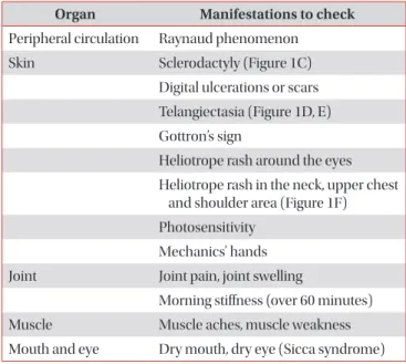
Table 1. Core manifestations in ILD patients to diagnose underlying CTD

66

Organ Manifestations to check Peripheral circulation Raynaud phenomenon

Skin Sclerodactyly (Figure 1C)

Digital ulcerations or scars Telangiectasia (Figure 1D, E) Gottron’s sign

Heliotrope rash around the eyes Heliotrope rash in the neck, upper chest

and shoulder area (Figure 1F) Photosensitivity

Mechanics’ hands Joint Joint pain, joint swelling

Morning stiffness (over 60 minutes)

Muscle Muscle aches, muscle weakness

Mouth and eye Dry mouth, dry eye (Sicca syndrome)

ILD: interstitial lung disease; CTD: connective tissue disease.

(4)

ally experiences respiratory failure while chest HRCT shows a ground glass opacity that is gradually progressing, the pos- sibility of CTD-ILD should be considered. Particularly, if the level of muscle enzymes (e.g., creatinine phosphokinase, and aldolase) increases, IIM should be suspected as the underly- ing disease

65

. Even in cases with chronic ILD, CTD could be the underlying disease. Hence, whether or not the patient presents any symptom or other manifestation suggestive of CTD, this possibility should be considered (Table 1)

66

. For instance, Raynaud’s phenomenon, dry eye and mouth, and muscle weakness tend to have low specificity, but the specific- ity of mechanic’s hands (Figure 1A) or Gottron’s papules (Fig- ure 1B) is high. Thus, if mechanic’s hands or Gottron’s papules are present, DM is highly likely to be an underlying disease

67

. Such systemic symptoms or other manifestations that raise a

suspicion of CTD often occur in women under age 50

68-71

. 2. Radiologic findings (chest HRCT findings)

It is not possible to differentiate IIP and CTD-ILD based on radiologic findings. CTD-ILD often manifests itself in NSIP or organizing pneumonia (OP), or LIP (albeit rare) (Figure 2A–

C), and IIP most often manifests itself in UIP (Figure 3). Thus, if a typical UIP is not found in an imaging test, CTD is likely to be the underlying disease

60

. An exception is that a radiologic finding of UIP is the most common in RA-ILD, and is often found in SSc-ILD, as well. The prognosis of RA-ILD is known to be poor in patients with a UIP pattern compared to those without UIP

60

.

The radiologic pattern of ILD in CTD-ILD patients dif-

Figure 1. (A) Mechanic’s hand, cracking and fissuring along the sides of the digits and palm. (B) Gottron’s papules, red and scaly papules that erupt on the metacarpophalangeal joints. (C) Sclerodactyly, fixed fingers in a semi-flexed position with tightened and wax like skin. (D) Digi- tal ulceration, an ulceration on the tip of index finger. (E) Telangiectasias, multiple dilated facial small vessels. (F) Heliotrope rash, violaceous erythema on the upper eyelids (The patient provided verbal consent for the picture).

Figure 2. (A) Radiologic pattern of non-specific interstitial pneumonia in a patient with systemic sclerosis. High resolution computed to-

mography (HRCT) image shows bilateral subpleural and basal predominant fine reticular pattern and ground-glass opacity. (B) Radiologic

pattern of organizing pneumonia in a patient with dermatomyositis. HRCT image shows multiple peripheral patch consolidations. (C) Radio-

logic pattern of lymphocytic interstitial pneumonia in a patient with Sjögren’s syndrome. HRCT image shows multifocal variable-sized, thin-

walled, cystic lesions on both lungs.

(5)

fers according to CTD type. In RA, UIP is the most common (50%–60%); in SSc, NSIP is the most common radiologic finding (80%–90%), although UIP is also found in as many as 10%–20%; in MCTD, NSIP is the most common; and, in PM and DM, NSIP, OP, UIP, and diffuse alveolar damage may be found

65

.

3. Surgical lung biopsy

It is still controversial whether surgical lung biopsy should be performed to confirm the diagnosis when CTD-ILD is suspected. The prognosis may differ depending on the patho- logical finding (UIP vs. others), and with biopsy, differential diagnoses of other diseases as well as ILD are possible. But, complications may occur following surgical lung biopsy, and thus the decision should be made by considering the pros and cons. In some patients, BAL is an alternative to biopsy, and us- ing this procedure, infection can be excluded and the number

of cells is estimated

65

.

Because a specific pathology does not exist in CTD-ILD, IIP and CTD-ILD cannot be completely differentiated based on pathological findings alone. However, because the proportion of NSIP is high in CTD-ILD, the presence or absence of under- lying CTD should be looked for if NSIP is found on pathologi- cal analysis

28,72-78

.

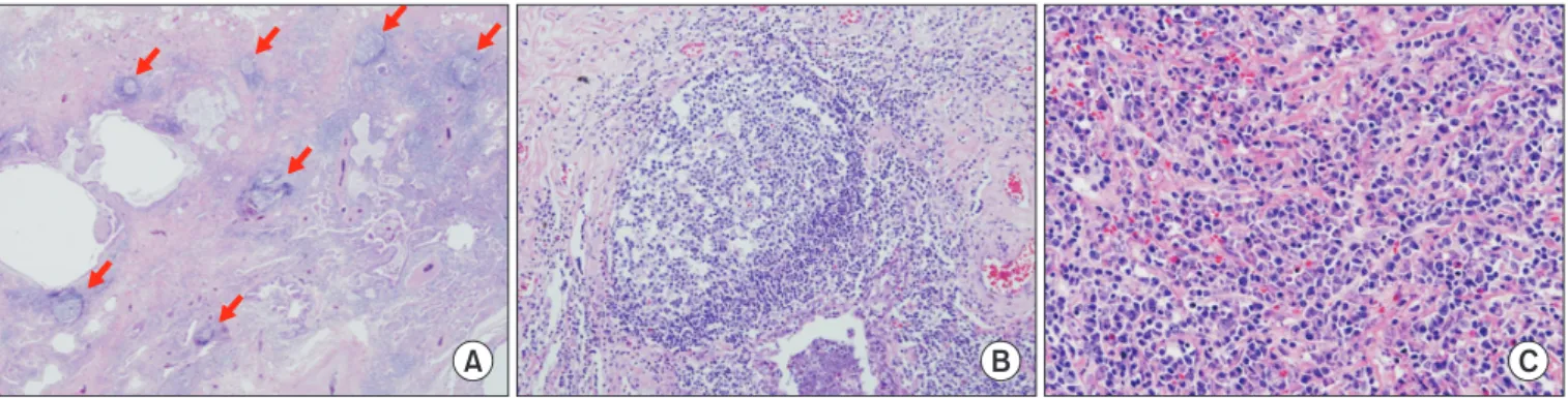
In addition, among those with CTD-ILD with a UIP pattern there were more germinal centers, lymphoid hyperplasia, plasma cells, and fewer fibroblastic foci and honeycombing, compared with cases of IIP with UIP. If the aforementioned features are found in conjunction with UIP, the presence or absence of CTD should be examined, because the co-occur- rence of lymphocytic or follicular bronchiolitis is common (Figure 4)

79-83

.

4. Autoantibodies

When a patient is diagnosed with ILD, autoantibody testing should be performed to determine the likelihood of CTD-ILD.

As shown in Table 2, cases exist in which an autoantibody is found but the clinical features do not correspond to the diag- nosis of CTD or do not meet specific CTD diagnostic criteria.

However, since such patients may present clinical features of CTD later, continuous follow-up is required and consultation with a rheumatologist for the symptoms is necessary

64,65

.

Treatment of CTD-ILD

1. Acute presentations of CTD-ILD

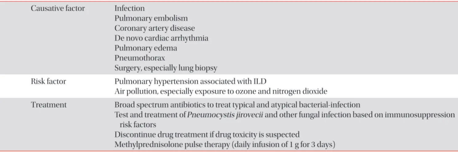
Acute presentation of CTD-ILD manifests itself in de novo acute interstitial pneumonia or an acute exacerbation of un- derlying CTD

65

. The treatment approach for acute exacerba- tion of CTD-ILD has not yet been established, but convention- ally, it is treated using intravenous methylprednisolone (1 g/

Figure 4. Histopathology of the lung in a patient with interstitial pneumonia with autoimmune features. (A) Usual interstitial with lymphoid follicles (×10). (B) Lymphoid bronchiolitis (×100). (C) lymphoplasmacytoid cell infiltrates (×200). Hematoxylin eosin saffron. Courtesy of Prof. Shim HS, Yonsei University.

Figure 3. Radiologic pattern of usual interstitial pneumonia in a

patient with rheumatoid-arthritis. High resolution computed to-

mography image shows bilateral subpleural honeycombing and

reticular opacity with traction bronchiectasis.

(6)

day intravenously for 3 days) followed by oral prednisolone (1 mg/kg/day). Cyclosporine (500–750 mg/m

2

intravenously) and cyclophosphamide (CYC) (2–3 mg/kg/day) may addi- tionally be administered (Table 3). Despite immunosuppres- sive therapy, patients presenting an acute exacerbation often need a ventilator and the mortality rate is high (83.3%)

65,84,85

. 2. Chronic presentations of ILD

The fundamental treatment of CTD-ILD is to tackle the primary cause of CTD. Evidence regarding treatment effect in

CTD-ILD is lacking, because comparison group studies have not been conducted with the exception of SSc-ILD. Consen- sus has not yet formed regarding treatment initiation for CTD- ILD, but conventionally, a steroid or immunosuppressive drug is administered in patients in whom clinical features are severe, symptoms have recently started to progress, or the du- ration of systemic disease has been short

86

. Lung transplanta- tion may be considered if the patient does not respond to drug therapy and ILD worsens.

Table 2. Types and significance of autoantibodies

65

Test Interpretation

Antinuclear antibody It is nonspecific, but the likelihood of CTD is high if the titer is high Rheumatoid factor It may increase in RA, but is nonspecific in other types of CTD

Scl-70 Associated with SSc

RNP Associated with MCTD

SSA, SSB Associated with Sjögren’s syndrome

Anti-CCP Associated with RA

ANCA It may be observed in pure ILD, though rare

Jo-1 Myositis-specific antibody

Tests associated with other types of myositis

Antisynthetase PL-7, PL-12, EJ, and OJ antibodies → all associated with ILD

MDA-5 Associated with invasive Gottron’s papules and severe ILD

PMScl Associated with polymyostis co-occurring with sclerosis

Ro-52 Associated with severe ILD

CPK, Aldolase Muscle enzymes. It may increase in myositis, but may be normal in clinically amyopathic dermatomyositis

CTD: connective tissue disease; RA: rheumatoid arthritis; SSc: systemic sclerosis; RNP: ribonucleoprotein; MCTD: mixed connective tissue disease; CCP: cyclic citrullinated peptide; ANCA: antineutrophil cytoplasmic antibody; ILD: interstitial lung disease.

Table 3. Summary of acute presentation of CTD-ILD Causative factor Infection

Pulmonary embolism Coronary artery disease De novo cardiac arrhythmia Pulmonary edema

Pneumothorax

Surgery, especially lung biopsy

Risk factor Pulmonary hypertension associated with ILD

Air pollution, especially exposure to ozone and nitrogen dioxide

Treatment Broad spectrum antibiotics to treat typical and atypical bacterial-infection

Test and treatment of Pneumocystis jirovecii and other fungal infection based on immunosuppression risk factors

Discontinue drug treatment if drug toxicity is suspected

Methylprednisolone pulse therapy (daily infusion of 1 g for 3 days)

CTD-ILD: connective tissue disease associated with interstitial lung disease.

(7)

1) Initial treatment of CTD-ILD

For the initial treatment of CTD-ILD, steroids are commonly used, but the recommendation as to which steroid is appropri- ate is undecided. The dose, route of administration, treatment duration, and dose tapering vary according to individual pa- tients’ clinical picture and the clinician’s impression (Table 4)

65

. As the most common ILD form in IIM-ILD patients is OP, pred- nisolone 1 mg/kg/day is often used, i.e., the dose at which clini- cians are relatively experienced in the treatment of cryptogenic organizing pneumonia (COP). However, treatment response to the therapy is reported to be low in the all forms of IIM-ILD except COP

87

.

Azathioprine (1–2 mg/kg/day) is often administered as combination therapy with a steroid to treat CTD-ILD, but evi- dence regarding monotherapy is insufficient

88

. A retrospective survival analysis conducted on 192 IIM-ILD patients showed lower mortality in the group administered with azathio- prine (hazard ratio, 0.35; 95% confidence interval, 0.16–0.74;

p=0.0064)

47

, but there is no evidence of an improvement in the survival rate in other forms of CTD-ILD.

CYC is the only immunosuppressive investigated in a ran- domized controlled trial. In SSc-ILD, FVC was improved after 1-year treatment in oral CYC (2 mg/kg/day) group compared with the control group

89

. Evidence on pulmonary function improvement is lacking in other types of CTD-ILD aside from SSc-ILD.

In a study with 125 patient cases with SSc-ILD, IIM-ILD, and RA-ILD, mycophenolate mofetil (MMF; 1.0–1.5 g bid) showed a reduction in the required dose of steroid and an improve- ment in FVC

90

. In addition, a recent randomized controlled trial with SSc-ILD patients demonstrated that the effect of MMF was similar to that of CYC

91

.

A retrospective study in IIM-ILD patients found that com- pared to the groups administered with CYC or cyclosporine in combination with a steroid, the group in whom tacrolimus was added had more favorable event-free survival

92

. The typi- cal dose of tacrolimus is 1–3 mg/day

65

.

Currently, a randomized controlled trial with rapidly pro- gressive CTD-ILD (including SSC-ILD, IIM-ILD, and MCTD- ILD) is under way to compare the effects of CYC and rituximab

with FVC improvement as the primary endpoint

93

.

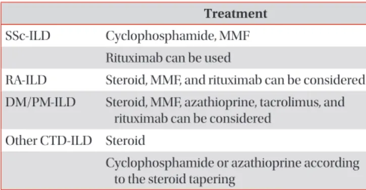
Table 5 summarizes treatment options for each CTD-ILD.

2) Treatment of SSc-ILD

(1) PICO for treatment of patients with SSc-ILD: In SSc- ILD, is CYC more effective compared with placebo?

(2) Summary of recommendations for the treatment of patients with SSc-ILD: We suggest that the use of CYC may be considered in consultation with an expert, in some SSc- ILD patients experiencing dyspnea and reduced pulmonary function (weak recommendation, level of evidence; low).

FVC, total lung capacity (TLC), dyspnea, and quality of life index improved in the patient group administered with oral CYC over 1 year compared to placebo. A 1-year follow up after treatment termination showed that the improvement in dys- pnea was maintained, but other indices returned to the levels prior to drug administration. Thus, evidence for long-term effectiveness of CYC is not sufficient, but it improved dyspnea and had a short-term effect in improving pulmonary function.

The effect of CYC in SSc-ILD patients has been reported in a considerable number of studies, but most of the studies were retrospective. Even the prospective studies tend to lack a comparison group or be observational. The authors con- ducted a systematic literature review on randomized placebo- controlled studies to investigate whether CYC has a positive effect on various SSc-ILD related indices such as the rate of pulmonary function decline, severity of dyspnea, quality of life, and functional ability. But the authors could not conduct meta-analysis because there was only one randomized pla- cebo-controlled trial on CYC, i.e., Tashkin et al.’s Scleroderma Lung Study (SLS)

89

published in N Engl J Med in 2006.

In SLS, 158 symptomatic SSc-ILD patients in whom disease activity was confirmed on either BAL or chest HRCT were randomly assigned to one of two groups, a group treated with CYC (2 mg/kg/day) for a year and a placebo group. During the study period, patients could be treated with prednisolone up

Table 4. Initial treatment of CTD-ILD

85

Classification Most types of CTD-ILD (with an exception of SSc-ILD) Dose Initial dose: prednisolone 0.5–1 mg/kg

Maintenance dose: tapered to prednisolone 10 mg/day or lower Steroid tapering guidance Oral or intravenous

cyclophosphamide or concurrent use of oral azathioprine

CTD-ILD: connective tissue disease associated interstitial lung dis- ease (ILD); SSc-ILD: systemic sclerosis associated ILD.

Table 5. Summary of treatment of CTD-ILD Treatment SSc-ILD Cyclophosphamide, MMF

Rituximab can be used

RA-ILD Steroid, MMF, and rituximab can be considered DM/PM-ILD Steroid, MMF, azathioprine, tacrolimus, and

rituximab can be considered Other CTD-ILD Steroid

Cyclophosphamide or azathioprine according to the steroid tapering

CTD-ILD: connective tissue disease associated interstitial disease

(ILD); SSc-ILD: systemic sclerosis associated ILD; MMF: mycophe-

nolate mofetil; RA-ILD: rheumatoid arthritis associated ILD; DM/PM-

ILD: dermatomyositis/polymyositis associated ILD.

(8)

to 10 mg/day. If an FVC reduction of 15% versus the baseline continued over 1 month from month 3 after treatment initia- tion, it was considered as treatment failure and the patient was unblinded and treated with CYC. The primary endpoint of the study was the FVC (expressed as a percentage of the predicted value) at 1 year after adjusting for baseline values and treatment group. Secondary endpoints were TLC, change in DLCO, disability index of health assessment questionnaire (HAQ), SF-36, transitional dyspnea index (TDI), and skin thickness score. Between-group difference in FVC change after 1 year of CYC administration was 2.53% (95% confidence interval, 0.28–4.79; p<0.03), indicating that treatment with CYC significantly slowed down the rate of FVC reduction. Of the secondary endpoints, TLC, HAQ, TDI, and skin thickness score were significantly improved in the CYC group in com- parison to the placebo group.

Side effects of CYC that occurred during the study were he- maturia (hemorrhagic cystitis), leukopenia, neutropenia, ane- mia, pneumonia, etc.

89,94

. Therefore, it is recommended to reg- ularly perform blood test and use mesna for the prevention of hemorrhagic cystitis, in patients treated with CYC. Long-term use of the drug is not recommended because of increased risk for bladder cancer and other malignant diseases.

After the completion of this SLS study, the researchers followed up the patients for an additional 1 year without ad- ministering a drug in order to investigate the duration of drug effect

94

. During the additional 1-year follow-up, if treatment failure (an FVC reduction over 15% compared to the baseline) occurred in either group, the patient was treated with CYC or an immunosuppressive according to the clinician’s decision.

During the 1-year follow-up, between-group differences in FVC and TLC were significant for the first 6 months, that is, the drug effect was maintained for a total of 18 months from the start of the study. But group difference disappeared by month 12 after treatment termination (that is, 24 months after the start of the study). An additional analysis showed a larger drug effect in patients with FVC less than 70%. On the other hand, the improvement of TDI (an index of dyspnea) was maintained up to month 12 after treatment termination

94

.

In summary, a year-long treatment with CYC administra- tion improved pulmonary function, dyspnea, and quality of life and the effects were maintained up to 6 months after treat- ment termination. One year after treatment termination, two groups still showed a significant difference in dyspnea but not in FVC or TLC. Based on the improvement of dyspnea that was maintained as long as 1 year after treatment termination and a short-term improvement of pulmonary function, CYC may be useful in some SSc-ILD patients. Thus, in SSc-ILD pa- tients experiencing dyspnea and reduced FVC, a short-term use of CYC may be considered in consultation with an expert.

Regarding drugs other than CYC, first, a study showed that steroid therapy (0.5–1 kg/kg of prednisolone) could be ex- pected to slow down pulmonary function decline

95

, though

the effect of steroid in treatment of SSc-ILD is controversial.

High dose steroid is not recommended because renal crisis can rapidly develop if the daily dose of prednisolone is over 10 mg

86

.

MMF has been known to suppress lymphoproliferation and to have an antifibrotic effect. A recently presented random- ized comparison group study, Scleroderma Lung Study II (SLS II), compared a patient group administered with MMF (1,500 mg bid) for 24 months and a patient group administered with CYC (2 mg/kg/day) for 12 months. There was little difference in the speed of FVC reduction between the MMF group and the CYC group, and the counts of leucocytes and platelets in the MMF group did not decrease as much as in the CYC group

91

. Based on the findings, MMF may be considered as initial and maintenance therapies in the treatment of SSc-ILD.

Lately, hematopoietic stem cell transplantation (HSCT) has been attempted in SSc-ILD patients. A randomized controlled trial that compared the effects of HSCT and CYC reported that HSCT helped improve long-term event-free survival and overall survival, but recurrence rate was higher

96

. Accordingly, HSCT may be considered in cases in which patients did not respond to immunosuppressive therapy or the disease had progressed.

In an open-label randomized controlled trial that investi- gated rituximab (a B cell depletion therapy) in SSc-ILD, the drug showed an effect of increasing FVC and DLCO 1 year af- ter commencement of therapy

97

. However, the study included only 12 patients in the sample, which is very small. So far, there is not enough evidence regarding rituximab, and large- scale randomized comparison group research is needed

98

. As for now, it is believed that patients not responding to the currently available immunosuppressives may be tested on rituximab in a limited manner.

Lung transplantation is the only treatment available to prolong survival of SSc-ILD patients not responding to drug therapy. To improve the prognosis following lung transplanta- tion, suitable patient selection is important. A recent study has proposed the following as lung transplantation exclusion criteria: uncontrolled active inflammatory myopathies, active digital ulcerations, severe SSc associated gastrointestinal dis- ease, cardiac arrhythmia, unstable renal function over the last 3 months, and high risk of SSc associated renal crisis

99

. How- ever, standardized criteria have not yet been established and further research is warranted.

3) Treatment of RA-ILD

(1) PICO for treatment of patients with RA-ILD: In RA- ILD, do steroids and immunosuppressants (MMF, rituximab) slow the worsening of FVC compared with placebo?

(2) Summary of recommendations for the treatment

of patients with RA-ILD: We suggest that, in clinically se-

vere cases, the use of the steroid, MMF, and rituximab may be

considered in consultation with an expert (recommendation;

(9)

expert opinion, level of evidence; low).

If RA-ILD deteriorates despite administration of disease modifying antirheumatic drugs (DMARDs: RA treatment) and tumor necrosis factor inhibitors, conventionally high-dose prednisolone therapy is administered

100

. But, the evidence for the effectiveness and safety of drug therapy is weak

100

and comparison group studies have never been conducted to evaluate drugs in RA-ILD patients.

The authors attempted a systematic literature review and meta-analysis on the effects of steroid and immunosuppres- sive therapy (MMF and rituximab) in RA-ILD, but could not perform comparative analysis because no study compared steroid or immunosuppressive therapy to placebo. Below are the findings reported in a few single-group studies that used steroid or immunosuppressive therapy such as MMF and rituximab in RA-ILD patients. In a retrospective cohort con- sisting of 40 RA-ILD patients, baseline FVC improved when prednisolone (1 mg/kg/day) was administered for 6 weeks and then tapered down to 10 mg/day while a DMARD was administered during a follow-up over 6–8 months

101

. A retro- spective single-group study was conducted with 18 CTD-ILD patients including some with RA-ILD and it was found that pulmonary function stabilized and prednisolone mainte- nance dose decreased during a median of 2.5-year follow-up after treatment with MMF

90

. A prospective cohort study

102

and two retrospective studies

103,104

with RA-ILD patients reported that rituximab stabilized pulmonary function. Also, it was re- ported that when rituximab (1,000 mg at day 1, 15, and again at weeks 24 and 26, with methotrexate) was used concomi- tantly with methotrexate, FVC was maintained in six out of seven RA-ILD patients after 48 weeks

102

. Also, some case stud- ies reported that azathioprine

105

, abatacept

106

, tocilizumab

107

, and infliximab

108

each stabilized pulmonary function in RA- ILD patients.

There is no clear consensus as to when to initiate treatment with the drugs mentioned above. Since no other drug has so far been demonstrated to be effective in RA-ILD, the use of steroids, MMF, or rituximab may be considered for clinically severe cases, in consultation with a specialist.

Prognosis of CTD-ILD

The prognosis of CTD-ILD patients varies according to the type of causative CTD. RA-ILD comprises 10%–20% of deaths among RA patients

109

, and 10% of deaths among all ILD pa- tients

7-9

. In SSc-ILD, it is reported that 10-year survival follow- ing diagnosis lies in the range 29%–69%. and that in 45%–55%

of the patients, pulmonary function deteriorates within the first 3 years, and in 16% severe restrictive pulmonary disease develops

1

. In Sjögren’s syndrome the 5-year survival is report- ed to be 84%

110

.

If ILD progresses in spite of immunosuppressive therapy,

lung transplantation may be considered. The prognosis of CTD-ILD after lung transplantation is reported to be not very different from that of lung transplantation in IIP patients.

Authors’ Contributions

Conceptualization: Choi SM, Lee HK. Formal analysis: Koo SM, Km SY, Choi SM, Lee HK. Data curation: Koo SM, Choi SM. Writing - original draft preparation: Koo SM, Kim SY. Writ- ing - review and editing: Choi SM, Lee HK. Approval of final manuscript: all authors.

Conflicts of Interest

No potential conflict of interest relevant to this article was reported.

Funding

No funding to declare.

References

1. Steen VD, Conte C, Owens GR, Medsger TA Jr. Severe re- strictive lung disease in systemic sclerosis. Arthritis Rheum 1994;37:1283-9.

2. Fewins HE, McGowan I, Whitehouse GH, Williams J, Mallya R. High definition computed tomography in rheumatoid arthritis associated pulmonary disease. Br J Rheumatol 1991;30:214-6.

3. Gabbay E, Tarala R, Will R, Carroll G, Adler B, Cameron D, et al. Interstitial lung disease in recent onset rheumatoid arthritis. Am J Respir Crit Care Med 1997;156(2 Pt 1):528-35.

4. McDonagh J, Greaves M, Wright AR, Heycock C, Owen JP, Kelly C. High resolution computed tomography of the lungs in patients with rheumatoid arthritis and interstitial lung disease. Br J Rheumatol 1994;33:118-22.

5. Cortet B, Perez T, Roux N, Flipo RM, Duquesnoy B, Delcam- bre B, et al. Pulmonary function tests and high resolution computed tomography of the lungs in patients with rheu- matoid arthritis. Ann Rheum Dis 1997;56:596-600.

6. Winstone TA, Assayag D, Wilcox PG, Dunne JV, Hague CJ, Leipsic J, et al. Predictors of mortality and progression in scleroderma-associated interstitial lung disease: a system- atic review. Chest 2014;146:422-36.

7. Cavagna L, Monti S, Grosso V, Boffini N, Scorletti E, Crepaldi G, et al. The multifaceted aspects of interstitial lung disease in rheumatoid arthritis. Biomed Res Int 2013;2013:759760.

8. de Lauretis A, Veeraraghavan S, Renzoni E. Review series:

(10)

aspects of interstitial lung disease: connective tissue dis- ease-associated interstitial lung disease: how does it differ from IPF? How should the clinical approach differ? Chron Respir Dis 2011;8:53-82.

9. Bongartz T, Nannini C, Medina-Velasquez YF, Achenbach SJ, Crowson CS, Ryu JH, et al. Incidence and mortality of in- terstitial lung disease in rheumatoid arthritis: a population- based study. Arthritis Rheum 2010;62:1583-91.

10. Assayag D, Lubin M, Lee JS, King TE, Collard HR, Ryerson CJ. Predictors of mortality in rheumatoid arthritis-related interstitial lung disease. Respirology 2014;19:493-500.

11. Saag KG, Kolluri S, Koehnke RK, Georgou TA, Rachow JW, Hunninghake GW, et al. Rheumatoid arthritis lung disease:

determinants of radiographic and physiologic abnormali- ties. Arthritis Rheum 1996;39:1711-9.

12. Turesson C. Extra-articular rheumatoid arthritis. Curr Opin Rheumatol 2013;25:360-6.

13. Doyle TJ, Lee JS, Dellaripa PF, Lederer JA, Matteson EL, Fischer A, et al. A roadmap to promote clinical and transla- tional research in rheumatoid arthritis-associated intersti- tial lung disease. Chest 2014;145:454-63.

14. Kelly CA, Saravanan V, Nisar M, Arthanari S, Woodhead FA, Price-Forbes AN, et al. Rheumatoid arthritis-related intersti- tial lung disease: associations, prognostic factors and physi- ological and radiological characteristics: a large multicentre UK study. Rheumatology (Oxford) 2014;53:1676-82.

15. Ytterberg AJ, Joshua V, Reynisdottir G, Tarasova NK, Ru- tishauser D, Ossipova E, et al. Shared immunological targets in the lungs and joints of patients with rheumatoid arthritis:

identification and validation. Ann Rheum Dis 2015;74:1772- 7.

16. Aubart F, Crestani B, Nicaise-Roland P, Tubach F, Bollet C, Dawidowicz K, et al. High levels of anti-cyclic citrullinated peptide autoantibodies are associated with co-occurrence of pulmonary diseases with rheumatoid arthritis. J Rheuma- tol 2011;38:979-82.

17. Luukkainen R, Saltyshev M, Pakkasela R, Nordqvist E, Huhtala H, Hakala M. Relationship of rheumatoid factor to lung diffusion capacity in smoking and non-smoking patients with rheumatoid arthritis. Scand J Rheumatol 1995;24:119-20.

18. Tuomi T, Heliovaara M, Palosuo T, Aho K. Smoking, lung function, and rheumatoid factors. Ann Rheum Dis 1990;49:753-6.

19. Lamblin C, Bergoin C, Saelens T, Wallaert B. Interstitial lung diseases in collagen vascular diseases. Eur Respir J Suppl 2001;32:69s-80s.

20. Desai SR, Veeraraghavan S, Hansell DM, Nikolakopolou A, Goh NS, Nicholson AG, et al. CT features of lung disease in patients with systemic sclerosis: comparison with idiopathic pulmonary fibrosis and nonspecific interstitial pneumonia.

Radiology 2004;232:560-7.

21. Walker UA, Tyndall A, Czirjak L, Denton C, Farge-Bancel D,

Kowal-Bielecka O, et al. Clinical risk assessment of organ manifestations in systemic sclerosis: a report from the EU- LAR Scleroderma Trials And Research group database. Ann Rheum Dis 2007;66:754-63.

22. Zhang XJ, Bonner A, Hudson M; Canadian Scleroderma Research Group, Baron M, Pope J. Association of gastro- esophageal factors and worsening of forced vital capacity in systemic sclerosis. J Rheumatol 2013;40:850-8.

23. Wells AU, Margaritopoulos GA, Antoniou KM, Denton C.

Interstitial lung disease in systemic sclerosis. Semin Respir Crit Care Med 2014;35:213-21.

24. Briggs DC, Vaughan RW, Welsh KI, Myers A, duBois RM, Black CM. Immunogenetic prediction of pulmonary fibrosis in systemic sclerosis. Lancet 1991;338:661-2.

25. Hu PQ, Fertig N, Medsger TA Jr, Wright TM. Correlation of serum anti-DNA topoisomerase I antibody levels with disease severity and activity in systemic sclerosis. Arthritis Rheum 2003;48:1363-73.

26. Raghu G, Collard HR, Egan JJ, Martinez FJ, Behr J, Brown KK, et al. An official ATS/ERS/JRS/ALAT statement: idiopathic pulmonary fibrosis: evidence-based guidelines for diagnosis and management. Am J Respir Crit Care Med 2011;183:788- 824.

27. Solomon JJ, Olson AL, Fischer A, Bull T, Brown KK, Raghu G.

Scleroderma lung disease. Eur Respir Rev 2013;22:6-19.

28. Bouros D, Wells AU, Nicholson AG, Colby TV, Polychro- nopoulos V, Pantelidis P, et al. Histopathologic subsets of fibrosing alveolitis in patients with systemic sclerosis and their relationship to outcome. Am J Respir Crit Care Med 2002;165:1581-6.

29. Moore OA, Goh N, Corte T, Rouse H, Hennessy O, Thak- kar V, et al. Extent of disease on high-resolution computed tomography lung is a predictor of decline and mortality in systemic sclerosis-related interstitial lung disease. Rheuma- tology (Oxford) 2013;52:155-60.

30. Steen VD, Medsger TA. Changes in causes of death in sys- temic sclerosis, 1972-2002. Ann Rheum Dis 2007;66:940-4.

31. Dellaripa PF, Fischer A, Flaherty KR. Pulmonary manifesta- tions of rheumatic disease: a comprehensive guide. New York: Springer; 2014.

32. Palm O, Garen T, Berge Enger T, Jensen JL, Lund MB, Aalok- ken TM, et al. Clinical pulmonary involvement in primary Sjogren’s syndrome: prevalence, quality of life and mortality:

a retrospective study based on registry data. Rheumatology (Oxford) 2013;52:173-9.

33. Ramos-Casals M, Solans R, Rosas J, Camps MT, Gil A, Del Pino-Montes J, et al. Primary Sjogren syndrome in Spain:

clinical and immunologic expression in 1010 patients. Med- icine (Baltimore) 2008;87:210-9.

34. Yazisiz V, Arslan G, Ozbudak IH, Turker S, Erbasan F, Avci

AB, et al. Lung involvement in patients with primary Sjo-

gren’s syndrome: what are the predictors? Rheumatol Int

2010;30:1317-24.

(11)

35. Uffmann M, Kiener HP, Bankier AA, Baldt MM, Zontsich T, Herold CJ. Lung manifestation in asymptomatic patients with primary Sjogren syndrome: assessment with high reso- lution CT and pulmonary function tests. J Thorac Imaging 2001;16:282-9.

36. Cain HC, Noble PW, Matthay RA. Pulmonary manifesta- tions of Sjogren’s syndrome. Clin Chest Med 1998;19:687- 99.

37. Parambil JG, Myers JL, Lindell RM, Matteson EL, Ryu JH.

Interstitial lung disease in primary Sjogren syndrome. Chest 2006;130:1489-95.

38. Sirianni FE, Milaninezhad A, Chu FS, Walker DC. Alteration of fibroblast architecture and loss of Basal lamina apertures in human emphysematous lung. Am J Respir Crit Care Med 2006;173:632-8.

39. Sharp GC, Irvin WS, Tan EM, Gould RG, Holman HR. Mixed connective tissue disease: an apparently distinct rheumatic disease syndrome associated with a specific antibody to an extractable nuclear antigen (ENA). Am J Med 1972;52:148- 59.

40. Tani C, Carli L, Vagnani S, Talarico R, Baldini C, Mosca M, et al. The diagnosis and classification of mixed connective tis- sue disease. J Autoimmun 2014;48-49:46-9.

41. Gunnarsson R, Aalokken TM, Molberg O, Lund MB, Mynarek GK, Lexberg AS, et al. Prevalence and severity of interstitial lung disease in mixed connective tissue dis- ease: a nationwide, cross-sectional study. Ann Rheum Dis 2012;71:1966-72.

42. Gunnarsson R, El-Hage F, Aalokken TM, Reiseter S, Lund MB, Garen T, et al. Associations between anti-Ro52 anti- bodies and lung fibrosis in mixed connective tissue disease.

Rheumatology (Oxford) 2016;55:103-8.

43. Fathi M, Dastmalchi M, Rasmussen E, Lundberg IE, Torn- ling G. Interstitial lung disease, a common manifestation of newly diagnosed polymyositis and dermatomyositis. Ann Rheum Dis 2004;63:297-301.

44. Lee CS, Chen TL, Tzen CY, Lin FJ, Peng MJ, Wu CL, et al. Id- iopathic inflammatory myopathy with diffuse alveolar dam- age. Clin Rheumatol 2002;21:391-6.

45. Chen IJ, Jan Wu YJ, Lin CW, Fan KW, Luo SF, Ho HH, et al.

Interstitial lung disease in polymyositis and dermatomyosi- tis. Clin Rheumatol 2009;28:639-46.

46. Hayashi S, Tanaka M, Kobayashi H, Nakazono T, Satoh T, Fukuno Y, et al. High-resolution computed tomography characterization of interstitial lung diseases in polymyositis/

dermatomyositis. J Rheumatol 2008;35:260-9.

47. Yu KH, Wu YJ, Kuo CF, See LC, Shen YM, Chang HC, et al.

Survival analysis of patients with dermatomyositis and polymyositis: analysis of 192 Chinese cases. Clin Rheumatol 2011;30:1595-601.

48. Ji SY, Zeng FQ, Guo Q, Tan GZ, Tang HF, Luo YJ, et al. Pre- dictive factors and unfavourable prognostic factors of interstitial lung disease in patients with polymyositis or

dermatomyositis: a retrospective study. Chin Med J (Engl) 2010;123:517-22.

49. Matsushita T, Hasegawa M, Fujimoto M, Hamaguchi Y, Komura K, Hirano T, et al. Clinical evaluation of anti-amino- acyl tRNA synthetase antibodies in Japanese patients with dermatomyositis. J Rheumatol 2007;34:1012-8.

50. Yoshifuji H, Fujii T, Kobayashi S, Imura Y, Fujita Y, Kawabata D, et al. Anti-aminoacyl-tRNA synthetase antibodies in clini- cal course prediction of interstitial lung disease complicated with idiopathic inflammatory myopathies. Autoimmunity 2006;39:233-41.

51. Tillie-Leblond I, Wislez M, Valeyre D, Crestani B, Rabbat A, Israel-Biet D, et al. Interstitial lung disease and anti-Jo-1 anti- bodies: difference between acute and gradual onset. Thorax 2008;63:53-9.

52. Kono M, Nakamura Y, Enomoto N, Hashimoto D, Fujisawa T, Inui N, et al. Usual interstitial pneumonia preceding col- lagen vascular disease: a retrospective case control study of patients initially diagnosed with idiopathic pulmonary fibrosis. PLoS One 2014;9:e94775.

53. Solomon J, Swigris JJ, Brown KK. Myositis-related interstitial lung disease and antisynthetase syndrome. J Bras Pneumol 2011;37:100-9.

54. Doyle TJ, Dellaripa PF. Lung manifestations in the rheumat- ic diseases. Chest 2017;152:1283-95.

55. Mittoo S, Fell CD. Pulmonary manifestations of sys- temic lupus erythematosus. Semin Respir Crit Care Med 2014;35:249-54.

56. Chua F, Higton AM, Colebatch AN, O’Reilly K, Grubnic S, Vlahos I, et al. Idiopathic inflammatory myositis-associated interstitial lung disease: ethnicity differences and lung func- tion trends in a British cohort. Rheumatology (Oxford) 2012;51:1870-6.

57. Eisenberg H, Dubois EL, Sherwin RP, Balchum OJ. Diffuse interstitial lung disease in systemic lupus erythematosus.

Ann Intern Med 1973;79:37-45.

58. ter Borg EJ, Groen H, Horst G, Limburg PC, Wouda AA, Kal- lenberg CG. Clinical associations of antiribonucleoprotein antibodies in patients with systemic lupus erythematosus.

Semin Arthritis Rheum 1990;20:164-73.

59. Ward MM, Polisson RP. A meta-analysis of the clinical mani- festations of older-onset systemic lupus erythematosus.

Arthritis Rheum 1989;32:1226-32.

60. Perez T, Farre JM, Gosset P, Wallaert B, Duquesnoy B, Voisin C, et al. Subclinical alveolar inflammation in rheumatoid arthritis: superoxide anion, neutrophil chemotactic activity and fibronectin generation by alveolar macrophages. Eur Respir J 1989;2:7-13.

61. Doyle TJ, Hunninghake GM, Rosas IO. Subclinical intersti- tial lung disease: why you should care. Am J Respir Crit Care Med 2012;185:1147-53.

62. Gochuico BR, Avila NA, Chow CK, Novero LJ, Wu HP, Ren P,

et al. Progressive preclinical interstitial lung disease in rheu-

(12)

matoid arthritis. Arch Intern Med 2008;168:159-66.

63. Launay D, Remy-Jardin M, Michon-Pasturel U, Mastora I, Hachulla E, Lambert M, et al. High resolution computed tomography in fibrosing alveolitis associated with systemic sclerosis. J Rheumatol 2006;33:1789-801.

64. Cottin V. Idiopathic interstitial pneumonias with connective tissue diseases features: a review. Respirology 2016;21:245- 58.

65. Mathai SC, Danoff SK. Management of interstitial lung disease associated with connective tissue disease. BMJ 2016;352:h6819.

66. Cottin V. Significance of connective tissue diseases features in pulmonary fibrosis. Eur Respir Rev 2013;22:273-80.

67. Mosca M, Tani C, Bombardieri S. A case of undifferentiated connective tissue disease: is it a distinct clinical entity? Nat Clin Pract Rheumatol 2008;4:328-32.

68. Kinder BW, Collard HR, Koth L, Daikh DI, Wolters PJ, Elicker B, et al. Idiopathic nonspecific interstitial pneumonia: lung manifestation of undifferentiated connective tissue disease?

Am J Respir Crit Care Med 2007;176:691-7.

69. Corte TJ, Copley SJ, Desai SR, Zappala CJ, Hansell DM, Nich- olson AG, et al. Significance of connective tissue disease features in idiopathic interstitial pneumonia. Eur Respir J 2012;39:661-8.

70. Nunes H, Schubel K, Piver D, Magois E, Feuillet S, Uzunhan Y, et al. Nonspecific interstitial pneumonia: survival is influ- enced by the underlying cause. Eur Respir J 2015;45:746-55.

71. Moua T, Zamora Martinez AC, Baqir M, Vassallo R, Limper AH, Ryu JH. Predictors of diagnosis and survival in idiopath- ic pulmonary fibrosis and connective tissue disease-related usual interstitial pneumonia. Respir Res 2014;15:154.

72. Park JH, Kim DS, Park IN, Jang SJ, Kitaichi M, Nicholson AG, et al. Prognosis of fibrotic interstitial pneumonia: idiopathic versus collagen vascular disease-related subtypes. Am J Respir Crit Care Med 2007;175:705-11.

73. Kim EJ, Elicker BM, Maldonado F, Webb WR, Ryu JH, Van Uden JH, et al. Usual interstitial pneumonia in rheumatoid arthritis-associated interstitial lung disease. Eur Respir J 2010;35:1322-8.

74. Tansey D, Wells AU, Colby TV, Ip S, Nikolakoupolou A, du Bois RM, et al. Variations in histological patterns of intersti- tial pneumonia between connective tissue disorders and their relationship to prognosis. Histopathology 2004;44:585- 96.

75. Kim DS. Interstitial lung disease in rheumatoid arthritis: re- cent advances. Curr Opin Pulm Med 2006;12:346-53.

76. Cottin V, Thivolet-Bejui F, Reynaud-Gaubert M, Cadranel J, Delaval P, Ternamian PJ, et al. Interstitial lung disease in amyopathic dermatomyositis, dermatomyositis and poly- myositis. Eur Respir J 2003;22:245-50.

77. Douglas WW, Tazelaar HD, Hartman TE, Hartman RP, Decker PA, Schroeder DR, et al. Polymyositis-dermatomy- ositis-associated interstitial lung disease. Am J Respir Crit

Care Med 2001;164:1182-5.

78. Cottin V. Pragmatic prognostic approach of rheumatoid arthritis-associated interstitial lung disease. Eur Respir J 2010;35:1206-8.

79. Fischer A, du Bois R. Interstitial lung disease in connective tissue disorders. Lancet 2012;380:689-98.

80. Antin-Ozerkis D, Rubinowitz A, Evans J, Homer RJ, Matthay RA. Interstitial lung disease in the connective tissue diseas- es. Clin Chest Med 2012;33:123-49.

81. Song JW, Do KH, Kim MY, Jang SJ, Colby TV, Kim DS. Patho- logic and radiologic differences between idiopathic and col- lagen vascular disease-related usual interstitial pneumonia.

Chest 2009;136:23-30.

82. Fischer A, West SG, Swigris JJ, Brown KK, du Bois RM. Con- nective tissue disease-associated interstitial lung disease: a call for clarification. Chest 2010;138:251-6.

83. Devouassoux G, Cottin V, Liote H, Marchand E, Frachon I, Schuller A, et al. Characterisation of severe obliterative bron- chiolitis in rheumatoid arthritis. Eur Respir J 2009;33:1053- 61.

84. Suda T, Kaida Y, Nakamura Y, Enomoto N, Fujisawa T, Imo- kawa S, et al. Acute exacerbation of interstitial pneumonia associated with collagen vascular diseases. Respir Med 2009;103:846-53.

85. Bradley B, Branley HM, Egan JJ, Greaves MS, Hansell DM, Harrison NK, et al. Interstitial lung disease guideline: the British Thoracic Society in collaboration with the Thoracic Society of Australia and New Zealand and the Irish Thoracic Society. Thorax 2008;63 Suppl 5:v1-58.

86. Wallace B, Vummidi D, Khanna D. Management of con- nective tissue diseases associated interstitial lung disease:

a review of the published literature. Curr Opin Rheumatol 2016;28:236-45.

87. Aggarwal R, Oddis CV. Therapeutic advances in myositis.

Curr Opin Rheumatol 2012;24:635-41.

88. Hoyles RK, Ellis RW, Wellsbury J, Lees B, Newlands P, Goh NS, et al. A multicenter, prospective, randomized, double- blind, placebo-controlled trial of corticosteroids and intra- venous cyclophosphamide followed by oral azathioprine for the treatment of pulmonary fibrosis in scleroderma.

Arthritis Rheum 2006;54:3962-70.

89. Tashkin DP, Elashoff R, Clements PJ, Goldin J, Roth MD, Furst DE, et al. Cyclophosphamide versus placebo in sclero- derma lung disease. N Engl J Med 2006;354:2655-66.

90. Fischer A, Brown KK, Du Bois RM, Frankel SK, Cosgrove GP, Fernandez-Perez ER, et al. Mycophenolate mofetil improves lung function in connective tissue disease-associated inter- stitial lung disease. J Rheumatol 2013;40:640-6.

91. Tashkin DP, Roth MD, Clements PJ, Furst DE, Khanna D,

Kleerup EC, et al. Mycophenolate mofetil versus oral cy-

clophosphamide in scleroderma-related interstitial lung

disease (SLS II): a randomised controlled, double-blind,

parallel group trial. Lancet Respir Med 2016;4:708-19.

(13)

92. Kurita T, Yasuda S, Amengual O, Atsumi T. The efficacy of calcineurin inhibitors for the treatment of interstitial lung disease associated with polymyositis/dermatomyositis. Lu- pus 2015;24:3-9.

93. Saunders P, Tsipouri V, Keir GJ, Ashby D, Flather MD, Par- frey H, et al. Rituximab versus cyclophosphamide for the treatment of connective tissue disease-associated interstitial lung disease (RECITAL): study protocol for a randomised controlled trial. Trials 2017;18:275.

94. Tashkin DP, Elashoff R, Clements PJ, Roth MD, Furst DE, Sil- ver RM, et al. Effects of 1-year treatment with cyclophospha- mide on outcomes at 2 years in scleroderma lung disease.

Am J Respir Crit Care Med 2007;176:1026-34.

95. Ando K, Motojima S, Doi T, Nagaoka T, Kaneko N, Aoshima M, et al. Effect of glucocorticoid monotherapy on pul- monary function and survival in Japanese patients with scleroderma-related interstitial lung disease. Respir Investig 2013;51:69-75.

96. Burt RK, Shah SJ, Dill K, Grant T, Gheorghiade M, Schroeder J, et al. Autologous non-myeloablative haemopoietic stem- cell transplantation compared with pulse cyclophospha- mide once per month for systemic sclerosis (ASSIST): an open-label, randomised phase 2 trial. Lancet 2011;378:498- 506.

97. Daoussis D, Liossis SN, Tsamandas AC, Kalogeropoulou C, Kazantzi A, Sirinian C, et al. Experience with rituximab in scleroderma: results from a 1-year, proof-of-principle study.

Rheumatology (Oxford) 2010;49:271-80.

98. Lam GK, Hummers LK, Woods A, Wigley FM. Efficacy and safety of etanercept in the treatment of scleroderma-associ- ated joint disease. J Rheumatol 2007;34:1636-7.

99. Launay D, Savale L, Berezne A, Le Pavec J, Hachulla E, Mouthon L, et al. Lung and heart-lung transplantation for systemic sclerosis patients: a monocentric experience of 13 patients, review of the literature and position paper of a multidisciplinary Working Group. Presse Med 2014;43(10 Pt 2):e345-63.

100. Assayag D, Lee JS, King TE Jr. Rheumatoid arthritis associ- ated interstitial lung disease: a review. Medicina (B Aires)

2014;74:158-65.

101. Rojas-Serrano J, Gonzalez-Velasquez E, Mejia M, Sanchez- Rodriguez A, Carrillo G. Interstitial lung disease related to rheumatoid arthritis: evolution after treatment. Reumatol Clin 2012;8:68-71.

102. Matteson EL, Bongartz T, Ryu JH, Crowson CS, Hartman TE, Dellaripa PF. Open-label, pilot study of the safety and clini- cal effects of rituximab in patients with rheumatoid arthritis- associated interstitial pneumonia. Open J Rheumatol Auto- immune Dis 2012;2:53-8.

103. Keir GJ, Maher TM, Ming D, Abdullah R, de Lauretis A, Wickremasinghe M, et al. Rituximab in severe, treatment- refractory interstitial lung disease. Respirology 2014;19:353- 9.

104. Chartrand S, Swigris JJ, Peykova L, Fischer A. Rituximab for the treatment of connective tissue disease-associated interstitial lung disease. Sarcoidosis Vasc Diffuse Lung Dis 2016;32:296-304.

105. Cohen JM, Miller A, Spiera H. Interstitial pneumonitis com- plicating rheumatoid arthritis. Sustained remission with azathioprine therapy. Chest 1977;72:521-4.

106. Mera-Varela A, Perez-Pampin E. Abatacept therapy in rheu- matoid arthritis with interstitial lung disease. J Clin Rheu- matol 2014;20:445-6.

107. Mohr M, Jacobi AM. Interstitial lung disease in rheumatoid arthritis: response to IL-6R blockade. Scand J Rheumatol 2011;40:400-1.

108. Dellaripa PF, Fry TA, Willoughby J, Arndt WF, Angelakis EJ, Campagna AC. The treatment of interstitial lung disease associated with rheumatoid arthritis with inflixima. Chest 2003;124:109S.

109. Marigliano B, Soriano A, Margiotta D, Vadacca M, Afeltra A.

Lung involvement in connective tissue diseases: a compre- hensive review and a focus on rheumatoid arthritis. Autoim- mun Rev 2013;12:1076-84.

110. Ito I, Nagai S, Kitaichi M, Nicholson AG, Johkoh T, Noma

S, et al. Pulmonary manifestations of primary Sjogren’s

syndrome: a clinical, radiologic, and pathologic study. Am J

Respir Crit Care Med 2005;171:632-8.

수치

Table 1. Core manifestations in ILD patients to diagnose  underlying CTD 66
Figure 2. (A) Radiologic pattern of non-specific interstitial pneumonia in a patient with systemic sclerosis
Figure 3. Radiologic pattern of usual interstitial pneumonia in a  patient with rheumatoid-arthritis
Table 3. Summary of acute presentation of CTD-ILD Causative factor Infection
+2

참조

관련 문서

Cryptogenic organizing pneumonia (COP) is a type of idiopathic interstitial pneumonia with an acute or subacute clinical course.. Bilateral lung consolidations located

Undifferentiated connective tissue disease (UCTD) associated with eosinophilic pleural effusion is a rare condition generally characterized by the presence of the signs

Respiratory bronchiolitis associated interstitial lung disease is a rare condition among current or ex - smokers, which has features consistent with interstitial lung

Due to the limited number of studies regarding the predic- tive factors of interstitial lung disease (ILD) and the progres- sion or patterns of lung function changes in

Predicting mortality in systemic sclerosis- associated interstitial lung disease using risk prediction models derived from idiopathic pulmonary fibrosis.. Prediction of progression

방사선학적 으로 평가된 간질성 폐렴의 종류는 통상적 간질성 폐렴 (usual interstitial pneumonia)이 57.8%으로 가장 많았으며, 비특이성 간질성 폐렴(nonspecific interstitial

DM, dermatomyositis; ESR, erythrocyte sedimentation rate; ILD, interstitial lung disease; CDM, classic der- matomyositis; CADM, clinically amyopathic dermatomyositis.... In

Mutations of the surfactant protein (SP)-C gene (SFTPC) have been associated with neonatal respiratory distress syndrome (RDS) and childhood interstitial lung disease (ILD).. If