• 검색 결과가 없습니다.

Intrapleural Corticosteroid Injection in Eosinophilic Pleural Effusion Associated with Undifferentiated Connective Tissue Disease

N/A
N/A
Protected

Academic year: 2021

Share "Intrapleural Corticosteroid Injection in Eosinophilic Pleural Effusion Associated with Undifferentiated Connective Tissue Disease"

Copied!
4
0
0

로드 중.... (전체 텍스트 보기)

전체 글

(1)

Intrapleural Corticosteroid Injection in

Eosinophilic Pleural Effusion Associated with Undifferentiated Connective Tissue Disease

Eunjung Kim, M.D., Changhwan Kim, M.D., Bokyung Yang, M.D., Mihee Kim, M.D., Jingu Kang, M.D., and Jiun Lee, M.D.

Department of Internal Medicine, Hallym University Kangdong Sacred Heart Hospital, Hallym University College of Medicine, Seoul, Korea

Eosinophilic pleural effusion (EPE) is defined as a pleural effusion that contains at least 10% eosinophils. EPE occurs due to a variety of causes such as blood or air in the pleural space, infection, malignancy, or an autoimmune disease.

Undifferentiated connective tissue disease (UCTD) associated with eosinophilic pleural effusion is a rare condition generally characterized by the presence of the signs and symptoms but not fulfilling the existing classification criteria.

We report a case involving a 67-year-old man with UCTD and EPE, who has been successfully treated with a single intrapleural corticosteroid injection.

Keywords: Pleural Effusion; Instillation; Methyprednisolone; Eosinophilia; Connective Tissue Diseases

Strauss syndrome, and ulcerative colitis have been reported as the causes)

1-8

. The treatment of EPE involves the treatment of the underlying disease. There are data to support the view that systemic corticosteroids are effective in the treatment of EPEs associated with an autoimmune disease or induced by drugs

9-11

. Patients who present with nonspecific clinical or se- rological abnormalities and do not meet the diagnostic criteria for a specific rheumatic disease can be classified as having undifferentiated connective tissue disease (UCTD). However, no cases of EPEs associated with UCTD have been described so far. Intrapleural corticosteroid administration has been re- ported in a few cases of SLE-related and posttraumatic EPEs, but the results are inconsistent

2,10

. We present a case of an EPE associated with UCTD that was successfully treated with in- trapleural corticosteroid administration.

Case Report

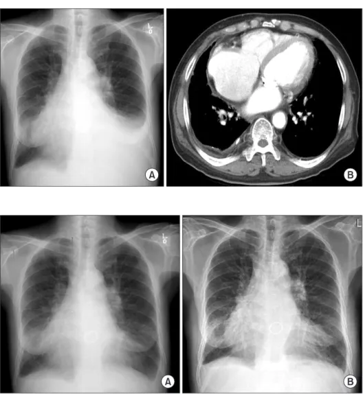
A 67-year-old man was admitted with a 1-month history of increasing dyspnea of modified Medical Research Council grade 3. The patient had undergone mitral valve replace- ment 20 years earlier and had been treated for heart failure, diabetes, and hypertension. His medications included war- farin, digoxin, furosemide, losartan, isosorbide mononitrate, Copyright © 2013

The Korean Academy of Tuberculosis and Respiratory Diseases.

All rights reserved.

Introduction

Eosinophilic pleural effusion (EPE), defined as an effusion containing 10% or more eosinophils, accounts for 5% to 16%

of exudative pleural effusions. It can be a manifestation of many different diseases, but one-third of the patients with EPE remain undiagnosed

1

. EPEs caused by an autoimmune disease are relatively rare (diseases such as rheumatoid arthri- tis, systemic lupus erythematosus [SLE], sarcoidosis, Churg-

CASE REPORT

http://dx.doi.org/10.4046/trd.2013.75.4.161

ISSN: 1738-3536(Print)/2005-6184(Online) • Tuberc Respir Dis 2013;75:161-164

161

Address for correspondence: Changhwan Kim, M.D.

Department of Pulmonary and Critical Care Medicine, Hallym University Kangdong Sacred Heart Hospital, Hallym University College of Medicine, 150 Seongan-ro, Gangdong-gu, Seoul 134-701, Korea

Phone: 82-2-2224-2561, Fax: 82-2-2224-0119 E-mail: [email protected]

Received: Apr. 30, 2013 Revised: Jul. 17, 2013 Accepted: Jul. 24, 2013

cc

It is identical to the Creative Commons Attribution Non-Commercial

License (http://creativecommons.org/licenses/by-nc/3.0/).

(2)

E Kim et al.

162 Tuberc Respir Dis 2013;75:161-164 www.e-trd.org

glimepiride, and thiotacid. He had no smoking history. At the time of hospitalization, the patient was alert and the following findings were noted in a physical examination: blood pres- sure, 140/60 mm Hg; pulse rate, 84 beats/min; respiratory rate, 20 breaths/min; and body temperature, 37.6

o

C. He had no his- tory of chest trauma or any prominent abnormalities on chest, abdominal, and lower-extremity examinations. Other results of the physical examination were non-specific.

On a physical examination, decreased breath sounds were auscultated on the left lower thorax. A chest radiograph showed blunting of the left costophrenic angle (Figure 1A).

Chest computed tomography images obtained after therapeu- tic thoracentesis revealed pleural thickening and calcification on the right posterior pleura but no nodule, mass, or lesion in the lung parenchyma showing in connective tissue disease (Figure 1B). Transthoracic echocardiography revealed en- largement of both atria, dilatation of the left-sided ventricle, and concentric left ventricular hypertrophy with left ventricle ejection fraction 57%. Mild mitral regurgitation and tricuspid regurgitation (grade II) were noted, but no definite change

could be observed. There was no pericardial effusion or peri- cardial thickening, and no valvular vegetations were seen.

Although a dose of diuretics had been added for several days, the amount of the left pleural effusion increased markedly, and the patient was referred to a pulmonologist. Laboratory examinations showed the following findings: white blood cell count, 9,300/mm

3

; hemoglobin, 11.7 g/dL; platelet count, 338,000/mm

3

; aspartate aminotransferase, 29 IU/L; alanine aminotransferase, 24 IU/L; total protein, 7.8 g/dL; blood urea nitrogen, 22 mg/dL; creatinine, 1.4 mg/dL; C-reactive protein, 8.3 mg/dL; and NT-pro B-type natriuretic peptide (BNP), 1,324 pg/mL. Arterial blood gas analysis in room air yielded results within normal ranges. Analysis of the pleural effusion revealed the following findings: color, yellow; pH, 7.4; specific gravity, 1.010; red blood cell count, 13,760/mm

3

; white blood cell count, 990/mm

3

(neutrophils, 2%; lymphocytes, 29%; eosino- phils, 34%); protein, 4.2 g/dL; lactate dehydrogenase, 384 U/L;

glucose, 117 mg/dL; carcinoembryogenic antigen, 0.2 ng/mL;

adenosine deaminase, 31 IU/L; negative acid-fast staining, culture, non-specific cytology, and negative results in real-time

Figure 1. (A) Baseline chest X-ray image shows blunting of the left costophrenic angle. (B) Chest computed tomography scan taken after therapeutic thoracente- sis reveals pleural thickening and calci- fication on the right posterior pleura but no nodule or mass in the parenchyma of the lungs.

Figure 2. (A) After 2 weeks of intrapleural

steroid injection, there is a significant

decrease in the pleural effusion. (B) At

1-year follow-up, no recurrence is ob-

served.

(3)

Intrapleural corticosteroid injection in eosinophilic pleural effusion

http://dx.doi.org/10.4046/trd.2013.75.4.161 163

www.e-trd.org

polymerase chain reaction for Mycobacterium tuberculosis.

Diagnostic thoracentesis revealed a pleural effusion appear- ing as an exudate with an elevated eosinophil count (34%). Se- rological tests for parasites (Clonorchis sinesis, Paragonimus westermani, Cysticercus cellulosae, Sparganum) yielded negative results. Serological test for rheumatologic diseases and vasculitides yielded positive results for the antinuclear antibody (>1:1,280, speckled pattern) and the anti-SS-A/Ro (179.09 EU) and anti-SS-B/La antibodies (208.98 EU). Sero- logical tests for rheumatoid factor (RF) (<20 IU/mL), anti-ds DNA, and complement (C3, C4) yielded negative results. Of the medications the patient was receiving, although warfarin was associated with EPE, he had been taking the drug without any side effects since the mitral valve replacement surgery.

Therefore, we considered that the possibility of warfarin-as- sociated EPE was low. The patient complained of xerostomia but not of xerophthalmia, Raynaud’s phenomenon, skin lesion, or arthralgia. The patient’s heart failure was well controlled; he showed no pitting edema, and his NT-pro BNP level had re- duced from 2,419 pg/mL to 1,324 pg/mL in a 1-month period.

EPE associated with UCTD was considered, and the patient was discharged after 1 L of excess fluid was removed by thera- peutic thoracentesis. About 2 weeks later, his dyspnea wors- ened and the left pleural effusion increased again. Repeated therapeutic thoracentesis temporarily relieved the symptoms, but they aggravated again within 2 weeks. The administration of systemic corticosteroid was considered but was contraindi- cated by chronic anticoagulation therapy, uncontrolled blood sugar levels, and the patient’s age.

Because we were reluctant to administer systemic cortico- steroid therapy, a single dose of 125 mg methylprednisolone was injected directly into the pleural space. A chest X-ray im- age obtained 2 weeks later showed a marked decrease in the pleural effusion (Figure 2A). No recurrence was observed dur- ing a 1-year follow-up, and serological tests for autoantibodies yielded similar results to those obtained in the previous year (Figure 2B).

Discussion

EPEs, defined as effusions with 10% or more eosinophils, typically account for 5% to 16% of exudative pleural effusions

1

. EPEs can be caused by various conditions, including air or blood in the pleural space, malignancy, infection, pulmonary embolism, drug reactions, and autoimmune diseases

7

. EPEs related to UCTD have not been reported in the literature so far.

UCTD has been defined as a condition characterized by the presence of the signs and symptoms of a connective tissue disease but not meeting the existing classification criteria

4

. Our patient had xerostomia but did not have xerophthalmia, skin rash, or arthralgia. The patient’s samples were strongly

positive for antinuclear, anti-SS-A/Ro, and anti-SS-B/La anti- bodies but were negative for RF (<20) and anti-ds DNA. The definition of UCTD includes a wide spectrum of diseases ranging from organ-dominant conditions to simplified condi- tions, to early CTDs or mild forms of CTDs

4

. Antibodies to nuclear antigens (ANAs) are detected in the serum of 50−97%

of the patients with UCTD. Most of them are directed to Ro/

SSA or to U1 ribonucleoprotein (RNP) antigens

12

. When the presence of ANA is required for the definition UCTD, the con- dition usually presents with a single antibody specificity, with anti-Ro/SSA and anti-RNP antibodies being most frequently observed (8−30% and 10−30% of the patients, respectively)

4

. In the present case, the patient’ symptoms and signs were not specific enough to make a definitive diagnosis of CTD. This condition can be classified as anti-Ro/SSa-positive UCTD be- cause of ANA with Ro/SSA positive antibodies, which do not fulfill the connective tissue disease classification. Therefore, he could be diagnosed with EPE related to UCTD.

The treatment of EPE involves the treatment of the under- lying disease

1

. Systemic corticosteroids are effective for the treatment of EPE associated with acute or chronic eosino- philic pneumonia and hypereosinophilic syndrome

1

. After the administration of 20 mg/day paramethasone, the pleural effu- sion decreased in progressive systemic sclerosis-polymyositis overlap syndrome with EPE

5

.

The therapeutic options for refractory massive pleural ef- fusion in SLE include 7 different types of therapy

10

. When refractory pleural effusion is related to lupus exacerbation, the treatment of choice is systemic (such as an immunosuppres- sive therapy with high-dose steroids and cyclophosphamide).

Intrapleural steroid injections were reported in 3 patients at the doses of 100 to 240 mg methylprednisolone or 80 mg triamcinolone, but they were ineffective. In contrast, 2 cases with post-traumatic pleural effusion were successfully treated with intrapleural steroids

2

. In the present case, we decided to administer intrapleural corticosteroid therapy because of the patient’s age and his history of diabetes and heart failure. As mentioned above, a single dose of 125 mg methylpredniso- lone was injected into the intrapleural space, which resulted in a dramatic improvement in the left pleural effusion.

The differentiation of UCTD into a specific connective tis- sue disease usually occurs within 5 years after the disease onset. UCTD may transform into SLE, systemic sclerosis, primary Sjogren’s syndrome, mixed connective tissue disease, systemic vasculitis, polydermatomyositis, or rheumatoid ar- thritis

4

. During follow-up, our patient remained asymptomatic and serological tests yielded positive results. We did not deter- mine whether UCTD in our patient transformed into any of connective tissue diseases.

To our knowledge, this is the first report of EPE in a patient with UCTD. After corticosteroid administration, the pleural ef- fusion rapidly decreased.

The incidence of EPE in patients with autoimmune diseases

(4)

E Kim et al.

164 Tuberc Respir Dis 2013;75:161-164 www.e-trd.org

is reported to be as low as 4%

5

. However, if the cause of EPE is unknown, it may be considered to be related to UCTD.

Intrapleural corticosteroid may be considered as one of the therapeutic options in patients in whom the administration of systemic corticosteroid is not possible.

References

1. Kalomenidis I, Light RW. Eosinophilic pleural effusions. Curr Opin Pulm Med 2003;9:254-60.

2. Ishiura Y, Fujimura M, Nakamura N, Jokaji H, Minami S, Matsuda T. Intrapleural corticosteroid injection therapy for post-traumatic eosinophilic pleural effusion. Respir Med 1996;90:501-3.

3. Rubins JB, Rubins HB. Etiology and prognostic significance of eosinophilic pleural effusions: a prospective study. Chest 1996;110:1271-4.

4. Mosca M, Tani C, Carli L, Bombardieri S. Undifferentiated CTD: a wide spectrum of autoimmune diseases. Best Pract Res Clin Rheumatol 2012;26:73-7.

5. Maeshima E, Nishimoto T, Yamashita M, Mune M, Yukawa S. Progressive systemic sclerosis-polymyositis overlap syn- drome with eosinophilic pleural effusion. Rheumatol Int 2003;23:252-4.

6. Ferreiro L, San Jose E, Gonzalez-Barcala FJ, Alvarez-Dobano JM, Golpe A, Gude F, et al. Eosinophilic pleural effusion: inci- dence, etiology and prognostic significance. Arch Bronconeu- mol 2011;47:504-9.

7. Oba Y, Abu-Salah T. The prevalence and diagnostic signifi- cance of eosinophilic pleural effusions: a meta-analysis and systematic review. Respiration 2012;83:198-208.

8. Krenke R, Nasilowski J, Korczynski P, Gorska K, Przybylowski T, Chazan R, et al. Incidence and aetiology of eosinophilic pleural effusion. Eur Respir J 2009;34:1111-7.

9. Samman YS, Wali SO, Abdelaal MA, Gangi MT, Krayem AB.

Chronic eosinophilic pneumonia presenting with recur- rent massive bilateral pleural effusion: case report. Chest 2001;119:968-70.

10. Breuer GS, Deeb M, Fisher D, Nesher G. Therapeutic options for refractory massive pleural effusion in systemic lupus ery- thematosus: a case study and review of the literature. Semin Arthritis Rheum 2005;34:744-9.

11. Felz MW, Haviland-Foley DJ. Eosinophilic pleural effusion due to dantrolene: resolution with steroid therapy. South Med J 2001;94:502-4.

12. Cavazzana I, Franceschini F, Belfiore N, Quinzanini M, Capo-

rali R, Calzavara-Pinton P, et al. Undifferentiated connective

tissue disease with antibodies to Ro/SSa: clinical features and

follow-up of 148 patients. Clin Exp Rheumatol 2001;19:403-9.

수치

Figure 1. (A) Baseline chest X-ray image  shows blunting of the left costophrenic  angle

참조

관련 문서

Histogram shows the difference of pleural effusion volume depending on the experimental conditions in freshwater group... Youn Shin

The stain field associated with a dislocation can in certain cases provide a favorable interaction with the strain field of the martensite nucleus, such that one of the

The benefits and harms of intravenous thrombolysis with recombinant tissue plasminogen activator within 6 h of acute ischaemic stroke (the third

These results of the histologic distribution of lamina propria, adipose tissue, and glandular tissue of the hard palatal mucosa and the topography of the greater palatine

environmental burdens associated with a product, process, or activity by identifying energy and materials used and. wastes released

In the tissue culture using leaf tissue of Orostachys japonicus , callus were induced most efficiently in 5-week-after culture in the MS medium supplemented with 10 μ BAP and 4

The Jacobi energy integral is one type of invariant of motion associated with

The stain field associated with a dislocation can in certain cases provide a favorable interaction with the strain field of the martensite nucleus, such that one of the