Address for correspondence : Kyung Ho Kang, M.D.
Division of Respiratory and Critical Care Medicine Department of Internal Medicine Guro Hospital #80, Guro-dong, Guro-gu, Seoul, Republic of Korea (152-703)
Phone : +82-2-818-6638 Fax : +82-2-865-9670 E-mail : [email protected]
Received : Aug. 22. 2005 Accepted : Sep. 15. 2005
고려대학교 의과대학 내과학교실, 고려대학교 의과대학 흉부외과학교실
이경주1, 김정하1, 하은실1, 정진용1, 이승현1, 김세중1, 이은주1, 허규영1, 정혜철1, 이승룡1, 김현구2, 이상엽1, 김제형1, 신 철1, 심재정1, 인광호1, 강경호1, 유세화1
A Case of Passive Smoking Induced Respiratory Bronchiolitis Associated Interstitial Lung Disease
Kyoung Ju Lee, M.D.1, Jung Ha Kim, M.D.1, Eun Sil Ha, M.D.1, Jin Yong Jung, M.D.1, Seung Hyeun Lee, M.D.1, Se Joong Kim, M.D.1, Eun Joo Lee, M.D.1, Gyu Young Hur, M.D.1, Hye Cheol Jung, M.D.1, Sung Yong Lee, M.D.1, Hyn Koo Kim, M.D.2, Sang Yeub Lee, M.D.1, Je Hyeong Kim, M.D.1, Chol Shin, M.D.1, Jae Jeong Shim, M.D.1, Kwang Ho In, M.D.1, Kyung Ho Kang, M.D.1, Se Hwa Yoo, M.D.1
1Department of Internal Medicine, 2Department of Chest Surgery, College of Medicine, Korea University, Seoul, Korea
Respiratory bronchiolitis associated interstitial lung disease is a rare condition among current or ex-smokers, which has features consistent with interstitial lung disease. The presentations are non-specific, but symptoms generally include a cough and dyspnea on exertion, and its pathology is characterized by the accumulation of pigmented macrophages within the respiratory bronchioles and adjacent air spaces, and is associated with mild thickening of the peribronchiolar interstitium. Recently, the case of a 54-year-old woman passive smoker, diagnosed as having respiratory bronchiolitis associated interstitial lung disease, was experienced at our institution.
(Tuberc Respir Dis 2005; 59: 541-545)
Key words : Respiratory bronchiolitis, Interstitial lung disease, Passive smoking
서 론
호흡 세기관지염은 1974년 Niewoehner 등이 무증 상의 젊은 흡연가들의 부검 폐에서 처음으로 기술한 이후1, 흡연가의 폐에서 흔히 보는 병리소견으로 막성 세기관지와 호흡 세기관지의 만성 염증과 섬유화를 특징으로 하고 있다. 이러한 병변은 흔히 만성 폐쇄성 폐질환의 발생과 밀접한 관련이 있는 것으로 여겨지 고 있다. 최근에는 흡연과 관련된 호흡 세기관지염이 임상적으로 간질성 폐질환의 증상 및 소견을 나타내 는 환자의 원인으로 밝혀지면서 하나의 증후군으로 서 호흡 세기관지염 연관 간질성 폐질환(respiratory bronchiolitis associated interstitial pneumonia, RB- ILD)이 기술되고 있다2. 현재까지 발표된 논문들은
대부분 현재 흡연을 하거나 혹은 이전에 흡연한 경력 이 있는 증례들이었다. 그러나 저자들은 직접 흡연의 경력이 없으나 직업적으로 간접 흡연에 노출된 후 발 생한 호흡 세기관지염 연관 간질성 폐질환 1예를 경 험하였기에 보고 하는 바이다.
증 례
환 자 : 허○이, 여자 54세 주 소 : 쉰 소리, 호흡 곤란
현병력 : 환자는 3개월 전부터 시작된 쉰 소리와 호 흡 곤란, 기침을 주소로 본원 호흡기 내과 외래 내원 하여 시행한 단순 흉부 X-선 촬영상 미세 결절 보여 촬영한 흉부 컴퓨터 단층촬영에서 특발성 폐섬유증, 아급성 과민성 폐장염 의심되어 TBLB 혹은 BAL 시행하기 위해 입원하였다.
과거력 : 갑상선 기능저하증으로 본원 내분비 내과에 서 투약 중이며 2003년 7월에는 우측 자발성 기흉으 로 대림성모병원에서 수술 받은 병력 있음.
사회력 : 3년 전부터 곰팡이 및 기름때 많은 장소(1 층)에서 호프집 경영하며, 흡연경력은 없으나 호프집
Figure 1. Chest X-ray showed fine nodularities were observed on both entire lung field.
B A
Figure 2. Chest CT showed poorly defined centrilobular micronodules in both lungs, subsegmental atelectasis in RLL anterior basal segment(A), which was improved after exposure was stop(B).
에서 간접흡연경력 있음.
가족력 : 없음
이학적 소견 : 혈압은 100/60 mmHg, 맥박은 70회/분, 호흡수는 20회/분, 체온 36.5˚C였다.
신체 검진상 피부 병변이나, 두경부 임파절 비대는 없 고 청진상 양측 폐 음은 깨끗하였고, 심음은 규칙적이 며 심잡음은 들리지 않았다. 그외 다른 이학적 검사상 이상 소견은 없었다.
검사실 소견 : 내원시 말초혈액검사에서 백혈구 4,500/
μL(Neut 46.8%, Lymp 36.7%, Eosi 7.4%), 혈색소 12.1g/dL, 혈소판 174,000/μL로 정상이었으며 그 외 일반화학검사. 혈액응고검사, 요검사 등에서는 이상 소견 보이고 있지 않았으며, 면역 혈청 검사상 FANA, ANCA, RF 등은 모두 음성이었다.
방사선학적 소견 : 내원 당시 촬영한 단순 흉부 X-선 촬영상에서는 양측 전폐부에 미세결절을 보여 아급 성 과민성 폐장염 의심되는 소견 관찰되고 있었고 (Fig 1), 외래에서 촬영한 흉부 컴퓨터 단층촬영 상 소엽중심성 결절이 양측 폐야에 균일하게 분포되어 있었고 지도양상 간유리 음영이 좌하엽에서 관찰되 고 있었다(Fig 2A).
폐기능 검사 : FVC 3.61L(예측치의 108%), FEV1
2.82L(예측치의 112%), FEV1/FVC 78%이었고, 폐확 산능은 DLCO 20.9mL/mmHg/min(예측치의 127%), DLCO/VA 107% 이었다.
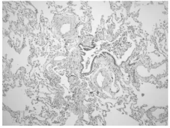
경 과 : 환자는 입원 후 아급성 과민성 폐장염, 간질 성 폐렴 등의 질환이 의심되어 조직검사 위해 흉부외 과로 전과 되어 흉강경(VATS)을 통해 폐 조직 생검 시행하였으며, 폐조직 생검 결과 호흡 세기관지 내에 대식세포의 침착이 관찰되고 섬유화 및 만성 염증소 견을 보이며 세기관지 주위의 폐포관과 폐포에서 입 방세포 증식도 관찰되어(Fig 3), 조직학적으로 호흡 세기관지염 및 임상적으로 호흡 세기관지염 연관 간 질성 폐질환으로 진단되었다.
Figure 3. Respiratory bronchiolitis pattern, with marked alveolar macrophage accumulation in and around bro
nchioles, mild chronic inflammation of bronchioles, fibrous thickening of the wall of bronchioles, cuboidal cell hyperplasia of alveolar ducts and alveoli adjacent to the bronchioles(H&E, x100).
진단 및 치료 : 환자는 간접 흡연으로 인한 호흡 세 기관지염 연관 간질성 폐질환으로 진단되었고, 간접 흡연으로부터 회피한 이후 환자는 증상이 호전되어 현재 외래에서 추적 관찰 중이며 외래에서 추적 검사 한 흉부 컴퓨터 단층촬영 상에서 양측 폐야에 관찰되 던 미세결절은 호전되는 소견 보이고 있다(Fig 2B).
고 찰
RB-ILD는 다량의 흡연력이 있는 사람에게서 발견 되는 증후군으로, 1987년 Myers2 등이 제한성 환기장 애를 가지면서 청진상 수포음을 동반하고, 단순 흉부 X-선 촬영상 간질성 폐질환 소견을 보이는 6명의 흡 연자를 대상으로 발표한 질환이다. 폐 조직 생검을 시 행한 결과 흡연과 관련된 호흡 세기관지염과 유사한 조직검사 결과를 보여주는 것으로 되어 있다. 주로 다량의 흡연력을 가진 30대에서 50대의 환자가 대부 분으로 남자가 여자보다 2배정도 많다. 증상은 서서 히 시작되는 호흡곤란과 지속적인 기침이 가장 흔하 며 객담은 흔하지 않다3-5. 이학적 검사상 양측 폐 기 저부에서 미세한 흡기말 수포음이 들리나 일부에서 는 정상소견을 보이기도 하며2, 곤봉지는 관찰되지 않는다. 본 증례의 환자는 50대의 여성으로 직업력 상 호프집에서 일하고 있으며 흡연력은 없으나 간접
흡연력이 있었다. 3개월동안 경도의 호흡곤란을 호소 하고 있었으나 이학적 검사상에서는 이상소견을 보이 지 않았다.
RB-ILD 의 방사선학적 소견으로는 단순 흉부 X- 선 촬영에서 미만성의 미세한 망상 결절형 병변, 기관 지 벽의 비후, 기관지 주위혈관의 확장, 주변의 작은 윤상 음영, 작은 규칙적 또는 불규칙적인 음영 등이 관찰3되지만 거의 정상에 가깝기 때문에 흉부 전산하 단층촬영이 중요한 진단적 가치를 가지게 된다6. 흉부 전산하 단층촬영상 미만성 또는 산발적인 간유리형 음영과 결절이 가장 흔한 소견이며 폐 허탈, 폐소엽내 및 폐소엽간 간질의 비대, 폐기종 등이 보일 수 있다7-9. 이러한 병변은 주로 양측 폐하부에 많이 관찰 되는 것이나 본 증례에서는 전폐야에 걸쳐 미세 결절이 관 찰되었다.
폐기능 검사에서는 제한성 소견과 폐쇄성 소견이 혼합되어 임상적인 상황에 따라 다양한 양상을 보이 게 된다2-4. 폐 확산능은 약간의 감소를 나타내거나 정 상이다. 경우에 따라서는 안정시 또는 운동시 약간의 저산소증을 보이기도 한다. 폐기종이 동반되어 있는 경우 FEV1/FVC, FEF25~75% 등에서는 폐쇄성 소견과 소기도 질환의 소견을 나타내는 경우가 많으나 다른 흡연가들 보다 심하지 않다. 이번 증례에서는 FEV1/ FVC 예측치의78%로 약간 감소된 것 외에는 이상소 견은 관찰되지 않았다.
RB-ILD는 궁극적으로 병리학적 소견으로 진단하 게 되기 때문에 폐생검이 중요한데, 병리학적으로 가 장 특징적인 점은 호흡 세기관지내와 인접 폐포, 그리 고 폐포관에 색소 침착을 동반한 대식세포의 축적이 다. 대식세포는 호산구성 세포질에 미세한 과립성의 갈색 색소 침착을 동반하게 되는데 이는 흡연가의 폐 에 일치하는 소견을 보여 주는 것이다3,10. 일반적으로 는 만성적인 염증세포가 세기관지와 폐포벽에 침착 하게 되며, 폐포 주위 막성 기관지와 호흡 세기관지의 점막하를 침범하는 섬유화가 인접 폐포와 폐포관의 벽에 뻗쳐 있는 양상이 관찰된다.
RB-ILD는 조직학적 혹은 방사선학적으로 다른 질 환과 감별을 요하게 되는데, 그 중 desquamative interstitial pneumonitis(DIP) 는 다량의 흡연가에게
호발하여 RB-ILD와 감별을 요하게 된다. 미만성의 폐포벽 비후 및 type II pneumocyte의 과증식과 폐포 내 대식세포의 침착을 특징으로 하는데11, RB-ILD와 의 가장 큰 차이는 RB-ILD에서 대식세포가 소엽중 심으로 침착 되는 것에 비해 DIP에서는 미만적으로 침착을 보이게 된다. 아급성 과민성 폐장염의 경우에 있어서는 본 증례에서와 마찬가지로 방사선학적으로 미세결절 등을 보여 RB-ILD와 감별을 요하는데, 이것 은 임상적으로 흡연가에게서 관찰되지는 않는다6,12.
RB-ILD의 임상 경과와 예후는 잘 알려져 있지 않 으나 흡연이 병인에 중요한 역할을 하므로 기능적으 로 심각한 손상을 가지고 있지 않은 상태에서는 담배 를 끊는 것으로 자발적으로 병의 회복이 가능한 것으 로 알려져 있으며 RB-ILD에 있어서 예후는 그다지 나쁘지 않은 것으로 되어 있다13. 경우에 따라 특발성 폐섬유증 일부 환자들에서 부신 피질 스테로이드나 면역 억제제가 폐기능과 흉부 x-ray, 증상의 호전에 도움이 될 수 있다고 보고되어 있으나 아직 명확히 밝혀진 바는 없다2,3.
본 환자는 직접 흡연을 한 병력은 없는 환자로 운 동시 호흡곤란과 지속적인 기침 호소하여 시행한 방 사선학적 검사상에서는 아급성 과민성 폐장염과 감 별을 요하는 상태로 폐 조직 생검으로 RB-ILD로 진 단한 증례이다. Niewoehner1등이 발표한 바에 따르면 무증상의 흡연가 혹은 과거력상 흡연 경력이 있는 자 에게서 호흡 세기관지염이 관찰되는 것으로 알려져 있었다. 그러나 본 증례에서는 직접 흡연을 하지 않으 나 폐 생검을 시행한 결과 조직학적인 검사에서 호흡 세기관지 내에 대식세포의 침착이 관찰되고 섬유화 및 만성 염증소견을 보이며 세기관지 주위의 폐포관 과 폐포에서 입방세포 증식도 관찰되어 RB-ILD의 조직학적인 소견과 일치하는 모습이 관찰되었다. 따 라서 환자의 직업력 상 간접 흡연의 노출이 RB-ILD 의 병인으로 생각된다. 이제까지 보고된 다른 증례에 서는8,14 흡연가에게서 관찰되는 질환이며 국내에서 보고된 첫 증례에서도 과다 흡연의 경력이 있는 자에 게서 관찰되는 질환이었으나15, 간접 흡연으로 발병 한 증례는 처음이다.
요 약
저자들은 간접 흡연에 노출되어 운동 시 호흡곤란 및 지속적인 기침을 호소하는 54세 여자 환자에서 폐 조직 생검상 호흡 세기관지염 연관 간질성 폐질환을 진단하였다. 간접 흡연력이 있는 자에서 RB-ILD를 진단한 증례는 국내에서 처음으로 이를 문헌 고찰과 함께 보고하는 바이다.
참 고 문 헌
1. Niewoehner DE, Kleinerman J, Rice DB. Pathologic changes in the peripheral airways of young cigarette smokers. N Engl J Med 1974;291:755-8.
2. Myers JL, Veal CF Jr, Shin MS, Katzenstein AL.
Respiratory bronchiolitis causing interstitial lung di
sease: a clinicopathologic study of six cases. Am Rev Respir Dis 1987;135:880-4.
3. Yousem SA, Colby TV, Gaensler EA. Respiratory bro
nchiolitis-associated interstitial lung disease and its relationship to desquamative interstitial pneumonia.
Mayo Clin Proc 1989;64:1373-80.
4. Moon J, du Bois RM, Colby TV, Hansell DM, Nicholson AG. Clinical significance of respiratory bronchiolitis on open lung biopsy and its relationship to smoking related interstitial lung disease. Thorax 1999;54:
1009-14.
5. King TE Jr. Respiratory bronchiolitis-associated inter
stitial lung disease. Clin Chest Med 1993;14:693-8.
6. Park JS, Brown KK, Tuder RM, Hale VA, King TE Jr, Lynch DA. Respiratory bronchiolitis-associated interstitial lung disease: radiologic features with clinical and pathologic correlation. J Comput Assist Tomogr 2002;26:13-20.
7. Kurumagawa T, Kobayashi H, Kanoh S, Nagata N, Aoki T, Aida S, et al. Respiratory bronchiolitis-ass
ociated interstitial lung disease. Nihon Kokyuki Gakkai Zasshi 1998;36:881-5.
8. Essadki O, Chartrand-Lefebvre C, Briere J, Grenier P. Respiratory bronchiolitis: radiographic and CT findings in a pathologically proven case. Eur Radiol 1998;8:1674-6.
9. Holt RM, Schmidt RA, Godwin JD, Raghu G. High resolution CT in respiratory bronchiolitis-associated interstitial lung disease. J Comput Assist Tomogr 1993;17:46-50.
10. Wesselius LJ, Flowers CH, Skikne BS. Alveolar ma
crophage content of isoferritins and transferrin: com
parison of nonsmokers and smokers with and without chronic airflow obstruction. Am Rev Respir Dis 1992;
145:311-6.
11. Liebow AA, Steer A, Billingsley JG. Desquamative Interstitial Pneumonia. Am J Med 1965;39:369-404.
12. Hansell DM, Wells AU, Padley SP, Muller NL. Hyper
sensitivity pneumonitis: correlation of individual CT patterns with functional abnormalities. Radiology 1996;199:123-8.
13. Goeckenjan G. Respiratory bronchiolitis-associated in
terstitial lung disease (RB-ILD). Pneumologie 2003;
57:278-87.
14. McWilliams AM, Lake FR. Respiratory bronchiolitis associated interstitial lung disease (RB-ILD) presenting with haemoptysis. Respirology 2000;5:385-7.
15. Ahn BH, Park HS, Do JH, Suh GY, Chung MP, Rhee CH, et al. A case of respiratory bronchiolitis-asociated interstitial lung disease. Tuberc Respir Dis 1999;46:
103-9.