• 검색 결과가 없습니다.

The Role of Innate and Adaptive Immune Cells in the Immunopathogenesis of Chronic Obstructive Pulmonary Disease

N/A
N/A
Protected

Academic year: 2021

Share "The Role of Innate and Adaptive Immune Cells in the Immunopathogenesis of Chronic Obstructive Pulmonary Disease"

Copied!
9
0
0

로드 중.... (전체 텍스트 보기)

전체 글

(1)

The Role of Innate and Adaptive Immune Cells in the Immunopathogenesis of

Chronic Obstructive Pulmonary Disease

Fariz Nurwidya, M.D., Ph.D., Triya Damayanti, M.D., Ph.D. and Faisal Yunus, M.D., Ph.D.

Department of Respiratory Medicine, Persahabatan General Hospital, University of Indonesia Faculty of Medicine, Jakarta, Indonesia

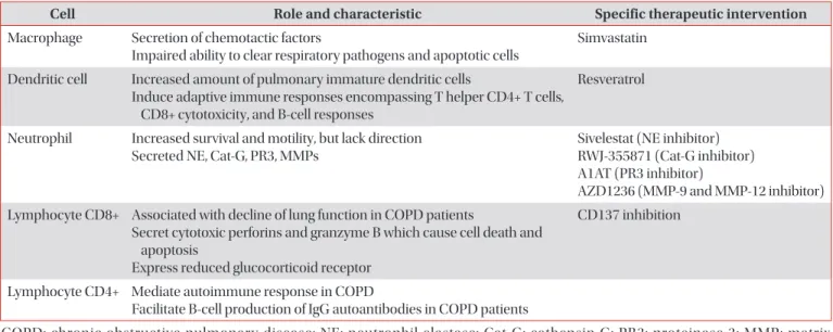
Chronic obstructive pulmonary disease (COPD) is a chronic and progressive inflammatory disease of the airways and lungs that results in limitations of continuous airflow and is caused by exposure to noxious gasses and particles. A major cause of morbidity and mortality in adults, COPD is a complex disease pathologically mediated by many inflammatory pathways. Macrophages, neutrophils, dendritic cells, and CD8+ T-lymphocytes are the key inflammatory cells involved in COPD. Recently, the non-coding small RNA, micro-RNA, have also been intensively investigated and evidence suggest that it plays a role in the pathogenesis of COPD. Here, we discuss the accumulated evidence that has since revealed the role of each inflammatory cell and their involvement in the immunopathogenesis of COPD. Mechanisms of steroid resistance in COPD will also be briefly discussed.

Keywords: Pulmonary Disease, Chronic Obstructive; Macrophages; Neutrophils; Dendritic Cells; Lymphocytes

For the population without COPD over the age of 40, the risk of developing COPD within the next 40 years was 12.7% for men and 8.3% for women

3

. In patients with very severe COPD, 26% died after 1 year of follow-up, whereas 2.8% died among the non-COPD subjects

3

.

COPD, a common and preventable disease, is characterized by persistent airflow limitation that is usually progressive and associated with an enhanced chronic inflammatory response in the airways and the lung to noxious particles or gasses

4

. The Global Initiative for Chronic Obstructive Lung Disease (GOLD) defines airflow obstruction as spirometry where the ratio of forced expiratory volume in the first second to forced vital capacity after bronchodilation is less than 0.70

5

.

Cigarette Smoke Exposure as a Model to Study COPD

Cigarette smoking (CS) is an established risk factor for COPD

6,7

, and study suggests that CS exposure could have a suppressive effect on host innate immunity including structural and functional changes in the respiratory ciliary epithelium, and immune cells such as alveolar macrophages (AMs), neutrophils, and lymphocytes

8

. Moreover, CS could

Introduction

In 2020, chronic obstructive pulmonary disease (COPD) will be the third leading cause of death worldwide (from sixth in 1990) and fifth leading cause of years lost through early mortality or handicap (disability-adjusted life years) from pre- viously 12th in 1990

1

. However, recent studies suggest COPD is already the third most common cause of death, worldwide

2

.

Address for correspondence: Fariz Nurwidya, M.D., Ph.D.

Department of Respiratory Medicine, Persahabatan General Hospital, University of Indonesia Faculty of Medicine, Jalan Persahabatan Raya No.1, Rawamangun, Jakarta 13220, Indonesia

Phone: 62-21-489-3536, Fax: 62-21-489-0744 E-mail: [email protected]

Received: Aug. 6, 2015 Revised: Sep. 1, 2015 Accepted: Oct. 12, 2015

cc It is identical to the Creative Commons Attribution Non-Commercial License (http://creativecommons.org/licenses/by-nc/4.0/).

Copyright © 2016

The Korean Academy of Tuberculosis and Respiratory Diseases.

All rights reserved.

(2)

also cause defect in the generation of adaptive immunity in the lung

9

. CS exposure is the most appropriate model to study the pathogenesis of emphysema in mice

10

. Following cigarette exposure for 26, 52, and 65 weeks, structural changes were observed and accompanied by altered lung function at 26 and 52 weeks

11

. After 13 weeks of CS exposure-free period, most biochemical, histopathological, and morphometrical altera- tions were restored, while emphysema was observed to per- sist in 18% of mice exposed to CS at 65 weeks. These findings suggest that the cigarette exposure induced emphysematous changes in the lungs, accompanied by altered lung function and inflammatory cell infiltration. There are data demonstrat- ing that smoking is associated with up- or down-regulation of many genes in the airway epithelium and, interestingly, ex- smokers continue to have persistent up- or down-regulation of many genes, despite smoking cessation

12

.

Early Changes in Airway Epithelium in the Pathogenesis of COPD

Most of the normal human airway is lined by a pseudostrati- fied epithelium of ciliated cells, secretory cells and 6%–30%

basal cells (BCs)

13

. In COPD, the remodeling of the airway epithelium, such as squamous metaplasia and mucous hyper- plasia that occur during injury, may considerably disturb the innate immune functions of the airway epithelium

14

. Although inflammatory reaction by immune processes plays a signifi- cant role in the pathogenesis of COPD, the earliest abnormali- ties in the COPD lung caused by smoking are hyperplasia of airway BC, the stem/progenitor cells of the ciliated and secre- tory cells that are central to pulmonary host defense

15

. Apart

from BC hyperplasia, smoking induces a number of COPD- relevant airway epithelial remodeling phenotypes that are likely initiated in the BC population, including mucous cell hy- perplasia, squamous cell metaplasia, epithelial-mesenchymal transition, altered ciliated and non-mucous secretory cell dif- ferentiation, and suppression of junctional barrier integrity

16

.

Inflammatory Cells in COPD

A smoking-induced inflammatory reactions in the airways and lung parenchyma have long been accepted to be the major cause of COPD in smokers

17

. Cigarette smoke activates innate immune cells by triggering pattern recognition recep- tors to release “danger signal” that act as ligands to Toll-like receptors, triggering the production of cytokines and inducing innate inflammation

18

.

Impaired immune function contributes to the develop- ment of COPD and disease progression is further exacerbated by infections due to impaired immune responses

19

. Sever- ity and course of acute exacerbations of COPD reflects the capacity of the adaptive immune system in modulating the innate response to pathogen (“the Goldilocks hypothesis”)

20

. Basically, this hypothesis states that there is no such thing as bad inflammation; it all depends on how and when

21

. Goldi- locks hypothesis is the state when the innate and adaptive immune response successfully eliminates the infection, and the response is mild and transient, not too much and not too little

20

. The immune inflammatory changes associated with COPD are linked to a tissue-repair and -remodeling process that increases mucus production and causes emphysematous destruction of the gas-exchanging surface of the lung

22

.

Table 1. The role of innate and adaptive immune cells in the pathogenesis of COPD

Cell Role and characteristic Specific therapeutic intervention

Macrophage Secretion of chemotactic factors

Impaired ability to clear respiratory pathogens and apoptotic cells

Simvastatin

Dendritic cell Increased amount of pulmonary immature dendritic cells

Induce adaptive immune responses encompassing T helper CD4+ T cells, CD8+ cytotoxicity, and B-cell responses

Resveratrol

Neutrophil Increased survival and motility, but lack direction Secreted NE, Cat-G, PR3, MMPs

Sivelestat (NE inhibitor) RWJ-355871 (Cat-G inhibitor) A1AT (PR3 inhibitor)

AZD1236 (MMP-9 and MMP-12 inhibitor) Lymphocyte CD8+ Associated with decline of lung function in COPD patients

Secret cytotoxic perforins and granzyme B which cause cell death and apoptosis

Express reduced glucocorticoid receptor

CD137 inhibition

Lymphocyte CD4+ Mediate autoimmune response in COPD

Facilitate B-cell production of IgG autoantibodies in COPD patients

COPD: chronic obstructive pulmonary disease; NE: neutrophil elastase; Cat-G: cathepsin G; PR3: proteinase 3; MMP: matrix

metalloproteinase; A1AT: antiprotease 1-antitrypsin.

(3)

The inflamed airways of COPD patients contain several inflammatory cells including neutrophils, macrophages, T lymphocytes, and dendritic cells (DCs)

23

(Table 1). Recent evidence also found increased population of goblet cells in COPD patient

24

, which cause the mucous overproduction and hypersecretion

25

. It seems likely that, only when all inflam- matory cell types (i.e., CD4+, CD8+, neutrophils, and macro- phages) are present in the lung, the airways remodeling and parenchymal destruction characteristic of COPD will ensue

17

. These cells release the reactive oxygen species, chemokines (e.g., interleukin [IL]-8), cytokines (e.g., tumor necrosis factor alpha [TNF-]) and proteases (e.g., neutrophil elastase [NE]

and matrix metalloproteinase [MMP]) that are instrumental in producing a chronic inflammatory state

26

. In the following sections, we will discuss each inflammatory cell that partici- pates in the immunopathogenesis of COPD.

Macrophage

AMs have been identified as one of the major cell types that plays a key role in orchestrating the inflammatory events as- sociated with the pathophysiology of COPD

27

. The number of AM is markedly increased in the lungs of patients with COPD as a result of increased recruitment, proliferation and survival

28

. One of the major functions of macrophages is the secretion of chemotactic factors and this function is markedly increased on exposure to CS

27

. However, it is also found that macrophage in COPD has impaired ability to clear respiratory pathogens and apoptotic cells

29

. The reduced phagocytic abil- ity of macrophage may drive the persistence of inflammation in COPD

30

.

Genome-wide analysis has underscored the heterogeneity and plasticity of macrophage phenotype which lead to the introduction of the term M1 (classical activation) and M2 (al- ternative activation)

31

. M1 phenotype macrophages express various pro-inflammatory mediators including TNF-, IL-1, IL-6, reactive nitrogen and oxygen intermediates, which have a strong microbicidal and tumoricidal activity; while M2 phenotype is involved in tissue remodeling and charac- terized with anti-inflammatory properties

32

. It is said that the surrounding pulmonary environment in COPD may generate a specific phenotype that is permanently pro-inflammatory (M1)

30

. Among COPD patients, M2 macrophages were re- duced in current smokers compared to ex-smokers which indicate that smoking cessation in COPD is associated with macrophage polarization towards an anti-inflammatory phenotype

33

. However another study suggests, rather than up-regulating the M1 polarization program as expected, CS induces in AM of COPD smokers the opposite phenotype, characterized by a substantial down-regulation of the M1- related genes

34

. Obviously, these conflicting results indicate that smoking induced a complex suppression of immune

response in the lung, including deactivation of AM inflam- matory and host defense function, and development of tissue remodeling.

The source of macrophage activation is still under intense investigation. Macrophage could be induced by TNF-

35

and IL-8

36

. In one study by Bozinovski et al.

37

, activation of innate cellular sources of IL-17A is an essential mediator of mac- rophage accumulation in CS-exposed lungs. Targeting non- conventional T cell sources of IL-17A may offer an alternative strategy to reduce pathogenic macrophages in COPD. How- ever, it has also been found that IL-17A contributes to normal lung homeostasis and does not mediate CS-induced loss of lung structure and pulmonary function

38

.

‘Macrophage-targeted therapy’ has been studied in many centers. It has been reported that simvastatin, member of statin which has immunomodulatory properties, reversed the IL-17A/IL-10 imbalance in the airways and reduced sputum macrophage but not neutrophil counts in patients with COPD

39

. Generally, simvastatin could provide substan- tial benefits in patients with COPD due to (1) reduction of cytokine secretion (TNF-, IL-6, and IL-8) and neutrophil infiltration into the lung; (2) attenuation of the fibrotic activity in the lung leading to small airways fibrosis and irreversible airflow limitation; (3) antioxidant and anti-inflammatory ef- fects on skeletal muscle; (4) reduced inflammatory response to pulmonary infection; and (5) inhibition of the epithelial- mesenchymal transition, a precursor event to lung cancer

40

. In one in vivo study, pretreatment with simvastatin prior to and continued throughout smoke exposure reduced the influx of macrophages into the lung and airways

41

. Molecular mecha- nism of simvastatin in attenuating CS-induced emphysema- tous abnormality in COPD has been described by Kim et al.

42

. In their in vitro study, simvastatin reversed CS-induced MMP- 9 expressions in AM. However, there are clinical trials that suggest simvastatin did not affect exacerbation rates or the time to a first exacerbation

43

, and did not reduce circulating inflammatory markers in patients with COPD

44

. Controversies about the use of statin in COPD still remain. Recently, positive result came from study conducted by Ingebrigtsen et al.

45,46

, who found that statin was associated with reduced odds of exacerbations in COPD patient, in particular those who have coexisting cardiovascular disease. This finding was consistent with other study, involving 1,584 patients, that confirm the ef- ficacy of statin in the reduction of COPD exacerbation

47

.

Dendritic Cells

DCs in the lung have an essential role in defense mecha-

nism based on their anatomical location that creates a func-

tional cellular interface between the external environment

and the internal lung microenvironment

48

. DCs are in charge

of capturing antigens in peripheral tissues, transporting them

(4)

to lymph nodes and presenting them on major histocompat- ibility complex molecules to T lymphocytes

49

. DCs are profes- sional antigen presenting cells linking innate and adaptive immune responses

50

. Cigarette smoke promotes DCs accu- mulation

51

, and induces the release of chemokines from DCs that play a role in the pathogenesis of COPD

52

. It has been re- ported that COPD patients have an increased amount of pul- monary immature DCs. Using CD83+ and DC-LAMP(+) cells as mature DC marker, Tsoumakidou et al.

53

, found that mature DCs were decreased in non-COPD subject when they actively smoke cigarette and increased when they ceased smoking.

They also found that COPD patients have a low mature DCs compared to never-smokers and ex-smokers non-COPD subject. Activated DCs induce adaptive immune responses encompassing T helper (Th1 and Th17) CD4+ T cells, CD8+

cytotoxicity, and B cell responses, which lead to the develop- ment of lymphoid follicles on chronic inflammation

54

. More- over, remarkable up-regulation of CD80 and CD86 and secre- tion of cytokines interferon (IFN)- were observed in DCs from COPD patients

55

. One of the drugs that has recently been found to inhibit dysfunction of DC is resveratrol

55

. Results of that study suggest that small doses of resveratrol are sufficient to inhibit phenotypic and functional TNF- effect on DC

56

.

Neutrophil

Neutrophils are also aberrant with increased survival and motility, but lack of direction could lead to more widespread destruction of host matrix during migration

30

. Neutrophil se- creted NE and the proteolytic activity of NE not only destroys pathogens but also degrades host matrix tissues by generat- ing a localized protease-antiprotease imbalance in COPD

57

. Although NE was identified as a therapeutic target for COPD more than 30 years ago, only Sivelestat (ONO-5046), an NE inhibitor from Ono Pharmaceutical, has been approved for clinical use in Japan, but not in United States

58

.

There are two similar neutrophil serine proteases that coex- ist with NE in COPD: cathepsin G (Cat-G); and proteinase 3 (PR3)

59

. Cat-G is a neutral proteinase originating from human neutrophils that displays a unique dual specificity (trypsin- and chymotrypsin-like); thus, its enzymatic activity is difficult to control

60

. Anti-inflammatory pharmacology of RWJ-355871, an inhibitor of Cat-G, has been tested in animal models of inflammation that represent COPD, and showed promising result in terms of reduction of smoke-induced neutrophilia

61

. Meanwhile, PR3 is a multifunctional serine proteinase mainly located in the azurophilic granules and on the cell surface of neutrophils and able to degrade many critical components of the extracellular matrix, including elastin, type IV collagen, fi- bronectin, and laminin

62

. In one report, PR3 is inhibited by the antiprotease 1-antitrypsin (A1AT)

63

.

MMPs have an NH2 terminal pro-domain, an active site

zinc atom, and a COOH terminal hemopexin domain that regulates the binding of the enzymes to their substrates

64

. In cigarette smoke-induced animal models of emphysema, MMP-12 appears to play a consistent and important role, whereas the data for other MMPs are controversial

65

. Other studies suggest MMP-9 is associated with acute exacerbations of COPD

66

, and airway remodeling

67

. A phase IIA study has been conducted to evaluate the effects of AZD1236, a selec- tive MMP-9 and MMP-12 inhibitor, in modifying differential cell count and in reducing TNF- level in induced sputum

68

. Although AZD1236 was generally well tolerated over 6 weeks in patients with moderate-to-severe COPD, no clinical efficacy of AZD1236 was demonstrated in the study.

Recently, Taylan et al.

69

reported that neutrophil-lympho- cyte ratio (NLR) was altered in patients with COPD and could be used for identifying the severity of inflammation and rec- ognition of acute exacerbation, similar to C-reactive protein and erythrocyte sedimentation rate. For an NLR cutoff of 3.29, sensitivity for detecting exacerbation of COPD was 80.8% and specificity was 77.7%

69

. Moreover, NLR values of the stable COPD patients were significantly higher than those of the healthy controls (p<0.001)

70

.

Lymphocyte CD8+

Decline in lung function in COPD patients has been corre-

lated to the number of CD8+ T cells present in the lung as well

as to a decline in the ratio of CD4+/CD8+ T cells

71

. Regardless

of smoking habits, CD8+ T-cell activation was found in COPD,

supporting the concept that this T-cell subset may play a role

in the pathogenesis of COPD

72

. Smokers with COPD had a

decreased ratio CD4+/CD8+ in the paratracheal lymph node

compared to smokers without COPD

73

. Moreover, CD8+ T

cells were found in both bronchial epithelium and airway lu-

men of COPD patients

74

. Lung CD8+ T cells might contribute

to progression of COPD indirectly via IFN- production or

directly via cytolysis, and in one in vitro study, stimulation of

lung CD8+ T cells with IL-18 plus IL-12 markedly increased

production of IFN- and TNF-, whereas IL-15 stimulation in-

duced increased intracellular perforin expression

75

. Cytotoxic

perforins and granzyme B, produced by CD8+ T cells, can

cause cell death and apoptosis which are feature of emphy-

sema pathology

76

. Other CD8+ T-cell product, IFN-inducible

protein-10, induces production of macrophage elastase

(MMP-12) that degrades elastin, both causing lung destruc-

tion directly and generating elastin fragments that serve as

monocyte chemokines augmenting macrophage-mediated

lung destruction

77

. In vitro CD8+/CD28– cells were increased

in both current- and ex-smoker COPD groups; under CS ex-

posure, these cells expressed significantly more interferon

IFN-, granzyme, and perforin compared with CS-exposed

CD8+/CD28+ T cells

78

. Moreover, CD8+/CD28– cells have

(5)

reduced glucocorticoid receptor which contributes to steroid resistance in COPD

79

. Recent evidence suggest that targeting CD137 expression in CD8+/CD28– lymphocyte T cells was associated with down-regulation of IFN-, TNF-, and gran- zyme B

80

.

Lymphocyte CD4+

CD4+ T cells are important in amplifying inflammatory responses by other immune effector cells by means of pro- moting the long-term survival of CD8+ T cells and activating antibody-elaborating B cells

81

. Autoimmune responses medi- ated by CD4+ T cells may contribute to the development of COPD

82

. CD4+ T cells might facilitate B cell production of IgG autoantibodies in COPD patients, because ~70% of COPD patients had circulating IgG autoantibodies against epithelial cells compared to 10% among non-smoking controls and 13%

of non-COPD cigarette smokers

83

. Increased CD4+/CD28–

T cells in COPD indicates that chronic antigen exposure, e.g., through contents of smoke, leads to loss of CD28 and up- regulation of natural killer cell receptors expression on T cells in susceptible patients

84

.

MicroRNA in COPD

MicroRNAs (miRNAs) are small non-coding RNA mol- ecules that negatively regulate gene expression at the post- transcriptional level

85,86

. Altered miRNA expression profiles may be associated with pathological processes within the lung and lead to the development of various pulmonary diseases, including inflammatory lung diseases

87

.

Among well studied miRNA, miR-15b, miR-223, and miR- 1274a were the most important miRNAs in patients with COPD

88

to both emphysematous and fibrotic areas and was differentially expressed according to the GOLD classifica- tion of COPD. miR-15b was increased in COPD samples compared with smokers without obstruction and localized to both emphysematous and fibrotic areas and was differentially expressed according to the GOLD classification of COPD.

Meanwhile, miR-223 and miR-1274a were the most affected miRNAs in subjects with COPD compared with smokers with- out obstruction. miRNA-34c is associated with emphysema severity

89

. Healthy subject showed higher expression of let-7c and miR-125b compared with COPD subject

90

. Target gene of let-7c is tumor necrotizing factor receptor II (TNFR-II), and the study confirmed that the concentration of TNFR-II was inversely correlated with the sputum levels of let-7c.

Mechanism of Steroid Resistance in COPD

Resistance to the anti-inflammatory effects of corticoste- roids is common in COPD

91

. Regular treatment with high dos- es of inhaled glucocorticoids does not significantly change the number of inflammatory cells in bronchial biopsies from pa- tients with moderate COPD

92

. Several molecular mechanisms of glucocorticoid resistance have now been identified, includ- ing activation of mitogen-activated protein kinase pathways by certain cytokines, excessive activation of the transcription factor activator protein 1, reduced histone deacetylase-2 (HDAC2) expression, raised macrophage migration inhibitory factor, and increased P-glycoprotein–mediated drug efflux

93

. One of the novel strategies for overcoming steroid resistance is to increase HDAC2 expression by theophylline or phos- phoinositide 3-kinase  inhibitors

94

.

Several studies indicate that increased expression of IFN-

has been correlated with macrophage-mediated steroid-resis- tant phenotype in chronic inflammatory disease

95,96

. IFN- is a cytokine produced by Th1 cell and may play an important role in inflammation in individuals with COPD

97

, because it has been demonstrated that COPD patients have increased level of IFN- signaling in their lung

98

. Moreover, it has been shown that inhibition of IFN- signaling is a potentially novel strategy to sensitize macrophage to steroid in COPD

99

. Despite these findings, conflicting evidence also suggest that IFN-

reversed steroid resistant in COPD through the restoration of dexamethasone-induced glucocorticoids-receptor  nuclear translocation in T cell

100

. Taken together, targeting IFN- as a mean to sensitize COPD patients to steroid need further investigations because of the wide variation of immune cells response to IFN-–targeted approach.

Lymphocyte has also been associated with steroid-resistant in COPD. One report suggests that T lymphocytes from the airways COPD patients display corticosteroid insensitivity via increased IFN- production

101

. Another work found that there is subset of lymphocyte population, CD8+/CD28– lympho- cyte cells, that is resistant to steroid and this subset of popula- tion was increased in COPD

79

. Other mechanisms of lym- phocyte-associated steroid resistance in COPD are reduced population of Treg cells (through reduced expression of IL-19 and vitamin D

3

) and increased population of IL17-producing Th17

94

.

Conclusion

COPD is a complex inflammatory disease of lung and expo- sure to CS is the most appropriate model to study the patho- physiology of COPD. The abnormalities in the earliest phase of smoking COPD are hyperplasia of airway BCs, the stem/

progenitor cells of the ciliated and secretory cells that are cen-

tral to pulmonary host defense. The airways of COPD patients

(6)

have inflammatory cells including neutrophils, macrophages, T lymphocytes, and DC. Despite the inflammation character- istic of COPD, treatment with steroid does not significantly change the number of inflammatory cells due to several resis- tance mechanisms. Further studies are required to verify the effect of biological intervention on the inflammatory cells and find the ways to overcome the steroid resistance in COPD.

Conflicts of Interest

No potential conflict of interest relevant to this article was reported.

References

1. Raherison C, Girodet PO. Epidemiology of COPD. Eur Respir Rev 2009;18:213-21.

2. Lozano R, Naghavi M, Foreman K, Lim S, Shibuya K, Aboy- ans V, et al. Global and regional mortality from 235 causes of death for 20 age groups in 1990 and 2010: a systematic analysis for the Global Burden of Disease Study 2010. Lan- cet 2012;380:2095-128.

3. Afonso AS, Verhamme KM, Sturkenboom MC, Brusselle GG. COPD in the general population: prevalence, incidence and survival. Respir Med 2011;105:1872-84.

4. Global Initiative for Chronic Obstructive Lung Disease (GOLD). Global strategy for the diagnosis, management, and prevention of chronic obstructive pulmonary disease.

Global Initiative for Chronic Obstructive Lung Disease;

2014.

5. Bellinger CR, Peters SP. Outpatient chronic obstructive pul- monary disease management: going for the GOLD. J Allergy Clin Immunol Pract 2015;3:471-8.

6. Salvi S. Tobacco smoking and environmental risk factors for chronic obstructive pulmonary disease. Clin Chest Med 2014;35:17-27.

7. Eisner MD, Anthonisen N, Coultas D, Kuenzli N, Perez- Padilla R, Postma D, et al. An official American Thoracic Society public policy statement: novel risk factors and the global burden of chronic obstructive pulmonary disease.

Am J Respir Crit Care Med 2010;182:693-718.

8. Mehta H, Nazzal K, Sadikot RT. Cigarette smoking and in- nate immunity. Inflamm Res 2008;57:497-503.

9. Lugade AA, Bogner PN, Thatcher TH, Sime PJ, Phipps RP, Thanavala Y. Cigarette smoke exposure exacerbates lung inflammation and compromises immunity to bacterial in- fection. J Immunol 2014;192:5226-35.

10. Rinaldi M, Maes K, De Vleeschauwer S, Thomas D, Ver- beken EK, Decramer M, et al. Long-term nose-only cigarette smoke exposure induces emphysema and mild skeletal muscle dysfunction in mice. Dis Model Mech 2012;5:333-

41.

11. Tsuji H, Fujimoto H, Lee KM, Renne R, Iwanaga A, Okubo C, et al. Characterization of biochemical, functional and structural changes in mice respiratory organs chronically exposed to cigarette smoke. Inhal Toxicol 2015;27:342-53.

12. Wang G, Wang R, Strulovici-Barel Y, Salit J, Staudt MR, Ahmed J, et al. Persistence of smoking-induced dysregula- tion of miRNA expression in the small airway epithelium despite smoking cessation. PLoS One 2015;10:e0120824.

13. Rock JR, Randell SH, Hogan BL. Airway basal stem cells: a perspective on their roles in epithelial homeostasis and re- modeling. Dis Model Mech 2010;3:545-56.

14. Puchelle E, Zahm JM, Tournier JM, Coraux C. Airway epi- thelial repair, regeneration, and remodeling after injury in chronic obstructive pulmonary disease. Proc Am Thorac Soc 2006;3:726-33.

15. Crystal RG. Airway basal cells. The “smoking gun” of chronic obstructive pulmonary disease. Am J Respir Crit Care Med 2014;190:1355-62.

16. Shaykhiev R, Crystal RG. Early events in the pathogenesis of chronic obstructive pulmonary disease: smoking-induced reprogramming of airway epithelial basal progenitor cells.

Ann Am Thorac Soc 2014;11 Suppl 5:S252-8.

17. Cosio MG, Majo J, Cosio MG. Inflammation of the airways and lung parenchyma in COPD: role of T cells. Chest 2002;121(5 Suppl):160S-5S.

18. Rovina N, Koutsoukou A, Koulouris NG. Inflammation and immune response in COPD: where do we stand? Mediators Inflamm 2013;2013:413735.

19. Keller IE, Vosyka O, Takenaka S, Kloss A, Dahlmann B, Wil- lems LI, et al. Regulation of immunoproteasome function in the lung. Sci Rep 2015;5:10230.

20. Curtis JL, Freeman CM, Hogg JC. The immunopathogenesis of chronic obstructive pulmonary disease: insights from re- cent research. Proc Am Thorac Soc 2007;4:512-21.

21. Soriano JB, Agusti A. The yin and yang of COPD: or balanc- ing repair (yang) and inflammation (yin). Eur Respir J 2008;

32:1426-7.

22. Hogg JC, Timens W. The pathology of chronic obstructive pulmonary disease. Annu Rev Pathol 2009;4:435-59.

23. Givi ME, Peck MJ, Boon L, Mortaz E. The role of dendritic cells in the pathogenesis of cigarette smoke-induced em- physema in mice. Eur J Pharmacol 2013;721:259-66.

24. Kim V, Oros M, Durra H, Kelsen S, Aksoy M, Cornwell WD, et al. Chronic bronchitis and current smoking are associat- ed with more goblet cells in moderate to severe COPD and smokers without airflow obstruction. PLoS One 2015;10:

e0116108.

25. Ramos FL, Krahnke JS, Kim V. Clinical issues of mucus accu- mulation in COPD. Int J Chron Obstruct Pulmon Dis 2014;9:

139-50.

26. Celli BR. Predictors of mortality in COPD. Respir Med 2010;

104:773-9.

(7)

27. Murugan V, Peck MJ. Signal transduction pathways linking the activation of alveolar macrophages with the recruitment of neutrophils to lungs in chronic obstructive pulmonary disease. Exp Lung Res 2009;35:439-85.

28. Barnes PJ. Alveolar macrophages in chronic obstructive pulmonary disease (COPD). Cell Mol Biol (Noisy-le-grand) 2004;50 Online Pub:OL627-37.

29. Hiemstra PS. Altered macrophage function in chronic ob- structive pulmonary disease. Ann Am Thorac Soc 2013;10 Suppl:S180-5.

30. Holloway RA, Donnelly LE. Immunopathogenesis of chron- ic obstructive pulmonary disease. Curr Opin Pulm Med 2013;19:95-102.

31. Gordon S, Martinez FO. Alternative activation of macro- phages: mechanism and functions. Immunity 2010;32:593- 604.

32. Liu YC, Zou XB, Chai YF, Yao YM. Macrophage polarization in inflammatory diseases. Int J Biol Sci 2014;10:520-9.

33. Kunz LI, Lapperre TS, Snoeck-Stroband JB, Budulac SE, Ti- mens W, van Wijngaarden S, et al. Smoking status and anti- inflammatory macrophages in bronchoalveolar lavage and induced sputum in COPD. Respir Res 2011;12:34.

34. Shaykhiev R, Krause A, Salit J, Strulovici-Barel Y, Harvey BG, O’Connor TP, et al. Smoking-dependent reprogramming of alveolar macrophage polarization: implication for patho- genesis of chronic obstructive pulmonary disease. J Immu- nol 2009;183:2867-83.

35. Churg A, Wang RD, Tai H, Wang X, Xie C, Dai J, et al. Mac- rophage metalloelastase mediates acute cigarette smoke- induced inflammation via tumor necrosis factor-alpha release. Am J Respir Crit Care Med 2003;167:1083-9.

36. Culpitt SV, Rogers DF, Shah P, De Matos C, Russell RE, Don- nelly LE, et al. Impaired inhibition by dexamethasone of cy- tokine release by alveolar macrophages from patients with chronic obstructive pulmonary disease. Am J Respir Crit Care Med 2003;167:24-31.

37. Bozinovski S, Seow HJ, Chan SP, Anthony D, McQualter J, Hansen M, et al. Innate cellular sources of interleukin-17A regulate macrophage accumulation in cigarette- smoke-in- duced lung inflammation in mice. Clin Sci (Lond) 2015;129:

785-96.

38. Voss M, Wolf L, Kamyschnikow A, Wonnenberg B, Hon- ecker A, Herr C, et al. IL-17A contributes to maintenance of pulmonary homeostasis in a murine model of cigarette smoke-induced emphysema. Am J Physiol Lung Cell Mol Physiol 2015;309:L188-95.

39. Maneechotesuwan K, Wongkajornsilp A, Adcock IM, Barnes PJ. Simvastatin suppresses airway IL-17 and upregulates IL- 10 in patients with stable COPD. Chest 2015;148:1164-76.

40. Young RP, Hopkins R, Eaton TE. Pharmacological actions of statins: potential utility in COPD. Eur Respir Rev 2009;18:

222-32.

41. Davis BB, Zeki AA, Bratt JM, Wang L, Filosto S, Walby WF,

et al. Simvastatin inhibits smoke-induced airway epithelial injury: implications for COPD therapy. Eur Respir J 2013;42:

350-61.

42. Kim SE, Thanh Thuy TT, Lee JH, Ro JY, Bae YA, Kong Y, et al. Simvastatin inhibits induction of matrix metallopro- teinase-9 in rat alveolar macrophages exposed to cigarette smoke extract. Exp Mol Med 2009;41:277-87.

43. Criner GJ, Connett JE, Aaron SD, Albert RK, Bailey WC, Casaburi R, et al. Simvastatin for the prevention of exacerba- tions in moderate-to-severe COPD. N Engl J Med 2014;370:

2201-10.

44. Kaczmarek P, Sladek K, Skucha W, Rzeszutko M, Iwaniec T, Dziedzina S, et al. The influence of simvastatin on selected inflammatory markers in patients with chronic obstructive pulmonary disease. Pol Arch Med Wewn 2010;120:11-7.

45. Ingebrigtsen TS, Marott JL, Nordestgaard BG, Lange P, Hal- las J, Vestbo J. Statin use and exacerbations in individuals with chronic obstructive pulmonary disease. Thorax 2015;

70:33-40.

46. Ingebrigtsen T, Marott J, Nordestgaard B, Lange P, Hallas J, Vestbo J. Statin use and risk of exacerbations in individuals with COPD: the Copenhagen general population study. Eur Respir J 2014;44(Suppl 58):426.

47. Wang MT, Lo YW, Tsai CL, Chang LC, Malone DC, Chu CL, et al. Statin use and risk of COPD exacerbation requiring hospitalization. Am J Med 2013;126:598-606.e2.

48. Condon TV, Sawyer RT, Fenton MJ, Riches DW. Lung den- dritic cells at the innate-adaptive immune interface. J Leu- koc Biol 2011;90:883-95.

49. Chabaud M, Heuze ML, Bretou M, Vargas P, Maiuri P, Solanes P, et al. Cell migration and antigen capture are an- tagonistic processes coupled by myosin II in dendritic cells.

Nat Commun 2015;6:7526.

50. Van Pottelberge GR, Bracke KR, Joos GF, Brusselle GG. The role of dendritic cells in the pathogenesis of COPD: liaison officers in the front line. COPD 2009;6:284-90.

51. Vassallo R, Walters PR, Lamont J, Kottom TJ, Yi ES, Limper AH. Cigarette smoke promotes dendritic cell accumulation in COPD: a Lung Tissue Research Consortium study. Respir Res 2010;11:45.

52. Givi ME, Redegeld FA, Folkerts G, Mortaz E. Dendritic cells in pathogenesis of COPD. Curr Pharm Des 2012;18:2329-35.

53. Tsoumakidou M, Bouloukaki I, Koutala H, Kouvidi K, Mi- trouska I, Zakynthinos S, et al. Decreased sputum mature dendritic cells in healthy smokers and patients with chronic obstructive pulmonary disease. Int Arch Allergy Immunol 2009;150:389-97.

54. Brusselle GG, Joos GF, Bracke KR. New insights into the im- munology of chronic obstructive pulmonary disease. Lan- cet 2011;378:1015-26.

55. Wang X, Zhang C, Huang G, Han D, Guo Y, Meng X, et al.

Resveratrol inhibits dysfunction of dendritic cells from

chronic obstructive pulmonary disease patients through

(8)

promoting miR-34. Int J Clin Exp Pathol 2015;8:5145-52.

56. Silva AM, Oliveira MI, Sette L, Almeida CR, Oliveira MJ, Bar- bosa MA, et al. Resveratrol as a natural anti-tumor necrosis factor-alpha molecule: implications to dendritic cells and their crosstalk with mesenchymal stromal cells. PLoS One 2014;9:e91406.

57. Tsai YF, Hwang TL. Neutrophil elastase inhibitors: a patent review and potential applications for inflammatory lung diseases (2010-2014). Expert Opin Ther Pat 2015;25:1145- 58.

58. Lucas SD, Costa E, Guedes RC, Moreira R. Targeting COPD:

advances on low-molecular-weight inhibitors of human neutrophil elastase. Med Res Rev 2013;33 Suppl 1:E73-101.

59. Guyot N, Wartelle J, Malleret L, Todorov AA, Devouassoux G, Pacheco Y, et al. Unopposed cathepsin G, neutrophil elastase, and proteinase 3 cause severe lung damage and emphysema. Am J Pathol 2014;184:2197-210.

60. Kosikowska P, Lesner A. Inhibitors of cathepsin G: a patent review (2005 to present). Expert Opin Ther Pat 2013;23:

1611-24.

61. Maryanoff BE, de Garavilla L, Greco MN, Haertlein BJ, Wells GI, Andrade-Gordon P, et al. Dual inhibition of cathepsin G and chymase is effective in animal models of pulmonary inflammation. Am J Respir Crit Care Med 2010;181:247-53.

62. Sinden NJ, Stockley RA. Proteinase 3 activity in sputum from subjects with alpha-1-antitrypsin deficiency and COPD. Eur Respir J 2013;41:1042-50.

63. Rooney CP, Taggart C, Coakley R, McElvaney NG, O’Neill SJ.

Anti-proteinase 3 antibody activation of neutrophils can be inhibited by alpha1-antitrypsin. Am J Respir Cell Mol Biol 2001;24:747-54.

64. Owen CA. Roles for proteinases in the pathogenesis of chronic obstructive pulmonary disease. Int J Chron Ob- struct Pulmon Dis 2008;3:253-68.

65. Churg A, Zhou S, Wright JL. Series “matrix metalloprotein- ases in lung health and disease”: matrix metalloproteinases in COPD. Eur Respir J 2012;39:197-209.

66. Papakonstantinou E, Karakiulakis G, Batzios S, Savic S, Roth M, Tamm M, et al. Acute exacerbations of COPD are associ- ated with significant activation of matrix metalloproteinase 9 irrespectively of airway obstruction, emphysema and in- fection. Respir Res 2015;16:78.

67. Grzela K, Litwiniuk M, Zagorska W, Grzela T. Airway re- modeling in chronic obstructive pulmonary disease and asthma: the role of matrix metalloproteinase-9. Arch Immu- nol Ther Exp (Warsz) 2015 Jun 28 [Epub]. http://dx.doi.org/

10.1007/s00005-015-0345-y.

68. Dahl R, Titlestad I, Lindqvist A, Wielders P, Wray H, Wang M, et al. Effects of an oral MMP-9 and -12 inhibitor, AZD1236, on biomarkers in moderate/severe COPD: a randomised controlled trial. Pulm Pharmacol Ther 2012;25:169-77.

69. Taylan M, Demir M, Kaya H, Selimoglu Sen H, Abakay O, Carkanat AI, et al. Alterations of the neutrophil-lymphocyte

ratio during the period of stable and acute exacerbation of chronic obstructive pulmonary disease patients. Clin Respir J 2015 Jun 19 [Epub]. http://dx.doi.org/10.1111/crj.12336.

70. Gunay E, Sarinc Ulasli S, Akar O, Ahsen A, Gunay S, Koyun- cu T, et al. Neutrophil-to-lymphocyte ratio in chronic ob- structive pulmonary disease: a retrospective study. Inflam- mation 2014;37:374-80.

71. Glader P, von Wachenfeldt K, Lofdahl CG. Systemic CD4+

T-cell activation is correlated with FEV1 in smokers. Respir Med 2006;100:1088-93.

72. Roos-Engstrand E, Ekstrand-Hammarstrom B, Pourazar J, Behndig AF, Bucht A, Blomberg A. Influence of smoking cessation on airway T lymphocyte subsets in COPD. COPD 2009;6:112-20.

73. Saetta M, Baraldo S, Turato G, Beghe B, Casoni GL, Bellet- tato CM, et al. Increased proportion of CD8+ T-lymphocytes in the paratracheal lymph nodes of smokers with mild COPD. Sarcoidosis Vasc Diffuse Lung Dis 2003;20:28-32.

74. Roos-Engstrand E, Pourazar J, Behndig AF, Blomberg A, Bu- cht A. Cytotoxic T cells expressing the co-stimulatory recep- tor NKG2 D are increased in cigarette smoking and COPD.

Respir Res 2010;11:128.

75. Freeman CM, Han MK, Martinez FJ, Murray S, Liu LX, Chensue SW, et al. Cytotoxic potential of lung CD8(+) T cells increases with chronic obstructive pulmonary disease severity and with in vitro stimulation by IL-18 or IL-15. J Im- munol 2010;184:6504-13.

76. Tetley TD. Inflammatory cells and chronic obstructive pul- monary disease. Curr Drug Targets Inflamm Allergy 2005;4:

607-18.

77. Maeno T, Houghton AM, Quintero PA, Grumelli S, Owen CA, Shapiro SD. CD8+ T cells are required for inflammation and destruction in cigarette smoke-induced emphysema in mice. J Immunol 2007;178:8090-6.

78. Hodge G, Mukaro V, Reynolds PN, Hodge S. Role of in- creased CD8/CD28(null) T cells and alternative co-stimu- latory molecules in chronic obstructive pulmonary disease.

Clin Exp Immunol 2011;166:94-102.

79. Hodge G, Jersmann H, Tran HB, Holmes M, Reynolds PN, Hodge S. Lymphocyte senescence in COPD is associated with loss of glucocorticoid receptor expression by pro- inflammatory/cytotoxic lymphocytes. Respir Res 2015;16:2.

80. Hodge G, Holmes M, Jersmann H, Reynolds PN, Hodge S.

Targeting peripheral blood pro-inflammatory cytotoxic lym- phocytes by inhibiting CD137 expression: novel potential treatment for COPD. BMC Pulm Med 2014;14:85.

81. Gadgil A, Duncan SR. Role of T-lymphocytes and pro- inflammatory mediators in the pathogenesis of chronic ob- structive pulmonary disease. Int J Chron Obstruct Pulmon Dis 2008;3:531-41.

82. Wang H, Peng W, Weng Y, Ying H, Li H, Xia D, et al. Imbal-

ance of Th17/Treg cells in mice with chronic cigarette

smoke exposure. Int Immunopharmacol 2012;14:504-12.

(9)

83. Feghali-Bostwick CA, Gadgil AS, Otterbein LE, Pilewski JM, Stoner MW, Csizmadia E, et al. Autoantibodies in patients with chronic obstructive pulmonary disease. Am J Respir Crit Care Med 2008;177:156-63.

84. Lambers C, Hacker S, Posch M, Hoetzenecker K, Pollreisz A, Lichtenauer M, et al. T cell senescence and contraction of T cell repertoire diversity in patients with chronic obstructive pulmonary disease. Clin Exp Immunol 2009;155:466-75.

85. Angulo M, Lecuona E, Sznajder JI. Role of MicroRNAs in lung disease. Arch Bronconeumol 2012;48:325-30.

86. De Smet EG, Mestdagh P, Vandesompele J, Brusselle GG, Bracke KR. Non-coding RNAs in the pathogenesis of COPD.

Thorax 2015;70:782-91.

87. Rupani H, Sanchez-Elsner T, Howarth P. MicroRNAs and respiratory diseases. Eur Respir J 2013;41:695-705.

88. Ezzie ME, Crawford M, Cho JH, Orellana R, Zhang S, Gelinas R, et al. Gene expression networks in COPD: microRNA and mRNA regulation. Thorax 2012;67:122-31.

89. Savarimuthu Francis SM, Davidson MR, Tan ME, Wright CM, Clarke BE, Duhig EE, et al. MicroRNA-34c is associated with emphysema severity and modulates SERPINE1 ex- pression. BMC Genomics 2014;15:88.

90. Van Pottelberge GR, Mestdagh P, Bracke KR, Thas O, van Durme YM, Joos GF, et al. MicroRNA expression in induced sputum of smokers and patients with chronic obstructive pulmonary disease. Am J Respir Crit Care Med 2011;183:

898-906.

91. Barnes PJ. Corticosteroid resistance in airway disease. Proc Am Thorac Soc 2004;1:264-8.

92. Di Stefano A, Caramori G, Ricciardolo FL, Capelli A, Adcock IM, Donner CF. Cellular and molecular mechanisms in chronic obstructive pulmonary disease: an overview. Clin

Exp Allergy 2004;34:1156-67.

93. Barnes PJ, Adcock IM. Glucocorticoid resistance in inflam- matory diseases. Lancet 2009;373:1905-17.

94. Barnes PJ. Corticosteroid resistance in patients with asthma and chronic obstructive pulmonary disease. J Allergy Clin Immunol 2013;131:636-45.

95. Yang M, Kumar RK, Foster PS. Pathogenesis of steroid-resis- tant airway hyperresponsiveness: interaction between IFN- gamma and TLR4/MyD88 pathways. J Immunol 2009;182:

5107-15.

96. Li JJ, Wang W, Baines KJ, Bowden NA, Hansbro PM, Gibson PG, et al. IL-27/IFN-gamma induce MyD88-dependent ste- roid-resistant airway hyperresponsiveness by inhibiting glu- cocorticoid signaling in macrophages. J Immunol 2010;185:

4401-9.

97. Barnes PJ. The cytokine network in asthma and chronic ob- structive pulmonary disease. J Clin Invest 2008;118:3546-56.

98. Torvinen M, Campwala H, Kilty I. The role of IFN-gamma in regulation of IFN-gamma-inducible protein 10 (IP-10) ex- pression in lung epithelial cell and peripheral blood mono- nuclear cell co-cultures. Respir Res 2007;8:80.

99. Southworth T, Metryka A, Lea S, Farrow S, Plumb J, Singh D. IFN-gamma synergistically enhances LPS signalling in alveolar macrophages from COPD patients and controls by corticosteroid-resistant STAT1 activation. Br J Pharmacol 2012;166:2070-83.

100. Goleva E, Li LB, Leung DY. IFN-gamma reverses IL-2- and IL-4-mediated T-cell steroid resistance. Am J Respir Cell Mol Biol 2009;40:223-30.

101. Kaur M, Smyth LJ, Cadden P, Grundy S, Ray D, Plumb J, et

al. T lymphocyte insensitivity to corticosteroids in chronic

obstructive pulmonary disease. Respir Res 2012;13:20.

수치

Table 1. The role of innate and adaptive immune cells in the pathogenesis of COPD

참조

관련 문서

2재화 2요소 헥셔-올린 모형에서는 어느 한 경제에서 어느 한 요소의 양이 증가하면, 그 요소를 집약적으로 사용하는 산업의 생산량은 증가하고 다른

웹 표준을 지원하는 플랫폼에서 큰 수정없이 실행 가능함 패키징을 통해 다양한 기기를 위한 앱을 작성할 수 있음 네이티브 앱과

_____ culture appears to be attractive (도시의) to the

다양한 번역 작품과 번역에 관한 책을 읽는 것은 단순히 다른 시대와 언어, 문화의 교류를 넘어 지구촌이 서로 이해하고 하나가

The index is calculated with the latest 5-year auction data of 400 selected Classic, Modern, and Contemporary Chinese painting artists from major auction houses..

Randomised, double blind, placebo controlled study of fluticasone propionate in patients with moderate to severe chronic obstructive pulmonary disease: the

1) Global Initiative for Chronic Obstructive Lung Disease (GOLD). Global strategy for the diagnosis, management and prevention of COPD. Yates, B.S., Jørgen Vestbo, M.D.,

1 John Owen, Justification by Faith Alone, in The Works of John Owen, ed. John Bolt, trans. Scott Clark, &#34;Do This and Live: Christ's Active Obedience as the