Original Article
Glucotoxicity in the INS-1 Rat Insulinoma Cell Line Is Mediated by the Orphan Nuclear Receptor Small
Heterodimer Partner
Keun-Gyu Park,1Kyeong-Min Lee,2 Hye-Young Seo,2Ji-Ho Suh,3Hye-Soon Kim,1 Li Wang,4
Kyu-Chang Won,5 Hyoung-Woo Lee,5Joong-Yeol Park,6Ki-Up Lee,6Jung-Guk Kim,2Bo-Wan Kim,2 Hueng-Sik Choi,3and In-Kyu Lee2
Prolonged elevations of glucose concentration have dele- terious effects on-cell function. One of the hallmarks of such glucotoxicity is a reduction in insulin gene expres- sion, resulting from decreased insulin promoter activity.
Small heterodimer partner (SHP; NR0B2) is an atypical orphan nuclear receptor that inhibits nuclear receptor signaling in diverse metabolic pathways. In this study, we found that sustained culture of INS-1 cells at high glucose concentrations leads to an increase in SHP mRNA expres- sion, followed by a decrease in insulin gene expression.
Inhibition of endogenous SHP gene expression by small interfering RNA partially restored high-glucose–induced suppression of the insulin gene. Adenovirus-mediated over- expression of SHP in INS-1 cells impaired glucose-stimu- lated insulin secretion as well as insulin gene expression.
SHP downregulates insulin gene expression via two mech- anisms: by downregulating PDX-1 and MafA gene expres- sion and by inhibiting p300-mediated pancreatic duodenal homeobox factor 1– and BETA2-dependent transcriptional activity from the insulin promoter. Finally, the pancreatic islets of diabetic OLETF rats express SHP mRNA at higher levels than the islets from LETO rats. These results collec- tively suggest that SHP plays an important role in the development of-cell dysfunction induced by glucotoxicity.
Diabetes56:431– 437, 2007
Insulin resistance and -cell dysfunction are two main features of type 2 diabetes (1). During insulin resistance, insulin secretion is increased to maintain glucose homeostasis. However, a prolonged in- crease in the demand for insulin secretion often leads to defects in insulin secretion, resulting in sustained hyper- glycemia (2). Chronic hyperglycemia exerts deleterious effects on-cell function, as shown in primary cultured rat and human islet cells as well as-cell lines (3–6). One of the hallmarks of such glucotoxicity is reduced insulin gene expression caused by decreased insulin promoter activity (4,5,7–10). The transcriptional activity of the insulin gene is mainly regulated by transcription factors that are prom- inently expressed in -cells. These include pancreatic duodenal homeobox factor 1 (PDX-1), BETA/NeuroD, and RIPE3b1/MafA (4,9 –14), and it has been demonstrated that impaired insulin gene expression resulting from prolonged exposure to elevated glucose levels is associated with diminished PDX-1 (7,15) and MafA (9,16,17) activity.
Small heterodimer partner (SHP; NR0B2) is an atypical orphan nuclear receptor that lacks a conventional DNA- binding domain. It consists only of a putative ligand- binding domain (18). SHP represses the transcriptional activity of a number of nuclear receptors (18 –29). Two recent studies have suggested that SHP plays an important role in glucose homeostasis by regulating hepatic glu- coneogensis (30,31). In one of these studies, Yamagata et al. (30) showed that the expression of gluconeogenic genes, including glucose-6-phosphatase, phosphoenol- pyruvate carboxykinase, and fructose 1,6-bis phosphatase, is inhibited by bile acids in an SHP-dependent fashion. The hypothesis that SHP plays a pivotal role in hepatic glu- coneogenesis was supported by our previous observations (31). Notably, SHP is also abundantly expressed in the pancreas (21,25,31,32), and we recently demonstrated that SHP directly interacts with BETA2 and suppresses the transcriptional activity of BETA2 (33). These results sug- gested that SHP could modulate insulin gene expression.
However, little is known about the role that SHP plays in insulin gene expression and/or secretion in pancreatic
-cells.
In the current study, we report that high glucose con- centrations induce SHP gene expression in the INS-1 rat insulinoma cell line and that SHP decreases the expression and transcriptional activity of -cell–enriched transcrip- tion factors as well as insulin gene expression and secre-
From the1Department of Internal Medicine, Keimyung University School of Medicine, Daegu, Republic of Korea; the2Department of Internal Medicine, and Biochemistry and Cell Biology, Kyungpook National University School of Medicine, Daegu, Republic of Korea; the3Hormone Research Center, School of Biological Sciences and Technology, Chonnam National University, Kwangju, Republic of Korea; the4Departments of Medicine and Pharmacol- ogy, University of Kansas Medical Center, Kansas; the5Department of Internal Medicine, Yeungnam University College of Medicine, Daegu, Republic of Korea; and the6Department of Internal Medicine, University of Ulsan College of Medicine, Seoul, Republic of Korea.
Address correspondence and reprint requests to In-Kyu Lee, MD, PhD, Department of Internal Medicine, Kyungpook National University Hospital, 50 Samduk-2ga, Jung-gu, Daegu, 700-721, Republic of Korea. E-mail: leei@knu.
ac.kr.
Received for publication 2 June 2006 and accepted in revised form 19 October 2006.
K.-G.P. and K.-M.L. contributed equally to this work.
Additional information for this article can be found in an online appendix at http://dx.doi.org/10.2337/db06-0753.
FBS, fetal bovine serum; GSIS, glucose-stimulated insulin secretion; KRBB, Krebs-Ringer bicarbonate buffer; PDX, pancreatic duodenal homeobox factor;
SHP, small heterodimer partner; siRNA, small interfering RNA.
DOI: 10.2337/db06-0753
© 2007 by the American Diabetes Association.
The costs of publication of this article were defrayed in part by the payment of page charges. This article must therefore be hereby marked “advertisement” in accordance with 18 U.S.C. Section 1734 solely to indicate this fact.
tion. These observations suggest that high-glucose–
induced-cell dysfunction is mediated, at least in part, by SHP.
RESEARCH DESIGN AND METHODS
The INS-1 rat insulinoma cell line was cultured at 5% CO2/95% air at 37°C in RPMI-1640 (Gibco BRL, Grand Island, NY) containing 11.2 mmol/l glucose and 2 mmol/lL-glutamine. The medium was supplemented with 10% fetal bovine serum (FBS), 1 mmol/l pyruvate, 10 mmol/l HEPES, 50mol/l 2-mercapto- ethanol, 100 units/ml penicillin, and 100g/ml streptomycin (INS-1 medium).
All experiments were performed using INS-1 cells between the 20th and 30th passage.
Male 40-week-old Otsuka Long-Evans Tokushima fatty (OLETF) rats and their lean nondiabetic counterparts, Long-Evans Tokushima Otsuka (LETO) rats, were supplied by Otsuka Pharmaceutical (Tokushima, Japan). Male 6- to 7-week-old Sprague-Dawley (SD) rats weighting 250 g were purchased from Hyochang (Daegu, Korea). All procedures were performed according to institutional guidelines for animal research.
Intraperitoneal glucose tolerance test. For the intraperitoneal glucose tolerance test, rats were fasted for 14 h, glucose was injected intraperitoneally (2 g/kg body wt), and blood was drawn from the tail vein every 30 min. Blood glucose levels were measured using glucose reagent strips and a glucose meter (Abbott, Bedford, MA).
Islet isolation.Islets were isolated from the pancreata of OLETF, LETO, and SD rats by a collagenase digestion technique. Briefly, the animals were anesthetized using Nembutal and then humanely killed by exsanguination.
Collagenase type V (Sigma, St. Louis, MO) was dissolved to a concentration of 1 mg/ml in Hank’s balanced salts solution and transfused into the pancreatic ducts via the common bile duct. The dissected pancreas was then incubated for 10 –15 min at 37°C in a water bath with shaking every 3 min. The islets were picked up under a stereoscope (15⫻ magnification) and placed in
Krebs-Ringer bicarbonate buffer (KRBB) containing 1% BSA, penicillin (100 units/ml), and streptomycin (0.1 mg/ml). Between 400 and 500 islets per rat were retrieved. The isolated islets were cultured overnight in RPMI-1640 containing 10% FBS and 5 mmol/l glucose to ensure optimal recovery, and 300 islets were used for each condition.
Preparation of recombinant adenovirus.The cDNA encoding full-length human SHP was inserted in the EcoRI/XhoI sites of the pAdTrack-CMV shuttle vector. Human SHP has 77% identity with rat SHP at the amino acid level. The resulting vector was then electroporated into BJ5138 cells containing the Adeasy adenoviral vector to produce the recombinant adenoviral plasmid. The recombinants were amplified in HEK-293 cells and isolated and purified using CsCl (Sigma) gradient centrifugation. The preparations were collected and desalted, and the titers were determined using Adeno-X Rapid titer (BD Bioscience, San Jose, CA) according to the manufacturer’s instructions. The efficiency of adenoviral infection was measured using an adenovirus contain- ing cDNA encoding full-length human SHP tagged with green fluorescence protein (supplementary Fig. 1, which can be found in the online appendix [available at http://dx.doi.org/10.2337/db06-0753]).
Construction of small interfering RNA for SHP.The small interfering RNAs (siRNAs) for SHP were chemically synthesized (Samchully Pharm, Seoul, Korea), deprotected, annealed, and transfected according to the manufacturer’s instructions. INS-1 cells were plated at a density of 3⫻ 106per 60-mm dish and subcultured for 2 days in INS-1 medium. Cells were trans- fected with 100 nmol/l of SiRNA-SHP oligos in Lipofectamine 2000 transfec- tion reagent. At 48 h after transfection, total RNA was isolated for Northern blot analysis. The siRNA and nonspecific control siRNA sequences are as follows: rat SiRNA-SHP, AAA GAU CUU GCU AGA GGA ACC tt (sense);
nonspecific control siRNA, GGA GUA CGC AUA CCU GAA AGG tt (sense).
The efficiency of SHP-specific siRNA (siRNA-SHP) on endogenous SHP mRNA expression was measured by RT-PCR (supplementary Fig. 2).
Northern blot analysis.INS-1 cells were plated at a density of 3⫻ 106per 60-mm dish and subcultured for 2 days in INS-1 medium. Cells were then FIG. 1. Chronic exposure to high glucose concentration induces glucotoxicity. A: Northern blot analysis of the effect of different glucose concentrations on insulin mRNA expression. INS-1 cells were incubated with the indicated glucose concentrations for the indicated times (first incubation). Cells were then additionally incubated with 5 mmol/l glucose (lanes 2, 4, 6, and 8) for 48 h (second incubation). B: Effects of chronic exposure to high glucose on GSIS and insulin content. INS-1 cells were incubated with 30 mmol/l glucose for 96 h, and then GSIS (left panel) and insulin content (right panel) were measured as described in RESEARCH DESIGN AND METHODS. Data are the means ⴞ SE of three separate measurements. *P < 0.01 compared with 5 mmol/l glucose at 0 h; **P < 0.01 compared with 5 mmol/l glucose at 0 h; #P < 0.01 compared with 25 mmol/l glucose at 0 h. C: Northern blot analysis of the effect of chronic exposure to high glucose on-cell–enriched transcription factor expression. INS-1 cells were incubated with 30 mmol/l glucose for the indicated times. The RNA levels at the different time points were normalized by the 18S rRNA levels. Data in the bar graph are the meansⴞ SE of three separate measurements (bottom panel). *P < 0.01, **P < 0.001, and
#P < 0.05 compared with 0 h (5 mmol/l glucose).
SHP INDUCES-CELL DYSFUNCTION
cultured for various times in media containing the indicated glucose concen- trations with or without adenovirus. The media were changed every 24 h. An MTT (3-[4,5-dimethylthiazol-2-yl]-2,5-diphenyl tetrazolium bromide) assay for cell viability was performed before RNA isolation. Total RNA was isolated from the cells using Trizol reagent (Invitrogen, Carlsbad, CA) according to the manufacturer’s instructions. We used 20g of total RNA from each sample.
The probes for SHP, insulin, PDX-1, MafA, and BETA2 were labeled with [␣-32P]dCTP using a random-primer DNA-labeling system (Amersham Bio- sciences, Little Chalfont, U.K.).
Western blot analysis. Cells were plated and subcultured as described above for Northern blot analysis. Cell lysates were prepared using IPH lysis buffer (50 mmol/l Tris, pH 8.0, 150 mmol/l NaCl, 5 mmol/l EDTA, 0.1 mmol/l phenylmethylsulfonyl fluoride, and 0.5% NP-40) containing proteinase inhibi- tors and dithiothreitol. The proteins were then electrotransferred to polyvi- nylidene difluoride membranes (Millipore, Bedford, MA). The membranes were blocked by incubation in blocking buffer; incubated with anti–PDX-1 antibody (Santa Cruz Biotechnology, Santa Cruz, CA), anti-MafA antibody (Bethyl Laboratories, Montgomery, TX), or anti-BETA2 antibody (Santa Cruz);
washed; and incubated with horseradish peroxidase– conjugated secondary antibody. Signals were visualized by chemiluminescent detection according to the manufacturer’s instructions (Amersham). The membrane was reblotted with anti-actin antibody to verify equal loading of protein in each lane.
Densitometric measurements of the bands were made by using the digitalized scientific software program UN-SCAN-IT (Silk Scientific, Orem, UT).
Real-time quantitative RT-PCR.Total RNA was obtained from the islets (n⫽ 300) of OLETF and LETO rats by using Trizol Reagent (Invitrogen).
cDNA was synthesized using a first-strand cDNA synthesis kit (Fermentas, Hanover, MD) and 2 g of total RNA according to the manufacturer’s instructions. The following primers were used: rat SHP 5⬘-CCC AAG GAG TAC GCA TAC CTG A-3⬘ (forward) and 5⬘-TCC AGG ACT TCA CAC AAT GCC-3⬘
(reverse), rat insulin 5⬘-TCT TCT ACA CAC CCA AGT CCC G-3⬘ (forward) and 5⬘-AGT GCC AAG GTC TGA AGA TCC C-3⬘ (reverse), and rat -actin 5⬘-CCC GCG AGT ACA ACC TTC T-3⬘ (forward) and 5⬘-CGT CAT CCA TGG CGA ACT-3⬘ (reverse). PCRs were carried out in a volume of 20 l containing 4 l of cDNA template (0.1g/l), 10 pmol/l of each primer, and SYBR Green PCR Master Mix (Applied Biosystems, Foster City, CA). The products were detected with a real-time PCR System 7000 (Applied Biosystems), and the cycle threshold (Ct) values were used to determine the cycle number at which a statistically significant increase in fluorescence intensity was first detected.
This value was normalized to the value for the control gene-actin to yield the
relative abundance. The ratio of the gene to-actin were then normalized to that of controls.
RT-PCR.Total RNA was obtained from the islets (n⫽ 300) of SD rats using Trizol Reagent (Invitrogen). The synthesis of cDNA was performed as mentioned above. PCRs were carried out under the following conditions using Taqpolymerase (Takara, Tokyo): 94°C for 5 min for 33 cycles; 94°C for 50 s;
60°C for 50 s; and 72°C for 1 min. The following primers were used for standard PCR: rat SHP 5⬘-ATG AGC TCC AGC CAA TCA TCA GGG GTC-3⬘
(forward) and 5⬘-TCA CTT CAA CAA AAG CAT GTC TTC-3⬘ (reverse), actin 5⬘-GGC ATC GTC ACC AAC TGG GAC-3⬘ (forward) and 5⬘-CGA TTT CCC GCT CCG TGG-3⬘ (reverse).
In vitro transient transfection and reporter assays.INS-1 cells were plated at a density of 3⫻ 105cell per well in a 12-well plate and subcultured for 2 days in INS-1 medium. COS-1 cells were plated at a density of 1⫻ 105in a 12-well plate and subcultured for 1 day in Dulbecco’s modified Eagle’s medium (Gibco BRL) containing 10% FBS. The promoter constructs (300 ng/well) and other DNAs were transiently transfected using Lipofectamine 2000 transfection reagent (Invitrogen).-Galactosidase plasmids were co- transfected as an internal control. Cells were transfected for 4 h, washed to remove plasmids, and then cultured in media containing the indicated glucose concentration (INS-1) or in culture medium alone (COS-1). Cells were harvested⬃24 h after transfection for luciferase and -galactosidase assays.
We analyzed 20l of cell lysate containing 15 g of protein using a luciferase assay system according to the manufacturer’s instructions (Promega, Madi- son, WI). Luciferase activity was detected using a SIRUS luminometer (Berthold, Pforzheim, Germany). Luciferase activity was normalized using
-galactosidase activity.
Measurement of insulin secretion and content.INS-1 cells were plated and subcultured as described above for the reporter assay. To examine the effects of high glucose on glucose-stimulated insulin secretion (GSIS) in INS-1, INS-1 cells were incubated with 30 mmol/l glucose for 96 h. The cells was then starved in medium containing 5 mmol/l glucose and 2% FBS for 5 h and subsequently incubated for 1 h at 37°C in modified KRBB solution (114 mmol/l NaCl, 4.4 mmol/l KCl, 1.28 mmol/l CaCl2, 1 mmol/l MgSO4, 29.5 mmol/l NaHCO3, 10 mmol/l HEPES, 5 mmol/l glucose, and 0.1% BSA, pH 7.4 [adjusted with NaOH]) with or without 25 mmol/l glucose. The supernatant (200l) was carefully collected and subjected to rat insulin radioimmunoassay (Linco Research, St. Charles, MO). To examine the effect of adenoviruses carrying SHPcDNA (Ad-SHP) on GSIS in INS-1 cells, INS-1 cells were infected with 6⫻ 106pfu (plaque-forming units) of SHP or null adenoviruses for 2 h. The culture FIG. 2. High glucose concentration stimulates SHP gene transcription. A: Northern blot analysis of the effect of high glucose concentration on SHPmRNA expression. INS-1 cells were incubated with 30 mmol/l glucose for the indicated time. RNA levels at the different time points were normalized by 18S rRNA levels, and data in the bar graph are the meansⴞ SE of three separate measurements (bottom panel). *P < 0.05 and
**P < 0.01 compared with 0 h (5 mmol/l glucose). B: SHP promoter activity. INS-1 cells were transfected with a SHP-promoter-luciferase construct (300 ng/well) and then stimulated with the indicated concentrations of glucose for 24 h. Data are the meansⴞ SE of three separate measurements. *P < 0.01 and **P < 0.001 compared with 3 mmol/l glucose; #P < 0.01 compared with 11.2 mmol/l glucose. C: RT-PCR of the effect of high glucose concentration on SHP mRNA expression in primary cultured rat pancreatic islets. Islets were incubated with 30 mmol/l glucose for the indicated time. RNA levels at the different time points were normalized by the-actin mRNA levels.
medium was then changed to RPMI-1640 containing 10% FBS, and the cells were cultured for 24 h. GSIS was measured as described above. To measure the GSIS in pancreatic islets of OLETF and LETO rats, 10 islets were cultured overnight in RPMI containing 10% FBS in a 24-well plate. Islets were then starved by cultivating them in medium containing 5 mmol/l glucose and 2%
FBS for 5 h, before incubating them for 1 h at 37°C in modified KRBB solution with or without 15 mmol/l glucose. Insulin secretion was measured as described above. To determine insulin content, the cells and islets were washed with RPMI-1640 and lysed in ice-cold lysis buffer (50 mmol/l HEPES, 0.1% Triton X-100, 1mol/l phenylmethylsulfonyl fluoride, 10 mol/l E-64, 10
mol/l pepstatin A, 10 mol/l TLCK [tosyl-L-lysine chloromethyl ketone], and 100 mol/l leupeptin, pH 8.0). After sonication and centrifugation, the supernatant was collected and subjected to rat insulin radioimmunoassay.
Statistical analyses. Data are the means ⫾ SE. ANOVA was used to determine significant differences in multiple comparisons and was performed using the Duncan test. P⬍ 0.05 was considered statistically significant. All experiments were performed at least three times.
RESULTS
Chronic exposure to high glucose concentrations induces -cell dysfunction. Induction of glucotoxicity was confirmed by measuring insulin mRNA levels, insulin content, and GSIS. Under the basal glucose concentration (5 mmol/l glucose), the INS-1 cells expressed insulin mRNA abundantly. Insulin mRNA expression under basal glucose and 11.2 mmol/l glucose (the concentration at which INS-1 cells are normally cultured) conditions was
sustained for 96 h (Fig. 1A, lane 2; and supplementary Fig.
3; respectively). However, in the presence of 30 mmol/l glucose, insulin mRNA expression decreased gradually in a time-dependent manner and was faint at 96 h (Fig. 1A, lanes 5and 7; and Fig. 1C). The initial decrease in insulin mRNA expression observed after 48 h could be partially restored by further incubating the cells with 5 mmol/l glucose (Fig. 1A, lanes 5 and 6); however, the expression level of insulin mRNA was still significantly decreased compared with the insulin levels of cells cultured in 5 or 11.2 mmol/l glucose for 96 h (Fig. 1A, lanes 2 and 4).
Moreover, after 96 h, the decrease in insulin mRNA expression could not be recovered by further incubating the cells in 5 mmol/l glucose (Fig. 1A, lane 8). In addition, incubation with 30 mmol/l glucose for 96 h impaired GSIS and significantly decreased insulin content (Fig. 1B). The expression of the transcription factors PDX-1 and MafA also decreased over time in the presence of 30 mmol/l glucose, whereas the expression of BETA2 was not af- fected (Fig. 1C).
High glucose concentrations stimulate SHP gene ex- pression. Unlike insulin mRNA expression, SHP is ex- pressed at low levels at basal glucose concentrations.
However, exposure to 30 mmol/l glucose induced SHP expression, and this was followed by a decrease in insulin gene expression. SHP mRNA expression peaked at 24 h and was sustained for 96 h (Fig. 2A) in medium containing 30 mmol/l glucose. Transient transfection showed that the upregulation of SHP mRNA expression induced by the high glucose concentration was caused by increased SHP gene transcription (Fig. 2B). In primary cultured rat islets, high glucose also stimulated SHP gene expression in a time-dependent manner (Fig. 2C).
siRNA-mediated inhibition of endogenous SHP ex- pression blocks the suppression of insulin gene ex- pression induced by high glucose concentrations.To determine whether the-cell dysfunction induced by high glucose concentrations is mediated by SHP, we downregu- lated endogenous SHP expression by transfecting INS-1 cells with siRNA-SHP and then incubating them in medium containing 11.2 or 30 mmol/l glucose. siRNA-SHP signifi- cantly inhibited the upregulation of endogenous SHP expression at the high glucose concentration. Moreover, it partially blocked high-glucose–induced suppression of in- sulin gene expression. These observations suggest that high-glucose–induced -cell dysfunction is mediated, at least in part, by endogenous SHP expression (Fig. 3).
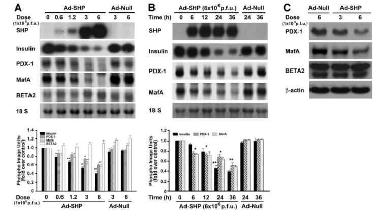
SHP overexpression induces -cell dysfunction. To elucidate the mechanism by which SHP mediates -cell dysfunction, INS-1 cells were infected with Ad-SHP at doses of 0.6, 1.2, 3, and 6⫻ 106pfu and then cultured in medium containing 5 mmol/l glucose. Ad-SHP downregu- lated insulin mRNA expression in a dose- and time- dependent manner (Fig. 4A and B). Moreover, Ad-SHP downregulated PDX-1 and MafA mRNA expression in a dose-dependent manner (Fig. 4A), and the decreases in PDX-1 and MafA mRNA expression preceded the de- creases in insulin mRNA expression (Fig. 4B). On the other hand, Ad-SHP had no effect on BETA2 expression (Fig. 4A). The disparate effects of SHP overexpression on PDX-1, MafA, and BETA2 expression were confirmed by Western blot analysis (Fig. 4C). Collectively, these results suggest that SHP decreases the expression of the insulin gene by inhibiting expression from the PDX-1 and MafA genes, but not from the BETA2 gene.
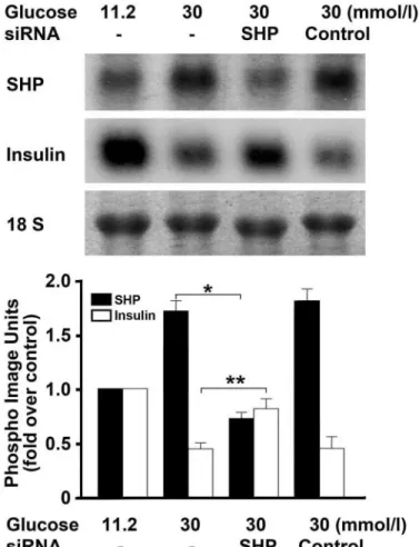
FIG. 3. Effect of siRNA-SHP on the high-glucose–induced decrease in insulin mRNA expression. INS-1 cells were transfected with 100 nmol/l of siRNA-SHP or control siRNA and then incubated with 11.2 or 30 mmol/l glucose for 48 h. The effect of siRNA-SHP on the high-glucose–
induced decrease in insulin mRNA expression was measured by North- ern blot analysis. RNA levels were normalized by the 18S rRNA levels.
Data in the bar graph are the meansⴞ SE of three separate measure- ments (bottom panel). *P < 0.01 and **P < 0.05 compared with 30 mmol/l glucose.
SHP INDUCES-CELL DYSFUNCTION
SHP represses p300-enhanced insulin transcription activity of PDX-1 and BETA2.PDX-1 and BETA2 inter- act in a p300 coactivator–mediated fashion, and this
interaction is critical for the activation of the insulin promoter (34). SHP also acts as a corepressor of BETA2 by competing with p300 for binding to BETA2 (33). Thus, we examined the effect of SHP on the coactivator activity of p300 in PDX-1 and BETA2 transactivation, using a human insulin promoter luciferase reporter assay. In the absence of SHP expression, the p300 expression vector dose- dependently increased the insulin transcriptional activity
FIG. 4. Effect of SHP overexpression on the expression of insulin and-cell–enriched transcription factors. A: Northern blot analysis of the effect of different doses of Ad-SHP on insulin, PDX-1, MafA, and BETA2 mRNA expression. INS-1 cells were infected with the indicated dose (pfu) of Ad-SHP or empty vector (Ad-Null) and cultured in 5 mmol/l glucose for 24 h. The RNA levels were normalized by the 18S rRNA levels. Data in the bar graph are the meansⴞ SE of three separate measurements (bottom panel). *P < 0.05, **P < 0.01, and ##P < 0.001 compared with Ad-SHP (0 pfu) and Ad-Null (6ⴛ 106pfu). B: Northern blot analysis of the time-dependent effect of Ad-SHP on insulin, PDX-1, and MafA mRNA expression. INS-1 cells were infected with 6ⴛ 106pfu of Ad-SHP or Ad-Null for the indicated times. RNA levels were normalized by the 18S rRNA levels. Data in the bar graph are the meansⴞ SE of three separate measurements (bottom panel). *P < 0.05 and **P < 0.01 compared with 0 h.
C: Western blot analysis of the effect of Ad-SHP on PDX-1, MafA, and BETA2 protein expression. INS-1 cells were infected with the indicated dose of Ad-SHP or Ad-Null for 24 h.
FIG. 5. SHP represses the p300-enhanced insulin transcription activity of PDX-1 and BETA2. COS-1 cells were cotransfected with the human insulin promoter (300 ng) and the expression vectors (pcDNA3) for PDX-1 (100 ng) and BETA2 (100 ng) together with the indicated amounts of p300 vector and increasing amounts of the SHP expression vector (pcDNA3) for 24 h. Data are the meansⴞ SE of three separate measurements. *P < 0.05, **P < 0.01, and ***P < 0.001 compared with cotransfection with the PDX-1 and BETA2 vectors without p300; #P <
0.05, #P < 0.01, and ###P < 0.001 compared with cotransfection with the PDX-1, BETA2, and p300 vectors without SHP.
FIG. 6. Expression of SHP in the pancreatic islets of OLETF rats. Islets were isolated from 40-week-old male OLETF and LETO rats. SHP and insulin mRNA expressions were measured by real-time RT-PCR. The results are the fold increases of the ratios of mRNA expression of SHP (or insulin) to actin relative to the ratio in LETO rats. Data are the meansⴞ SE of three separate measurements. *P < 0.05 compared with the equivalent LETO rat values.
of PDX-1 and BETA2 (Fig. 5). When the SHP expression vector was contransfected with 500 ng of the p300 expres- sion vector, SHP repressed the insulin transcription activ- ity of PDX-1, and BETA2 increased by p300 expression alone (Fig. 5). These data suggest that the inhibitory effect of SHP on insulin transcription may be partially attribut- able to its ability to inhibit the coactivation activity of p300.
SHPgene expression is upregulated in the pancreatic islets of OLETF rats.Next, we examined whether SHP gene expression is upregulated in an animal model of diabetes. The expression of SHP in the pancreatic islets of the two rat types was examined by real-time RT-PCR after confirming by intraperitoneal glucose tolerance test that the OLETF rats had developed hyperglycemia (supplemen- tary Fig. 4). The pancreatic islets of OLETF rats showed significantly increased SHP expression compared with the islets from LETO rats. Conversely, the islets of OLETF rats showed decreased insulin gene expression compared with the islets from LETO rats (Fig. 6). The GSIS and insulin content of the islets from OLETF rats were decreased compared with those of LETO rats (supplementary Fig. 5).
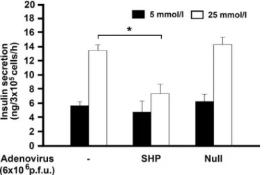
SHP inhibits GSIS. Finally, we tested whether SHP impairs GSIS in INS-1 cells. Static stimulation with 25 mmol/l glucose significantly increased the insulin secre- tion of INS-1 cells. This glucose-mediated stimulation of insulin secretion was significantly inhibited by Ad-SHP but not by empty vector (Ad-Null) (Fig. 7).
DISCUSSION
In this study, we found that a high glucose concentration increased SHP expression and that this was followed by decreased insulin enhancer– driven promoter activation as well as by decreases in insulin gene expression and secretion. SHP appears to downregulate insulin gene ex- pression by two mechanisms. First, it downregulates the gene expression of the insulin transcription enhancers PDX-1 and MafA, and, second, it inhibits the p300-upregu- lated transcriptional activity of PDX-1 and BETA2 on the insulin promoter. These observations collectively suggest that SHP plays an important role in the development of
-cell dysfunction induced by glucotoxicity.
The regulation of insulin gene expression in response to physiological stimuli enables-cells to maintain adequate stores of intracellular insulin when there is a sustained increase in the demand for insulin secretion. Glucose is the major physiological regulator of insulin gene expres- sion. However, prolonged elevation of glucose concentra- tions contributes to-cell dysfunction in type 2 diabetes, partly because it inhibits insulin gene expression (4,5,7–9).
Indeed, in this study, we observed that chronic exposure of INS-1 cells to a high glucose concentration decreased insulin gene expression in a time-dependent manner. We then found that adenovirus-mediated overexpression of SHP in INS-1 cells impaired both GSIS and insulin gene expression. Moreover, when endogenous SHP gene ex- pression in INS-1 cells was inhibited by SHP-specific siRNA, the insulin gene expression suppressed by high glucose concentrations was partially restored. In addition, SHPmRNA expression was higher in the pancreatic islets of diabetic OLETF rats than in the islets of the nondiabetic counterpart rats; the OLETF rat islets also expressed less insulin mRNA. Collectively, these data suggest that SHP could play a role in mediating the-cell dysfunction that arises from the metabolic stress conferred by chronic hyperglycemia.
In our previous study, we found that SHP acts as corepressor for BETA2 because it competes with the p300 coactivator for binding to BETA2 (33). However, our previous study did not fully explain how SHP downregu- lates insulin gene expression, since BETA2 activity on its own does not significantly stimulate insulin promoter activity (34). Transcriptional regulation of the insulin gene is a very complex process that requires the cooperation of a number of transcription factors, including PDX-1, BETA2, and MafA (34,35). The impairment of insulin gene expression after prolonged exposure to high glucose con- centrations is associated with diminished PDX-1 and MafA gene expression (7,9,15,16). Indeed, we found that INS-1 cells exposed chronically to a high glucose concentration showed decreased PDX-1 and MafA expression but no change in BETA2 expression. Like the effect of chronic high glucose levels, Ad-SHP downregulated the expression of PDX-1 and MafA but not BETA2. These data strongly suggest that SHP mediates the high-glucose–induced sup- pression of insulin gene expression by downregulating PDX-1and MafA gene expression.
We showed that SHP can act as a BETA2 corepressor by competing with the p300 coactivator. Next, we examined whether this activity contributes to the mechanism through which SHP downregulates insulin gene expres- sion. A recent study by Qiu et al. (34) demonstrated that the interaction between PDX-1 and BETA2 proteins, which is mediated by p300, plays a critical role in activating insulin promoter activity. More recently, Zhao et al. (35) demonstrated that MafA, PDX-1, and BETA2 cooperate in activating insulin gene transcription. Indeed, in this study, we found that p300, PDX-1, and BETA2 cooperate together to activate the insulin promoter in COS-1 cells. Moreover, we observed that SHP inhibited this cooperation. How- ever, this inhibition did not result from a block in the activation of the insulin promoter mediated by PDX-1 and MafA (data not shown). Taken together, these observa- tions as well as those of previous studies strongly suggest that SHP inhibits insulin promoter activity by preventing p300-mediated PDX-1 and BETA2 complex formation.
Thus, on being upregulated by a high glucose concen- tration, SHP impairs insulin gene expression by two
FIG. 7. Effect of SHP on GSIS by INS-1 cells. INS-1 cells were infected with 6ⴛ 106pfu of Ad-SHP or empty vector (Ad-Null) for 24 h, starved in 5 mmol/l glucose for 5 h, and then incubated in KRBB with 5 or 25 mmol/l glucose for 1 h. Insulin secretion (i.e., insulin concentration in the supernatant) was detected with a radioimmunoassay kit. Data are the meansⴞ SE of three separate measurements. *P < 0.01.
SHP INDUCES-CELL DYSFUNCTION
mechanisms that affect transactivators of insulin gene expression.
In conclusion, this study suggests that -cell dysfunc- tion induced by chronic high-glucose exposure is mediated by the abnormal regulation of SHP, although it remains to be determined whether SHP plays the same role in vivo.
Our findings suggest that SHP downregulates insulin gene expression by inhibiting the direct physical interactions between insulin enhancers as well as by repressing their gene expression. To more clearly elucidate the role SHP plays in the development of-cell dysfunction, it would be useful to generate animals in which SHP expression in the pancreatic-cells is conditionally knocked out.
ACKNOWLEDGMENTS
This study was supported by the Korea Science and Engineering Foundation through the NRL program (grant M106 00000271-06J000-27110). K.-M.L. and H.-Y.S. were supported by the Brain Korea 21 project in 2006.
We thank Prof. David D. Moore (Baylor College of Medicine, Houston, TX) for helpful comments and a critical review of the manuscript.
REFERENCES
1. Saltiel AR, Kahn CR: Insulin signalling and the regulation of glucose and lipid metabolism. Nature 414:799 – 806, 2001
2. Koh EH, Kim MS, Park JY, Kim HS, Youn JY, Park HS, Youn JH, Lee KU:
Peroxisome proliferator-activated receptor (PPAR)-alpha activation pre- vents diabetes in OLETF rats: comparison with PPAR-gamma activation.
Diabetes52:2331–2337, 2003
3. Tanaka Y, Tran PO, Harmon J, Robertson RP: A role for glutathione peroxidase in protecting pancreatic beta cells against oxidative stress in a model of glucose toxicity. Proc Natl Acad Sci U S A 99:12363–12368, 2002 4. Laybutt DR, Sharma A, Sgroi DC, Gaudet J, Bonner-Weir S, Weir GC:
Genetic regulation of metabolic pathways in beta-cells disrupted by hyperglycemia. J Biol Chem 277:10912–10921, 2002
5. Laybutt DR, Kaneto H, Hasenkamp W, Grey S, Jonas JC, Sgroi DC, Groff A, Ferran C, Bonner-Weir S, Sharma A, Weir GC: Increased expression of antioxidant and antiapoptotic genes in islets that may contribute to beta-cell survival during chronic hyperglycemia. Diabetes 51:413– 423, 2002 6. Efanova IB, Zaitsev SV, Zhivotovsky B, Kohler M, Efendic S, Orrenius S, Berggren PO: Glucose and tolbutamide induce apoptosis in pancreatic beta-cells: a process dependent on intracellular Ca2⫹concentration. J Biol Chem273:33501–33507, 1998
7. Olson LK, Redmon JB, Towle HC, Robertson RP: Chronic exposure of HIT cells to high glucose concentrations paradoxically decreases insulin gene transcription and alters binding of insulin gene regulatory protein. J Clin Invest92:514 –519, 1993
8. Olson LK, Qian J, Poitout V: Glucose rapidly and reversibly decreases INS-1 cell insulin gene transcription via decrements in STF-1 and C1 activator transcription factor activity. Mol Endocrinol 12:207–219, 1998 9. Poitout V, Olson LK, Robertson RP: Chronic exposure of betaTC-6 cells to
supraphysiologic concentrations of glucose decreases binding of the RIPE3b1 insulin gene transcription activator. J Clin Invest 97:1041–1046, 1996
10. Robertson RP, Zhang HJ, Pyzdrowski KL, Walseth TF: Preservation of insulin mRNA levels and insulin secretion in HIT cells by avoidance of chronic exposure to high glucose concentrations. J Clin Invest 90:320 – 325, 1992
11. Petersen HV, Serup P, Leonard J, Michelsen BK, Madsen OD: Transcrip- tional regulation of the human insulin gene is dependent on the homeodo- main protein STF1/IPF1 acting through the CT boxes. Proc Natl Acad Sci U S A91:10465–10469, 1994
12. Kaneto H, Sharma A, Suzuma K, Laybutt DR, Xu G, Bonner-Weir S, Weir GC: Induction of c-Myc expression suppresses insulin gene transcription by inhibiting NeuroD/BETA2-mediated transcriptional activation. J Biol Chem277:12998 –13006, 2002
13. Marshak S, Totary H, Cerasi E, Melloul D: Purification of the beta-cell glucose-sensitive factor that transactivates the insulin gene differentially in normal and transformed islet cells. Proc Natl Acad Sci U S A 93:15057–
15062, 1996
14. Olbrot M, Rud J, Moss LG, Sharma A: Identification of beta-cell-specific
insulin gene transcription factor RIPE3b1 as mammalian MafA. Proc Natl Acad Sci U S A99:6737– 6742, 2002
15. Olson LK, Sharma A, Peshavaria M, Wright CVE, Towle HC, Robertson RP, Stein R: Reduction of insulin gene transcription in HIT-T15 cells chroni- cally exposed to a supraphysiologic glucose concentration is associated with loss of STF-1 transcription factor expression. Proc Natl Acad Sci U S A92:9127–9131, 1995
16. Sharma A, Olson LK, Robertson RP, Stein R: The reduction of insulin gene transcription in HIT-T15 cells chronically exposed to high glucose concen- tration is associated with the loss of RIPE3b1 and STF-1 transcription factor expression. Mol Endocrinol 9:1127–1134, 1995
17. Harmon JS, Stein R, Robertson RP: Oxidative stress-mediated, post- translational loss of MafA protein as a contributing mechanism to loss of insulin gene expression in glucotoxic beta cells. J Biol Chem 280:11107–
11113, 2005
18. Seol W, Choi HS, Moore DD: An orphan nuclear hormone receptor that lacks a DNA binding domain and heterodimerizes with other receptors.
Science272:1336 –1339, 1996
19. Seol W, Chung M, Moore DD: Novel receptor interaction and repression domains in the orphan receptor SHP. Mol Cell Biol 17:7126 –7131, 1997 20. Seol W, Hanstein B, Brown M, Moore DD: Inhibition of estrogen receptor
action by the orphan receptor SHP (short heterodimer partner). Mol Endocrinol12:1551–1557, 1998
21. Johansson L, Thomsen JS, Damdimopoulos AE, Spyrou G, Gustafsson JA, Treuter E: The orphan nuclear receptor SHP inhibits agonist-dependent transcriptional activity of estrogen receptors ER␣ and ER. J Biol Chem 274:345–353, 1999
22. Johansson L, Bavner A, Thomsen JS, Farnegardh M, Gustafsson JA, Treuter E: The orphan nuclear receptor SHP utilizes conserved LXXLL- related motifs for interactions with ligand-activated estrogen receptors.
Mol Cell Biol20:1124 –1133, 2000
23. Lee YK, Dell H, Dowhan DH, Hadzopoulou-Cladaras M, Moore DD: The orphan nuclear receptor SHP inhibits hepatocyte nuclear factor 4 and retinoid X receptor transactivation: two mechanisms for repression. Mol Cell Biol20:187–195, 2000
24. Gobinet J, Auzou G, Nicolas JC, Sultan C, Jalaguier S: Characterization of the interaction between androgen receptor and a new transcriptional inhibitor, SHP. Biochemistry 40:15369 –15377, 2001
25. Sanyal S, Kim JY, Kim HJ, Takeda J, Lee YK, Moore DD, Choi HS:
Differential regulation of the orphan nuclear receptor small heterodimer partner (SHP) gene promoter by orphan nuclear receptor ERR isoforms.
J Biol Chem277:1739 –1748, 2002
26. Lee YK, Moore DD: Dual mechanisms for repression of the monomeric orphan receptor liver receptor homologous protein-1 by the orphan small heterodimer partner. J Biol Chem 277:2463–2467, 2002
27. Borgius LJ, Steffensen KR, Gustafsson JA, Treuter E: Glucocorticoid signaling is perturbed by the atypical orphan receptor and corepressor SHP. J Biol Chem 277:49761– 49766, 2002
28. Brendel C, Schoonjans K, Botrugno OA, Treuter E, Auwerx J: The small heterodimer partner interacts with the liver X receptor␣ and represses its transcriptional activity. Mol Endocrinol 16:2065–2076, 2002
29. Ourlin JC, Lasserre F, Pineau T, Fabre JM, Sa-Cunha A, Maurel P, Vilarem MJ, Pascussi JM: The small heterodimer partner interacts with the pregnane X receptor and represses its transcriptional activity. Mol Endo- crinol17:1693–1703, 2003
30. Yamagata K, Daitoku H, Shimamoto Y, Matsuzaki H, Hirota K, Ishida J, Fukamizu A: Bile acids regulate gluconeogenic gene expression via small heterodimer partner-mediated repression of hepatocyte nuclear factor 4 and Foxo1. J Biol Chem 279:23158 –23165, 2004
31. Kim JY, Kim HJ, Kim KT, Park YY, Seong HA, Park KC, Lee IK, Ha H, Shong M, Park SC, Choi HS: Orphan nuclear receptor small heterodimer partner represses hepatocyte nuclear factor 3/Foxa transactivation via inhibition of its DNA binding. Mol Endocrinol 18:2880 –2894, 2004
32. Lee HK, Lee YK, Park SH, Kim YS, Park SH, Lee JW, Kwon HB, Soh J, Moore DD, Choi HS: Structure and expression of the orphan nuclear receptor SHP gene. J Biol Chem 273:14398 –14402, 1998
33. Kim JY, Chu K, Kim HJ, Seong HA, Park KC, Sanyal S, Takeda J, Ha H, Shong M, Tsai MJ, Choi HS: Orphan nuclear receptor small heterodimer partner, a novel corepressor for a basic helix-loop-helix transcription factor BETA2/neuroD. Mol Endocrinol 18:776 –790, 2004
34. Qiu Y, Guo M, Huang S, Stein R: Insulin gene transcription is mediated by interactions between the p300 coactivator and PDX-1, BETA2, and E47Mol. Cell Biol 22:412– 420, 2002
35. Zhao L, Guo M, Matsuoka TA, Hagman DK, Parazzoli SD, Poitout V, Stein R: The islet beta cell-enriched MafA activator is a key regulator of insulin gene transcription. J Biol Chem 280:11887–11894, 2005