Salmonella Typhimurium SL1344 Utilizing Human Transferrin-bound Iron as an Iron Source Regardless of Siderophore-mediated Uptake
Yunjeong Choe1, Ah Young Yoo1, Sam Woong Kim2, Jihwan Hwang1 and Ho Young Kang1*
1Department of Microbiology, College of Natural Sciences, Pusan National University, Busan 46241, Korea
2Swine Science and Technology Center, Gyeongnam National University of Science and Technology, Jinju 52725, Korea Received January 17, 2017 /Revised January 19, 2017 /Accepted January 20, 2017
Inorganic iron is essential for various metabolic processes, including RNA synthesis, electron trans- port, and oxygen detoxification in microorganisms. Many bacterial pathogens compete for iron acquis- ition in diverse environmental condition such as host. Salmonella Typhimurium SL1344 also requires inorganic iron as a cofactor for growth. When a M9 minimal liquid medium was supplemented with ethylenediamine di-o-hydroxyphenylactic acid (EDDA) which acts as an iron-chelating agent, growth of Salmonella Typhimurium SL1344 in the supplemented medium was completely arrested by deficient of useful iron under iron-depleted condition. However, a number of siderophores, which are small, high-affinity iron chelating compounds secreted by microorganisms such as bacteria and fungi, were produced for utilization of restricted iron under iron-depleted condition. A M9 minimal liquid me- dium complemented with human transferrin (hTf)-iron complex turned completely off production of siderophores, but growth of Salmonella Typhimurium SL1344 maintained level similar to compare one complemented with iron (III) chloride (FeCl3). This means that human transferrin (hTf)-bound iron can utilize via directly interaction with Salmonella Typhimurium SL1344 without productions of siderophores. Through construction and analysis of negative mutant for utilization of human trans- ferrin (hTf)-bound iron, we confirm that the bacterium can directly use human transferrin (hTf)-bound iron without extracellularly intermediated carriers such as siderophores.
Key words : Iron, S. Typhimurium, siderophore, streptonigrin, Tn phoA, transferrin
*Corresponding author
*Tel : +82-51-510-2266, Fax : +82-51-513-4532
*E-mail : [email protected]
This is an Open-Access article distributed under the terms of the Creative Commons Attribution Non-Commercial License (http://creativecommons.org/licenses/by-nc/3.0) which permits unrestricted non-commercial use, distribution, and reproduction in any medium, provided the original work is properly cited.
Journal of Life Science 2017 Vol. 27. No. 1. 72~77 DOI : https://doi.org/10.5352/JLS.2017.27.1.72
Introduction
Inorganic iron was required by microorganisms as a co- factor for various metabolic processes, including RNA syn- thesis, electron transport, and oxygen detoxification [4].
Although iron is plentiful in nature, it readily oxidizes and precipitates to form biologically unavailable under oxic con- ditions and at a neutral pH. In the mammalian host, intra- cellular pools for most iron stores are found in heme com- pounds, ferritin, and metalloprotein complexes [18], while extracellular sources are sequestered by iron binding pro- teins, such as transferrin (in serum; Tf) and lactoferrin (at mucosal surfaces and in neutrophils; Lf). In order to over- come these low iron concentrations which may be as low as 10-18 M [5], pathogenic bacteria have developed a variety
of iron acquisition mechanisms [17]. One common way of acquiring iron is via the secretions of siderophores, which are high-affinity iron binding molecules and deliver the bound iron directly to the cell by means of a specific fer- ric-siderophore receptor [17].
Loss of the ability to produce siderophores is correlated with loss of virulence from Erwinia chrysanthemi in plants [10], Vibrio anguillarum in fish [8], and Pseudomonas aeruginosa [7, 14], Yersinia enterocolitica [12], and Escherichia coli [19] in mice. The direct interation with host iron-binding glyco- proteins such as Tf and Lf at the bacterial cell surface pro- vides an alternative to siderophores as a method to gain ac- cess to host iron supplies such as Tbp (transferrin binding protein) system in Neisseria gonorrhoeae [3, 20].
In this study, we exhibit that S. Typhimurium SL1344 can use transferrin-bound iron under condition lacking bio- syntheses of siderophores by treatment of EDDA. This con- firmed through construction of Tf-binding negative mutant that iron can’t be entered by direct interaction between trans- ferrin and specific sites on cell surface at S. Typhimurium SL1344.
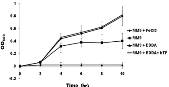
Fig. 1. Growth curves of S. Typhimurium in the M9 media con- taining various sources of iron. Rectangle is growth curve of S. Typhimurium incubated in M9 medium, diamond in M9 me- dium with FeCl3, triangle in M9 medium with EDDA, and re- mained symbol in M9 medium with EDDA/hTf. FeCl3, EDDA, and hTf were added to 30, 50, and 400 μM, respectively. X-axis indicates incubated times and Y-axis marks optical density at 600 nm in wavelength.
Materials and Methods
Media and reagents
S. Typhimurium SL1344 was used to investigate uptake of Tf-bound iron. Tn phoA [13] was used for construction of insertional mutation to a gene encoding an acceptor, which assumed to play a role as an acceptor for Tf, to inter- act with Tf-bound iron. S. Typhimurium SL1344 was in- cubated in Luria-Bertani (LB), M9 or Curtiss minimal (CM) media described previously [9, 15]. When required, anti- biotics were used by 100 μg/ml ampicillin (Am), 50 μg/ml kanamycin (Km), 50 μg/ml streptomycin (Sm) and streptoni- grin (Sn) to indicated concentration, etc. FeCl2 and hTf were purchased from sigma-aldrich and used to 30 μM and 400 μg/ml, respectively.
Analyses of growth curves for S. Typhimurium in MM9 media containing various iron sources
M9 medium was used for analyses of growth curves to S. Typhimurium. FeCl3 or hTf (human transferrin) (Sigma- Aldrich, U.S.) as iron sources were mixed with M9 medium.
Otherwise, free iron was arrested by chelating agent such as EDDA. S. Typhimurium was grown for overnight at 37℃
in LB broth. This broth was washed three times with M9 medium, diluted to 1:100 in prewarmed fresh M9 or M9 me- dia differentially supplemented with each component as shown in Fig. 1, 2 and 4, and incubated until indicated times.
Measurements of growth were analyzed by optical density at 600 nm in wavelength.
Detection and identification of siderophores changed by Tf-bound iron
Chrome azurol S (CAS) agar for assessment of side- rophores produced from S. Typhimurium was prepared as described earlier [1]. The CAS universal siderophore de- tection assay [16] was used to monitor productions of side- rophores by Salmonella cells grown in iron-depleted M9 me- dia by measuring the decrease of optical density at 630 nm in wavelength due to the CAS dye reaction as reported pre- viously [1]. In order to investigate productions of side- rophores when complemented with Tf, Tf was directly add- ed in M9 media or submerged in M9 media after sealing in dialysis bag.
Mutant isolation via streptonigrin enrichment culture S. Typhimurium was randomly mutated by conjugation to use Tn phoA (pRT733). Constructed mutants were selected on LB supplemented with Km and Sm, collected, and then resuspended to cell mass of 5×10⁹ CFU (colony forming unit)/ml. Fifty micro liter (2.5×10⁸ cells) pool of mutant were inoculated in 10 ml M9 media complemented with 400 μg/
ml hTf and 100 μg/ml Am, and then incubated for 4 hr at 37℃. The grown cells were harvested and washed twice with buffered saline-gelatin (BSG). Am-enriched cells were incubated in 10 ml LB medium for 2 hr at 37℃ by shaking to 200 rpm (revolution per min). Each 2 ml cultured broth was subcultured without Sn, with 4 μg/ml, and with 6 μg/
ml Sn for 12 hr at 37℃ by shaking. The cultured broths in Sn-treatments were patched onto CM agar medium contain- ing Km with and without EDDA/hTf. Mutated colonies were obtained by selection of isolates to show no growth on medium containing Km with EDDA/hTf.
Results and Discussion
S. Typhimurium is able to use Tf-bound iron regard- less of siderophores
Almost iron in a mammalian host is found intracellularly in the forms of heme, hemosiderin, ferritin or complexed with metalloproteins [18], whereas extracellular iron is stor- ed by Tf, Lf, and hemoglobin. Sequestration of iron by these iron-binding proteins and compounds effectively lowers the concentration of available soluble ferric iron to a level in- sufficient for bacterial growth. To overcome this limitation, pathogenic microorganisms have evolved to alternative mechanisms to acquire iron from these iron-bound com-
Fig. 2. Productions of siderophores by S. Typhimurium grown in M9 media containing differentially supplemented hTf.
Cell free culture supernatants of bacteria were tested by using the CAS assay. In or out indicates the location of supplemented Tf; In, inside of dialysis bag; Out, outside of dialysis tubing bag.
Fig. 3. Model for the effects of streptonigrin on bacteria. SNG, Streptonigrin; SNGH, reduced streptonigrin.
pounds [5].
S. Typhimurium was investigated whether alternative iron uptake pathway is or not, except for system utilizing siderophores. As shown in Fig. 1, growth of S. Typhimurium in M9 media supplemented with EDDA was completely ar- rested for indicated times. When compared to M9 medium, Salmonella growth in M9 supplemented with FeCl3 and EDDA/hTf were not nearly different to that of M9 media until 3 hr from inoculums, but, after its time, showed gradu- ally growth better than that of M9 medium. A medium add- ed with only EDDA was to completely inhibit growth of S. Typhimurium, whereas an hTf supplementation to M9
medium with EDDA showed recovery of growth as much as FeCl3. These results indicated that S. Typhimurium could directly use iron from Tf-bound iron without siderophores.
S. Typhimurium is a mechanism to directly use hTf-
bound iron
Many bacterial species grown in iron-deficient media causes the expression of so-called iron-regulated genes scat- tered throughout the genome. Among the enteric bacteria, this process is controlled at the transcriptional level by the regulatory protein Fur, a product of the chromosomal fur (ferric uptake regulation) gene [2, 11]. Some of the genes controlled by Fur encode high-affinity iron transport sys- tems mediated by siderophores, low-molecular-mass iron (III) chelators which scavenge iron in the external medium and make it available to bacteria.
It has been known that S. Typhimurium raises pro- ductions of siderphores under iron-depleted condition, but lowers under iron rich medium. Our results also showed that siderophores were highly expressed by M9 medium re- stricted with iron, but very low when complemented with iron (Fig. 2). These results indicate that productions of side- rophores are regulated by iron. Interestingly, when hTf was added by sealing in dialysis bag, productions of side- rophores were highly increased, whereas when added di- rectly into the M9 medium, its production shut off com- pletely (Fig. 2). Therefore, it was suggested that hTf-bound iron shuts off production of siderophores and is directly in-
Table 1. Results of streptonigrin enrichment cultures Streptonigrin
(μg/ml)
Initial cells (CFU)
After 12 hr culture (CFU) 0
4 6
3.2×10⁸ 3.2×10⁸ 3.2×10⁸
9.4×10⁹ 2.8×10³ 1.6×101
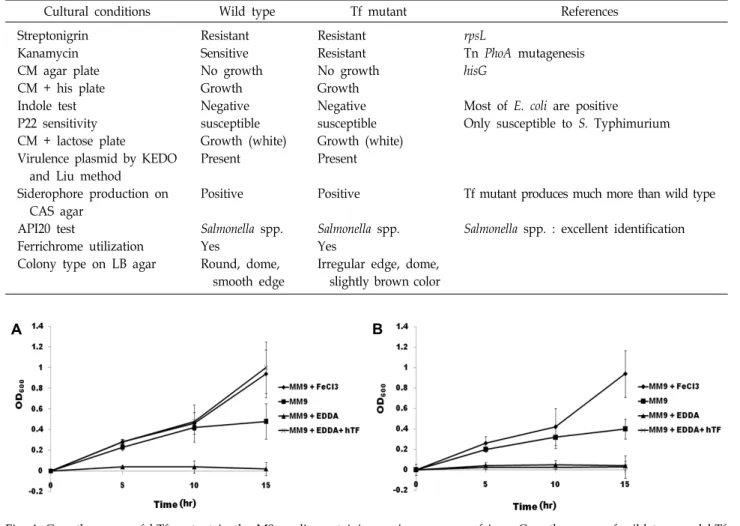
Table 2. Biochemical and genetic characterization of Tf mutant
Cultural conditions Wild type Tf mutant References
Streptonigrin Kanamycin CM agar plate CM + his plate Indole test P22 sensitivity CM + lactose plate
Virulence plasmid by KEDO and Liu method
Siderophore production on CAS agar
API20 test
Ferrichrome utilization Colony type on LB agar
Resistant Sensitive No growth Growth Negative susceptible Growth (white) Present
Positive
Salmonella spp.
Yes
Round, dome, smooth edge
Resistant Resistant No growth Growth Negative susceptible Growth (white) Present
Positive
Salmonella spp.
Yes
Irregular edge, dome, slightly brown color
rpsL
Tn PhoA mutagenesis hisG
Most of E. coli are positive
Only susceptible to S. Typhimurium
Tf mutant produces much more than wild type
Salmonella spp. : excellent identification
A B
Fig. 4. Growth curves of hTf mutant in the M9 media containing various sources of iron. Growth curves of wild type and hTf mutant in the M9 media containing various sources of iron were indicated by A and B, respectively. Open circle is results grown in M9 media complemented with EDDA. Remained marks were indicated by the same symbols as shown in Fig. 1.
teraction with any materials on cell surface of S. Typhimu- rium for iron utilization.
A mutant impossible of interacting directly with hTf-bound iron is unable to grow at existence of hTF Numerous mutations in a single chromosome are gen- erated by Sn treatment, and it has also the great potential to identify iron transport mutants. This chemical has been used in Neisseria and Bordetella species to identify iron uptake mutant. The quinine antibiotic Sn is believed to kill bacteria
by promoting formation of oxygen radical in the presence of iron [6] (Fig. 3).
For construction of mutants to iron transport system(s), the first random mutagenesis was performed by conjugation between S. Typhimurium and E. coli host containing Tn phoA (pRT733). The mutated colonies were selected on LB added with Km and Sm, and then pooled by total volume 5 ml (5×10⁹ CFU/ml). Am-enrichment culture performed to in- crease yield of mutant selection as follows. When mutant pools were incubated by M9 media supplemented with hTf and Am, hTf negative mutants could not grow due to defi- ciency of available iron. However, wild type or other mu- tants, which unrelated with hTf utilization, would grow rap- idly and then be killed due to Am. Mutant pools were in- cubated in M9 media with hTf and Am for 4 hr at 37℃.
The cultured broths were washed twice with BSG, re- suspended in 10 ml LB and incubated for 2 hr at 37℃.
Streptonigrin enrichment cultures were performed by M9 liquid media with EDDA, hTf, Km, and Sn for 12 hr at 37℃.
The results showed that 2800 CFU (colony forming unit) was observed by treatment of 4 μg/ml streptonigrin and 16 CFU by 8 μg/ml (Table 1). The next isolated 500 colonies were patched onto CM agar plates containing Km with and with- out EDDA/hTf. One of 500 patched colonies was unable to grow on CM agar with Km, EDDA, and hTf. The analyses of growth of mutant were performed by each factor for dem- onstration of mutant as shown in Table 2. The mutant didn’t grow under existence of hTf for indicated times (Fig. 4). As shown in Table 2, biochemical characterizations were the same as wild type S. Typhimurium SL1344. However, nu- cleotide sequencing results revealed that insertion site of Tn phoA is located on completely unrelated gene with iron transport system (data not shown). Therefore, it was as- sumed that the hTf mutant was obtained by accumulation of stress followed treatment of streptonigrin. Although we didn’t know to bind at any places, these results demon- strated that hTf-bound iron is possible of utilization through directly interaction with S. Typhimurium without ex- tracellularly intermediated carriers such as siderophores.
Acknowledgement
This work was supported by a 2-Year Research Grant of Pusan National University.
References
1. Armstrong, S. K. and Clements, M. O. 1993. Isolation and characterization of Bordetella bronchiseptica mutants deficient in siderophore activity. J. Bacteriol. 175, 1144-1152.
2. Bagg, A. and Neilands, J. B. 1987. Ferric uptake regulation protein acts as a repressor, employing iron (II) as a cofactor to bind the operator of an iron transport operon in Escherichia coli. Biochemistry 26, 5471-5477.
3. Boulton, I. C., Yost, M. K., Anderson, J. E. and Cornelissen, C. N. 2000. Identification of discrete domains within gon- ococcal transferrin-binding protein A that are necessary for ligand binding and iron uptake functions. Infect. Immun. 68, 6988-6996.
4. Briat, J. 1992. Iron assimilation storage in prokaryotes.
Microbiology 138, 2475-2483.
5. Bullen, J., Rogers, H. J. and Griffiths, E. 1978. Role of iron
in bacterial infection, pp. 1-35, In Anonymous Current top- ics in microbiology and immunology, Springer.
6. Cohen, M. S., Chai, Y., Britigan, B. E., McKenna, W., Adams, J., Svendsen, T., Bean, K., Hassett, D. J. and Sparling, P.
F. 1987. Role of extracellular iron in the action of the qui- none antibiotic streptonigrin: mechanisms of killing and re- sistance of Neisseria gonorrhoeae. Antimicrob. Agents Chemother.
31, 1507-1513.
7. Cox, C. D. 1982. Effect of pyochelin on the virulence of Pseudomonas aeruginosa. Infect. Immun. 36, 17-23.
8. Crosa, J. H. 1980. A plasmid associated with virulence in the marine fish pathogen Vibrio anguillarum specifies an iron-sequestering system.
9. Curtiss, S. R. 3rd. 1965. Chromosomal Aberrations Associated with Mutations to Bacteriophage Resistance in Escherichia Coli. J. Bacteriol. 89, 28-40.
10. Enard, C., Diolez, A. and Expert, D. 1988. Systemic virulence of Erwinia chrysanthemi 3937 requires a functional iron as- similation system. J. Bacteriol. 170, 2419-2426.
11. Hantke, K. 1984. Cloning of the repressor protein gene of iron-regulated systems in Escherichia coli K12. Mol. Gen.
Genet. 197, 337-341.
12. Heesemann, J., Hantke, K., Vocke, T., Saken, E., Rakin, A., Stojiljkovic, I. and Berner, R. 1993. Virulence of Yersinia en- terocolitica is closely associated with siderophore production, expression of an iron‐repressible outer membrane poly- peptide of 65 000 Da and pesticin sensitivity. Mol. Microbiol.
8, 397-408.
13. Manoil, C. and Beckwith, J. 1985. TnphoA: a transposon probe for protein export signals. Proc. Natl. Acad. Sci. US A 82, 8129-8133.
14. Meyer, J. M., Neely, A., Stintzi, A., Georges, C. and Holder, I. A. 1996. Pyoverdin is essential for virulence of Pseudomo- nas aeruginosa. Infect. Immun. 64, 518-523.
15. Miller, J. H. 1992. A short course in bacterial genetics: a labo- ratory manual and handbook for Escherichia coli and related bacteria.
16. Schwyn, B. and Neilands, J. 1987. Universal chemical assay for the detection and determination of siderophores. Anal.
Biochem. 160, 47-56.
17. Weinberg, E. D. 2009. Iron availability and infection. Biochi- mica et Biophysica Acta (BBA)-General Subjects. 1790, 600-605.
18. Weinberg E. D. 1978. Iron and infection. Microbiol. Rev. 42, 45-66.
19. Williams, P. H. 1979. Novel iron uptake system specified by ColV plasmids: an important component in the virulence of invasive strains of Escherichia coli. Infect. Immun. 26, 925- 932.
20. Yost-Daljev, M. K. and Cornelissen, C. N. 2004. Determination of surface-exposed, functional domains of gonococcal trans- ferrin-binding protein A. Infect. Immun. 72, 1775-1785.
초록:
Salmonella
Typhimurium SL1344의 사람의 트렌스페린(hTf)에 부착된 철 이용에 관한 연구최윤정1․유아영1․김삼웅2․황지환1․강호영1*
(1부산대학교 미생물학과, 2경남과학기술대학교 양돈과학기술센터)
S. Typhimurium SL1344는 성장을 위한 보조인자로 무기철이 요구된다. 철 킬레이트제인 ethylenediamine di-o-hydroxyphenylactic acid (EDDA)가 첨가 된 M9 최소배지에서 S. Typhimurium은 성장에 있어 철 이용이 완전하게 억제된다. 하지만, 세균이나 곰팡이와 같은 미생물들은 철이 부족한 환경에서 제한된 철을 이용하기 위 해 사이드로포어를 생산한다. 사람에서 유래한 트랜스페린(hTf)-철 복합체를 M9 배지에 첨가한 조건에서 S.
Typhimurium의 사이드로포어 생산은 완전하게 중단되었다. 반면, S. Typhimurium의 성장은 염화철(FeCl3)을 첨 가한 조건과 동일한 수준으로 유지되었다. 이 결과는 사이드로포어의 생산 없이도 S. Typhimurium이 hTf에 부착 된 철을 직접적으로 이용할 수 있다는 것을 알 수 있다. 돌연변이주의 구축과 이를 이용한 분석을 통하여 우리는 세균이 hTf-철 복합체를 직접적으로 이용할 수 있다는 것을 확인할 수 있었다.