Regulation of Matrix Metalloproteinase-1 Expression by the Homeodomain Transcription Factor Caudal in Drosophila Intestine
Shin-Hae Lee
1†, Mi-Sun Hwang
1‡, Yoon-Jeong Choi
1¶, Young-Shin Kim
2and Mi-Ae Yoo
1*
1
Department of Molecular Biology, Colleges of Natural Sciences, Pusan National University, Busan 609-735, Korea
2
Research Institute of Genetic Engineering, Pusan National University, Busan 609-735, Korea
Received October 30, 2012 /Revised December 11, 2012 /Accepted December 11, 2012The matrix metalloproteinase (MMP) family plays essential roles in physiological processes such as embryonic development, angiogenesis, wound healing, and tissue homeostasis as a consequence of MMPr capacity for breaking down many types of extracellular matrix proteins. Imbalanced regulation of MMP expression can also lead to pathological conditions such as tumor progression. We recently reported that the Drosophila Mmp1 gene is highly expressed in the digestive tract and is required for the maintenance of intestinal homeostasis such as by restriction of uncontrolled intestinal stem cell proliferation. However, the regulatory mechanisms of MMP gene expression in the intestine remain unclear. In this study, we determined that the expression of Mmp1 is regulated by the homeodomain transcription factor Caudal. Experiments using the targeted expression of Caudal under the regulation of Gal4-UAS system indicated that endogenous Caudal is required for the Mmp1 gene expression in the adult Drosophila intestine and that exogenous Caudal induces Mmp1 expression. Transient trans- fection experiments indicated that Caudal can activate the promoter activity of Mmp1 and that several putative Caudal binding sites in the 5´-flanking region of the Mmp1 gene may be critical to the upre- gulation by Caudal. Our data suggest that Mmp1 is one of the target genes of Caudal in physiological normal condition and in tumorigenesis.
Key words : Matrix metalloprotease-1, Caudal, Drosophila intestine
*Corresponding author
*Tel:+82-51-510-2278, Fax:+82-51-513-9258
*E-mail : [email protected]
†Present address : Department of Biological Sciences, Inha University, Incheon 402-751, Korea
‡Present address : Department of Pharmacology and Pharmacogenomics Research Center, Inje University, College of Medicine, Busan 633-165, Korea
¶Present address : Research Center for Human Natural Defense System, Yonsei University, College of Medicine, Seoul 120-752, Korea
This is an Open-Access article distributed under the terms of the Creative Commons Attribution Non-Commercial License (http://creativecommons.org/licenses/by-nc/3.0) which permits unrestricted non-commercial use, distribution, and reproduction in any medium, provided the original work is properly cited.
Journal of Life Science 2012 Vol. 22. No. 12. 1600~1607 DOI : http://dx.doi.org/10.5352/JLS.2012.22.12.1600
Introduction
Extracellular proteases, such as matrix metalloproteinase (MMP), are crucial regulators for cell functions. The essential roles of MMP within physiological processes such as embry- onic development, angiogenesis, wound healing, and tissue homeostasis result from their capacity to break down all kinds of extracellular matrix (ECM) proteins [22]. As a result of their potent proteolytic activity, imbalanced regulation of MMP can lead to excessive ECM degradation and patho-
logical conditions such as rheumatoid arthritis and tumor progression [5,22]. As the importance of MMP, the activity of MMP is tightly regulated at various steps: transcription, post-translational activation through the cleavage of inactive prodomain, and interaction with tissue inhibitors of metal- loproteinases [23,31]. However, almost investigations for the regulatory mechanisms of MMP expression are limited to the stressed and pathological conditions. The regulatory mechanisms of MMP in physiological normal condition are not fully understood.
In Drosophila genome, only two MMPs have been identi-
fied as having conserved domain structures, which differ
from the 26 MMPs found in mammals [17,18]. Both fly
MMPs have the canonical MMP domain structure, with a
signal sequence, prodomain, catalytic domain, hinge and he-
mopexin domain [7]. Although there are not clear ortholo-
gous relationships between the Drosophila and vertebrate
MMPs [7], fly Mmp1 represents the typical secreted form
of MMP and fly Mmp2 represents the membrane anchored
form of MMP on the basis of their structure [17,18]. Recently,
we reported that the Drosophila Mmp1 gene is highly ex-
pressed in the digestive tract in both third instar larvae and
adult flies and is required for the maintenance of intestinal
homeostasis such as the restriction of uncontrolled intestinal stem cell proliferation, and the maintenance of intestinal ar- chitecture [15]. It was reported that Mmp1 expression is regulated by JNK and Fos to induce invasive tumor [28].
However, the underlying mechanisms of Drosophila Mmp1 gene expression in the intestine remain unknown.
Caudal-related homeobox genes play crucial roles in axial patterning of embryonic development and intestinal devel- opment and differentiation [20,21,26]. The Drosophila caudal gene contributes to defining the anteroposterior axis during development, patterning of the posterior segments, and gut development [16,30]. In addition, caudal gene was recently reported to be involved in the innate immune system in the gut [24,25]. Moreover, CDX1 and CDX2, mammalian homo- logues of Drosophila caudal, are highly expressed in intestinal epithelium, and are required for the maintenance of in- testinal integrity in the adult intestine [11]. Abnormal Cdx expression have been well known to be associated with in- testinal tumorigenesis [8].
In this study, we investigated whether the expression of Mmp1 is regulated by Caudal transcription factor in Drosophila. We found that endogenous Caudal is required for the Mmp1 gene expression in the adult Drosophila intes- tine and exogenous Caudal expression can activate Mmp1 promoter in vitro and in vivo.
Materials and Methods Immunohistochemistry
Immunostaining was performed as previously described [3]. Briefly, the fly guts and larval epidermis were fixed in 4% formaldehyde (Sigma-Aldrich, MO, USA) for 1 hr at 25°C, washed in PBS/0.1% Triton X-100 (PBST) and in- cubated with primary antibodies in 1% bovine serum albu- min (BSA, Sigma-Aldrich) overnight at 4°C. The samples were then washed in PBST, incubated with secondary anti- bodies for 45 min at 25°C, washed and mounted with Vectashield (Vector Labs, CA, USA). The images were ana- lyzed using a Zeiss AxioSkop 2Plus microscope (Carl Zeiss Inc., Germany). Primary antibodies were diluted as fol- lows: mouse anti-Mmp1 1:50 (Developmental Studies Hybridoma Bank, IA, USA); rabbit anti-GFP 1:500 (Molecular Probes, OR, USA). Secondary antibodies used in- cluded Cy3-conjugated goat anti-mouse and FITC-con- jugated goat anti-rabbit (Jackson ImmunoResearch, PA, USA); all were diluted 1:300.
Western blot
Proteins were extracted from whole third instar larvae us- ing PRO-PREP
™protein extract solution (iNtRON Biotech- nology, Korea) and separated by SDS-PAGE. Proteins were then transferred onto polyvinylidene fluoride (PVDF) mem- branes (GE Healthcare, UK), and incubated with primary an- tibodies for 16 hr at 4°C. After washing with Tris-based buf- fered saline (TBS)/0.1% Tween 20 (TBST), membranes were incubated with secondary antibodies for 45 min at 25°C.
Bound antibodies were then detected using the WEST-One
™Western Blot detection system (iNtRON Biotechnology).
Mouse anti-Mmp1, 1:20; mouse anti-α-tubulin, 1:10,000 (BioGenex, CA, USA) were used in combination with HRP-conjugated anti-mouse IgG secondary antibodies (1:5,000, Santa Cruz Biotechnology Inc., CA, USA).
Reverse-transcription polymerase chain reaction (RT-PCR)
Total RNA was isolated from whole third instar larvae with TRIzol reagent (Molecular Research Center Inc., OH, USA), according to manufacturer’s instructions, and 1 μg to- tal RNA was reverse-transcribed using M-MLV reverse tran- scriptase (Promega, WI, USA). Samples underwent 30 PCR cycles, were separated on 2% agarose gels, and stained with ethidium bromide (Sigma-Aldrich). All oligonucleotides were chemically synthesized. Oligonucleotide sequences for the analysis of Mmp1 [15], cad [12], and rp49 [1] were pre- viously reported.
Construction of reporter plasmids
To construct reporter plasmids of Mmp1, the 5´-flacking region of Mmp1 contained 2,345 bp fragments spanning from 1,980 bp upstream and 365 bp downstream with respect to the transcription initiation site was PCR-amplified from Kc genomic DNA and cloned into pGL2-basic (Promega), and the sequences were confirmed by sequencing. The primers for the promoter region of Mmp1 containing linker se- quences recognized by 5´ and 3´ -KpnI were as follow:
Forward, 5´-CCG GGT ACC AGA TGG CTT GCA CAA GCA-3´; Reverse, 5´-CCG GGT ACC TTC CAG CTC TCT CTG CCA TT-3´. The deletion constructs were generated by digestion of pMmp1-luc with SmaI, SmaI-KpnI and HindIII.
The resulting deletion constructs were designated
-1627pMmp1-luc, -708pMmp1-luc and -570pMmp1-luc,
respectively. Additional two deletion constructs were cloned
(-1119pMmp1-luc and -170pMmp1-luc). All oligonucleotides
were chemically synthesized.
DNA transfection and luciferase assay
Drosophila S2 cells were grown at 25°C in Schneider me- dium (Sigma-Aldrich) supplemented with 10% fetal bovine serum (FBS) and 1% penicillin-streptomycin (PS, GIBCO- BRL, MD, USA). All plasmids for transfection were prepared by using Qiagen Plasmid Midi Kit (Qiagen, CA, USA).
Dimethyldioctadecyl ammonium bromide (DDAB) mediated transfection of Drosophila cultured cells were performed us- ing 6-, 12-, or 24-well tissue culture plates as described pre- viously [9]. Each transfection mixture contained 100 ng of the appropriate reporter plasmids and 300 ng of pAc-lacZ internal control plasmids. For co-transfection experiments, 300-900 ng of effector plasmids and pAc-HisA for the control of effector plasmids were used. Forty-eight hr after trans- fection, cell extracts were prepared by adding 50-200 μl of Cell Culture Lysis Reagent (Promega) to each well after re- moving the media by aspiration. The well plate was shaken gently on a rotary shaker for 2-5 min and cell extracts were transferred to a microcentrifuge tube. After centrifugation for 5 min, these supernatant were subjected to β -galactosidase and luciferase assays. Luciferease assay was performed with a luminometer (TD-20/20, Turner Designs, CA, USA). Normalized luciferase activity was calculated by determining the luciferase/β-galactosidase activity ratio and by averaging the values from 3-8 experiments and the aver- age values, and standard errors were calculated.
Drosophila stocks
Drosophila stocks were consistently maintained at 25°C on standard cornmeal-sugar-yeast medium under a 12 hr/12 hr light/dark cycle. The food consisted of 79.2% water, 1% agar, 7% cornmeal, 2% yeast, 10% sucrose, 0.3% bokinin, and 0.5%
propionic acid. To avoid larval overpopulation in culture vi- als, 25-30 adult flies were cultured in a vial and transferred to new vials containing fresh food every 2-3 days. tub-Gal80
tsand hs-Gal4 fly strains were obtained from the Bloomington Drosophila Stock Center (Indiana University, IN, USA).
Temperature-inducible differentiated enteroyte-specific Myo1A-Gal80
tsflies were obtained from B. A. Edgar [13], and UAS-cad
RNAiflies from W. J. Lee [10]. The fly stocks of UAS-cad were described previously [12].
Results
Requirement of Caudal for Mmp1 expression in the adult Drosophila intestine
To understand the regulatory mechanism of Mmp1 ex-
pression, we assessed whether the homeodomain tran- scription factor Caudal is a potential regulator of Mmp1 gene in the adult gut. To investigate whether the endogenous Caudal can regulate Mmp1 expression in the adult intestine, we used GAL4/UAS system [6]. The GAL4/UAS ectopic ex- pression system is valuable machinery for the over- expression or knockdown of a transgene under specific con- ditions, such as cell-, tissue-, temperature-, and stage-specific conditions in Drosophila [6]. When the GAL4 transcription factor is expressed under the control of various promoters, GAL4 can drive the expression of a transgene under an up- stream activating sequence (UAS) [6]. We crossed the flies UAS-cad
RNAi, which expressing the interference RNAi (RNAi) against to the Caudal (cad
RNAi), with the heat-shock in- ducible enterocytes-specific Myo1A-Gal80
tsflies. Since these flies contained UAS-GFP, the cells expressing cad
RNAicon- struct were detected via the expression of GFP protein (Fig.
1A). When cad
RNAiwas expressed in the enterocyte of the fly guts, the cell expressing cad
RNAidecreased the Mmp1 lev- el detected by anti-Mmp1 antibody (inset in Fig. 1B). These
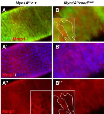
Fig. 1.
Mmp1
expression is decreased bycad
RNAiexpression in the enterocytes of adult intestine. The flies expressingcad
RNAi under enterocyte-specificMyo1A
-Gal80
ts driver (Myo1A
ts>cad
RNAi, B-B”) and driver alone (Myo1A
ts>+
, A-A”) were cultured at 29°C for 4 days and the guts were stained with anti-GFP (green), anti-Mmp1 (red), and DAPI (blue). Dashed line indicatescad
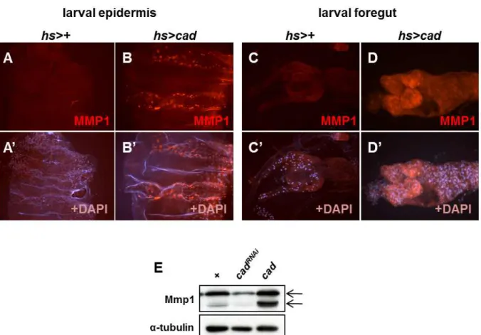
RNAi ex- pression region.Fig. 2.
Mmp1
expression is increased bycad
expression in the larval gut and epidermis. A-D. The third instar larvae expressingcad
under heat shock-induciblehs-Gal4
driver (hs>cad
) and driver alone (hs>+
) were incubated at 25°C for 24 hr after heat shock and the epidermis (A and B) and guts (C and D) were stained with anti-Mmp1 (red), and DAPI (blue). E. Mmp1 expression level was dependent withcad
expression in larvae. The third instar larvae expressingcad
RNAiorcad
under heat shock-induciblehs-Gal4
driver (hs>cad
RNAiorhs>cad
) and driver alone (hs>+
) were incubated at 25°C for 24 hr after heat shock and the whole larvae extracts were blotted with anti-Mmp1 antibody. Upper arrow indicates full length, inactive form of Mmp1. Lower arrow indicates cleaved, activated form of Mmp1. Alpha-tubulin was used as internal control.results indicate that Caudal is required for the maintenance of Mmp1 levels in the adult midgut.
Caudal overexpression induces ectopic Mmp1 expression in larval gut
To know whether the ectopic expression of Caudal can increases Mmp1 expression, we crossed the flies UAS-cad, which expressing the wild type Caudal protein, with the heat-shock inducible hs-Gal4 driver. We previously reported that ectopic overexpression of Caudal under hs-Gal4 driver induce melanotic tumors in the epidermis and guts of the third instar larvae [12]. As reported previously, the third in- star larvae expressing Caudal under hs-Gal4 showed mela- notic tumor in their epidermis and guts. We found that the expression of Mmp1 was drastically increased in the epi- dermis and guts having melanotic tumors (Fig. 2A-D).
Furthermore, the regulation of Mmp1 by Caudal was con- firmed at protein level using Western blot (Fig. 2E). These results indicate that ectopic Caudal expression increased Mmp1 expression.
Transcriptional regulation of Mmp1 by Caudal transcription factor
To understand whether Caudal transcriptionally regulate Mmp1 expression, we analyzed the mRNA level of Mmp1 in cad expressing flies. In the third instar larvae expressing Caudal protein under hs-Gal4, the mRNA level of Mmp1 was increased compared to control (Fig. 3A).
Next, to determine the effect of Caudal protein on Mmp1
promoter activity, we investigated whether the Caudal pro-
tein can regulate the activity of Mmp1 promoter, using a
pMmp1-luc reporter plasmid containing the promoter region
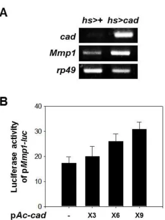
Fig. 3.
Mmp1
expression was regulated by cad expression. A.Increase in
Mmp1
mRNA level bycad
expression in the third instar larvae. The third instar larvae expressingcad
under heat shock-induciblehs-Gal4
driver (hs>cad
) and driver alone (hs>+) were incubated at 25°C for 24 hr after heat shock and the mRNA extract from whole lar- vae was analyzed by RT-PCR.Ribosomal protein 49
(rp49
) was used as internal control. B. Activation ofMmp1
pro- moter bycad
expression in S2 cells. The luciferase activ- ity of pMmp1-luc
was increased by expression ofcad
. The reporter plasmid pMmp1-luc
(100 ng) was co-trans- fected with the indicated amount of pAc-cad
intoDrosophila
S2 cells and the luciferase activities were nor- malized with co-transfected β-galactosidase activity.Average values obtained from 3 independent experi- ments with ±SE values are given.
(-1,980 to +365) of the Mmp1 gene fused with the luciferase reporter gene which was constructed as described in the Materials and Methods. Effect of Caudal on Mmp1 gene pro- moter activity was examined in transient transfection experi- ments using pMmp1-luc reporter plasmids and a pAc-cad ex- pression plasmid in the Drosophila S2 cell line. Mmp1 pro- moter was transactivated depending on the amount of pAc-cad, up to nearly 2-fold (Fig. 3B). These results indicate that Mmp1 expression is directly regulated at transcriptional level by the Caudal protein.
Putative Caudal recognition sites in the 5'-flanking region of the Drosophila Mmp1 gene
Within the promoter region of Mmp1 gene, we found eight putative Caudal binding sites similar to the Caudal binding consensus sequences, A/CTTTATA/G, named cad0, 1, 2, 3, 4, 5, 6 and 7 in the 5'-flanking region of the Mmp1 gene (Fig. 4A). To investigate the potential of Caudal bind- ing sites in Mmp1 gene, deletion constructs of pMmp1-luc were constructed as described in the Materials and Methods and named from their sites deleted (Fig. 4A).
The promoter activity of -1627pMmp1-luc reporter was not significantly decreased compared to control -1850pMmp1-luc reporter, in which three most distal Caudal binding sites (cad5-7) were deleted (Fig. 4A). The promoter activity of -1119pMmp1-luc reporter dramatically decreased compared to control, in which further three distal Caudal binding sites (cad2-4) were deleted and possessed only two putative Caudal binding sites (cad0, cad1) (Fig. 4A). The reporter lines -708, -570 and -170pMmp1-luc contained only one Caudal binding sites (cad0) in the upstream of transcrip- tional initiation site did not have significant promoter activ- ities (Fig. 4A). Since the marked reduction of Mmp1 pro- moter activity between the -1627 to -1119 regions, the pos- itive regulator might be a function in the promoter region of Mmp1 gene between the -1627 to -1119 regions.
To assess whether the this promoter region is responsible for Caudal, co-transfection experiments were carried out with the wild type or deleted reporter plasmids and Caudal expressing pAc-cad plasmid. The wild-type promoter re- porter activity was activated by Caudal expression; while the deletion reporter -1119pMmp1-luc did not responded to Caudal expression (Fig. 4B). In addition, the other deleted reporter lines also were not activated by Caudal (Fig. 4B).
Since the promoter activity of -1627pMmp1-luc was not sig- nificantly altered compared with -1850pMmp1-luc, the Caudal binding sites cad2-4 may be critical in regulating Mmp1 expression.
These results indicate that Caudal increases the promoter activity of the Mmp1 gene and the Caudal binding sites cad2-4 may be critical in the upregulation of Mmp1 ex- pression by Caudal.
Discussion
In the present study, we documented that the expression
of Mmp1 gene is regulated by the homeodomain tran-
Fig. 4. Caudal responsible region in the
Mmp1
promoter. A. Promoter activity of the deletion constructs of pMmp1
-luc
. Red triangles indicate putative Caudal binding sites in theMmp1
promoter region. Putative 8 Caudal binding sites were existed in theMmp1
promoter region. Wild type pMmp1-luc
or deletion reporter plasmids (100 ng) were transiently transfected into S2 cells and the luciferase activities were normalized with co-transfected β-galactosidase activity. Average values obtained from 3 independent experiments with ±SE values are given. B. Effect of Caudal on the activity ofMmp1
deletion reporter plasmids.Wild type p
Mmp1-luc
or deletion reporter plasmids (100 ng) were co-transfected with the pAc-cad
(600 ng) into S2 cells and the luciferase activities were normalized with co-transfected β-galactosidase activity. Average values obtained from three independent experiments with ±SE values are shown.scription factor Caudal in Drosophila.
Recently, we reported that the Drosophila Mmp1 gene is highly expressed in the digestive tract in both third instar larvae and adult flies and is required for the maintenance of intestinal homeostasis such as the restriction of uncon- trolled intestinal stem cell proliferation, and the maintenance of intestinal architecture [15]. The Drosophila cad gene encod- ing the homeodomain transcription factor Caudal and its mammalian counterpart Cdx genes are well known to be a master regulator of intestinal development [21,30].
Interestingly, mammalian Cdx1 has been known to be ex-
pressed in the intestinal crypt, where stem cells and progeni-
tor cells are resided, and Cdx2 is expressed in the differ-
entiated enterocytes [8] as similar with the expression of
Drosophila Mmp1 in intestinal enterocytes [15]. It was re-
ported the extinction of CDX1 in human colorectal tumors,
suggesting a tumor suppression function of Cdx1 [19,29]. In
addition, Cdx2 has been well known to function as tumor
suppressor gene in the intestine [2,14]. From these facts, our
results suggest that the regulation of MMP expression by
endogenous CDX in physiological normal condition may be
associated with the role of CDX as tumor suppressor.
Furthermore, previous our report showed that the ex- pression of Mmp1 is increased with age and exposure to oxi- dative stress [15]. Caudal was also reported to be accumu- lated in the midguts with age and oxidative stress, which is mediated with NFκB [4]. Thus, the increase of MMPs ex- pression with age and exposure to oxidative stress may also be regulated by the Caudal transcription factor, although further investigations are required. The overexpression of Mmp1 also induces intestinal stem cell proliferation [15] as consistent with a great deal of reports to show the tumori- genic function of MMP in mammal. The ectopic expression of Drosophila Caudal has been reported to induce melanotic tumor in third instar larvae [12] and an increase of cad ex- pression to induce hyperproliferation of the adult midgut [4]. Ectopic expression of human CDX1 has been known to be associated with intestinal metaplasia in gastric and esoph- ageal tumors [27], suggesting that Cdx1 has an oncogenic potential. From these studies, it will be interesting to further investigate the specific functions of Caudal/CDX and MMPs in regulating ISC proliferation and tumorigenesis.
Although further investigations including rescue experi- ment to assess the involvement of Mmp1 in the Caudal-regu- lated intestinal homeostasis was needed, our data suggests that Mmp1 is one of the target genes of Caudal in physio- logical normal condition and tumorigenesis
Acknowledgement
This work was supported by a 2-Year Research Grant of Pusan National University.
References
1. Biteau, B., Hochmuth, C. E. and Jasper, H. 2008. JNK activity in somatic stem cells causes loss of tissue homeostasis in the aging
Drosophila
gut.Cell Stem Cell
3, 442-455.2. Bonhomme, C., Duluc, I., Martin, E., Chawengsaksophak, K., Chenard, M. P., Kedinger, M., Beck, F., Freund, J. N.
and Domon-Dell, C. 2003. The Cdx2 homeobox gene has a tumour suppressor function in the distal colon in addition to a homeotic role during gut development.
Gut
52, 1465-1471.3. Choi, N. H., Kim, J. G., Yang, D. J., Kim, Y. S. and Yoo, M. A. 2008. Age-related changes in
Drosophila
midgut are associated with PVF2, a PDGF/VEGF-like growth factor.Aging Cell
7, 318-334.4. Choi, Y. J., Hwang, M. S., Park, J. S., Bae, S. K., Kim, Y.
S. and Yoo, M. A. 2008. Age-related upregulation of
Drosophila caudal
gene via NF-kappaB in the adult posteriormidgut.
Biochim. Biophys. Acta.
1780, 1093-1100.5. Deryugina, E. I. and Quigley, J. P. 2006. Matrix metal- loproteinases and tumor metastasis.
Cancer Metastasis Rev.
25, 9-34.
6. Duffy, J. B. 2002. GAL4 system in
Drosophila
: a fly genet- icist's Swiss army knife.Genesis
34, 1-15.7. Glasheen, B. M., Kabra, A. T. and Page-McCaw, A. 2009.
Distinct functions for the catalytic and hemopexin domains of a
Drosophila
matrix metalloproteinase.Proc. Natl. Acad.
Sci. USA
106, 2659-2664.8. Guo, R. J., Suh, E. R. and Lynch, J. P. 2004. The role of Cdx proteins in intestinal development and cancer.
Cancer Biol. Ther.
3, 593-601.9. Han, K. 1996. An efficient DDAB-mediated transfection of
Drosophila
S2 cells.Nucleic Acids Res.
24, 4362-4363.10. Han, S. H., Ryu, J. H., Oh, C. T., Nam, K. B., Nam, H. J., Jang, I. H., Brey, P. T. and Lee, W. J. 2004. The moleskin gene product is essential for Caudal-mediated constitutive antifungal Drosomycin gene expression in
Drosophila
epithelia.Insect Mol. Biol.
13, 323-327.11. Hryniuk, A., Grainger, S., Savory, J. G. and Lohnes, D. 2012.
Cdx function is required for maintenance of intestinal iden- tity in the adult.
Dev. Biol.
363, 426-437.12. Hwang, M. S., Kim, Y. S., Choi, N. H., Park, J. H., Oh, E.
J., Kwon, E. J., Yamaguchi, M. and Yoo, M. A. 2002. The caudal homeodomain protein activates
Drosophila
E2F gene expression.Nucleic Acids Res.
30, 5029-5035.13. Jiang, H., Patel, P. H., Kohlmaier, A., Grenley, M. O., McEwen, D. G. and Edgar, B. A. 2009. Cytokine/Jak/Stat signaling mediates regeneration and homeostasis in the
Drosophila
midgut.Cell
137, 1343-1355.14. Kawai, H., Tomii, K., Toyooka, S., Yano, M., Murakami, M., Tsukuda, K. and Shimizu, N. 2005. Promoter methylation downregulates CDX2 expression in colorectal carcinomas.
Oncol. Rep.
13, 547-551.15. Lee, S. H., Park, J. S., Kim, Y. S., Chung, H. Y. and Yoo, M. A. 2012. Requirement of matrix metalloproteinase-1 for intestinal homeostasis in the adult
Drosophila
midgut.Exp.
Cell Res.
318, 670-681.16. Lengyel, J. A. and Iwaki, D. D. 2002. It takes guts: the
Drosophila
hindgut as a model system for organogenesis.Dev. Biol.
243, 1-19.17. Llano, E., Adam, G., Pendas, A. M., Quesada, V., Sanchez, L. M., Santamaria, I., Noselli, S. and Lopez-Otin, C. 2002.
Structural and enzymatic characterization of
Drosophila
Dm2-MMP, a membrane-bound matrix metalloproteinase with tissue-specific expression.J. Biol. Chem.
277, 23321-23329.18. Llano, E., Pendas, A. M., Aza-Blanc, P., Kornberg, T. B. and Lopez-Otin, C. 2000. Dm1-MMP, a matrix metalloproteinase from
Drosophila
with a potential role in extracellular matrix remodeling during neural development.J. Biol. Chem.
275, 35978-35985.19. Lynch, J., Keller, M., Guo, R. J., Yang, D. and Traber, P.
2003. Cdx1 inhibits the proliferation of human colon cancer cells by reducing cyclin D1 gene expression.
Oncogene
22,초록:초파리 장조직에서 Caudal 전사조절인자에 의한 matrix metalloproteinase-1 발현 조절 이신해
1․황미선
1․최윤정
1․김영신
2․유미애
1*
(
1분자생물학과 부산대학교,
2유전공학연구소 부산대학교)
Matrix metalloproteinase (MMP)는 세포외골격의 주요 조절효소로, 배아발생, 혈관생성, 상처치료 및 조직 재 생과정에 중요한 인자로 알려져 있다. MMP의 조절 이상은 비정상적 세포외골격 분해로 인해 암 전이와 같은 질병을 일으킨다. 따라서, MMP의 발현과 활성은 엄격하게 조절되고 있다. 최근, 초파리 Mmp1이 소화기관에서 강하게 발현되며, 장줄기세포의 비정상적인 활성을 억제하여 장의 항상성 유지에 중요함을 밝혔다. 하지만, 장조 직에서 Mmp1의 발현 조절 기전은 아직 밝혀지지 않았다. 본 연구에서는, 장조직에서 Mmp1의 발현이 장 발생과 항상성 유지에 중요한 Caudal homeobox 유전자에 의해 조절되는지를 연구하였다. GAL4/UAS 조절계를 이용하 여 장조직 특이적으로 Caudal의 발현을 감소시켰을 때, Mmp1의 발현이 감소함을 확인하였으며, Caudal을 과발 현 시켰을 때, Mmp1의 발현이 증가함을 in vitro와 in vivo 실험 모두에서 확인하였다. 또한, Mmp1 promoter에 Caudal 전사인자 결합 부위가 존재하며, 이 부위가 Mmp1 발현에 중요한 역할을 함을 확인하였다. 이상의 본 연구는, 정상적 혹은 암화 과정에서 Mmp1이 Caudal의 표적 유전자일 수 있음을 의미한다.
6395-6407.
20. Maulbecker, C. C. and Gruss, P. 1993. The oncogenic poten- tial of deregulated homeobox genes.
Cell Growth Differ.
4, 431-441.21. McGinnis, W. and Krumlauf, R. 1992. Homeobox genes and axial patterning.
Cell
68, 283-302.22. Morrison, C. J., Butler, G. S., Rodriguez, D. and Overall, C. M. 2009. Matrix metalloproteinase proteomics: substrates, targets, and therapy.
Curr. Opin. Cell Biol.
21, 645-653.23. Nagase, H. 1997. Activation mechanisms of matrix metalloproteinases.
Biol. Chem.
378, 151-160.24. Ryu, J. H., Kim, S. H., Lee, H. Y., Bai, J. Y., Nam, Y. D., Bae, J. W., Lee, D. G., Shin, S. C., Ha, E. M. and Lee, W.
J. 2008. Innate immune homeostasis by the homeobox gene
caudal
and commensal-gut mutualism inDrosophila
.Science
319, 777-782.25. Ryu, J. H., Nam, K. B., Oh, C. T., Nam, H. J., Kim, S. H., Yoon, J. H., Seong, J. K., Yoo, M. A., Jang, I. H., Brey, P.
T. and Lee, W. J. 2004. The homeobox gene
Caudal
regulates constitutive local expression of antimicrobial peptide genes inDrosophila
epithelia.Mol. Cell Biol.
24, 172-185.26. Scott, M. P., Tamkun, J. W. and Hartzell, G. W. 3rd. 1989.
The structure and function of the homeodomain.
Biochim.
Biophys. Acta.
989, 25-48.27. Silberg, D. G., Furth, E. E., Taylor, J. K., Schuck, T., Chiou, T. and Traber, P. G. 1997. CDX1 protein expression in nor- mal, metaplastic, and neoplastic human alimentary tract epithelium.
Gastroenterology
113, 478-486.28. Uhlirova, M. and Bohmann, D. 2006. JNK- and Fos-regu- lated Mmp1 expression cooperates with Ras to induce in- vasive tumors in
Drosophila
.EMBO J.
25, 5294-5304.29. Wong, N. A., Britton, M. P., Choi, G. S., Stanton, T. K., Bicknell, D. C., Wilding, J. L. and Bodmer, W. F. 2004. Loss of CDX1 expression in colorectal carcinoma: promoter meth- ylation, mutation, and loss of heterozygosity analyses of 37 cell lines.
Proc. Natl. Acad. Sci. USA
101, 574-579.30. Wu, L. H. and Lengyel, J. A. 1998. Role of caudal in hindgut specification and gastrulation suggests homology between
Drosophila
amnioproctodeal invagination and vertebrate blastopore.Development
125, 2433-2442.31. Yong, V. W. 1999. The potential use of MMP inhibitors to treat CNS diseases.