ISSN 2234-3806 • eISSN 2234-3814
https://doi.org/10.3343/alm.2017.37.6.526
Clinical Usefulness of Human Epididymis Protein 4 in Lung Cancer
Soo In Choi, M.D.*, Mi-Ae Jang, M.D.*, Byung Ryul Jeon, M.D., Hee Bong Shin, M.D., You Kyoung Lee, M.D., and Yong-Wha Lee, M.D.
Department of Laboratory Medicine & Genetics, Soonchunhyang University Bucheon Hospital, Soonchunhyang University College of Medicine, Bucheon, Korea
Human epididymis protein 4 (HE4) has been suggested as a useful new biomarker of lung cancer; however, few relevant large-scale studies have been published. In this study, we evaluated the utility of serum HE4 for lung cancer detection. HE4 levels were mea- sured in serum samples from 100 lung cancer patients, 57 patients with benign lung dis- eases, and 274 healthy controls by using a chemiluminescent immunoassay, and varia- tions in HE4 levels were analyzed by clinical status such as lung cancer, benign lung dis- ease, and healthy condition, Tumor, Lymph Nodes, Metastasis (TNM) stage, tumor score, and histological cancer type. Lung cancer patients had significantly higher serum HE4 levels than patients with benign lung diseases and healthy controls (P <0.0001). The area under the ROC curve for HE4 was 0.84 (95% confidence interval, 0.78–0.89; P <0.0001) between lung cancer patients and healthy controls. Serum HE4 levels were significantly higher in patients with advanced disease (according to TNM stage) than in healthy con- trols (P <0.0001). HE4 levels were significantly elevated in patients with tumors of all types, those of different histological subgroups, and those with the smallest tumors (P = 0.002). This report supports the potential of serum HE4 as an ancillary diagnostic marker for lung cancer detection.
Key Words: HE4, Lung cancer, Tumor marker
Received: September 27, 2016 Revision received: March 23, 2017 Accepted: June 21, 2017
Corresponding author: Yong-Wha Lee Department of Laboratory Medicine and Genetics, Soonchunhyang University Bucheon Hospital, Soonchunhyang University College of Medicine, 170 Jomaru-ro, Bucheon 14584, Korea Tel: +82-32-621-5943
Fax: +82-32-621-5944 E-mail: [email protected]
* These authors contributed equally to this manuscript.
© Korean Society for Laboratory Medicine This is an Open Access article distributed under the terms of the Creative Commons Attribution Non-Commercial License (http://creativecom- mons.org/licenses/by-nc/4.0) which permits unrestricted non-commercial use, distribution, and reproduction in any medium, provided the original work is properly cited.
Lung cancer exhibits the highest mortality rate of all cancers [1].
As lung cancers do not exhibit specific early symptoms, it is dif- ficult to use routine clinical procedures to screen for and diag- nose early disease; early detection is crucial for improving sur- vival. Several lung cancer-screening methods are currently available including annual low-dose computed tomography combined with an annual chest X ray and sputum cytology.
However, current evidence does not support screening for lung cancer using these methods [2]. In contrast, cancer detection using a serum marker affords many advantages, including tech- nical simplicity, a relatively lower cost, non invasiveness, no ra- diological damage, and the possibility of continuous monitoring.
Several serum biomarkers, including carcinoembryonic antigen,
serum cytokeratin 19 fragment, and progastrin-releasing pep- tide, are elevated in the serum lung cancer patients [3, 4]. How- ever, they are not recommended for the diagnosis of early-stage lung cancer because their sensitivities and specificities are rela- tively low.
Human epididymis protein 4 (HE4) is one of the most inten- sively studied novel biomarkers for early diagnosis and monitor- ing of ovarian cancer [5, 6]. Abnormal HE4 immunoreactivity was first detected in tissue microarrays from lung cancer pa- tients [7], and HE4 levels were closely associated with the oc- currence, development, and prognosis of lung cancer [8]. We focused on HE4 levels, as opposed to other tumor markers, be- cause it is important to identify new, sensitive, and specific bio-
2017-03-16 https://crossmark-cdn.crossref.org/widget/v2.0/logos/CROSSMARK_Color_square.svg
markers beneficial for cancer patient detection. There were sev- eral studies on the association of serum HE4 with lung cancer in patients who were confirmed pathologically as lung cancer and those who were diagnosed as benign lung disease. How- ever, this is the first study of patients who have not identified the nature of lung mass or their respiratory symptoms. We com- pared whether HE4 could classify lung cancer and benign lung disease among patients suspected of having lung cancer and healthy control and lung cancer patients. In this way, we evalu- ated the clinical usefulness of serum HE4 levels for lung cancer detection.
The study adhered to all tenets of the Declaration of Helsinki and was approved by the Institutional Review Board of Soonc- hunhyang University Bucheon Hospital, Soonchunhyang Uni- versity College of Medicine, Korea (approval no. SCHBC 2015- 07-014-004). All patients provided written informed consent.
Because healthy control groups were retrospectively analyzed through the medical record review, consent acquisition about them was omitted.
Serum samples were collected at the time of diagnosis from 157 newly admitted patients with lung masses or solitary pul- monary nodules (100 cases of lung cancer and 57 cases of pathologically confirmed benign lung diseases [e.g., pneumonia and tuberculosis]) between August 1, 2015 and January 21, 2016. Thus, all samples were collected before any form of treat- ment was administered. Moreover, samples were collected from 274 healthy donors who visited the healthcare center of our hospital between July 5, 2015 and January 21, 2016. The gen- der distribution of each group was as follows: lung cancer, 80 men and 20 women; benign lung disease, 34 men and 23 women; and healthy controls, 81 men and 193 women. The median and range of age of each group was as follows; 60 yr (range 34–91 yr), 60 yr (40–87 yr), and 59 yr (30–88 yr), re- spectively.
Samples were held for 1 hr at room temperature and then sera were isolated by centrifugation (1,500g) and stored at –20°C. Tumors were classified by using the criteria of the 7th edition of Tumor, Lymph Nodes, Metastasis (TNM) Classification of Malignant Tumors [9]. Cancer histological types were deter- mined according to the criteria of the WHO and the International Union against Cancer TNM staging system [10].
HE4 levels were measured by using an Abbott Architect i2000 analyzer running the Architect HE4 assay (Abbott Diag- nostics, Chicago, IL, USA). This is a two-step immunoassay that quantitatively measures HE4 levels in human serum using che- miluminescent microparticle technology. During the first step,
HE4 in a specimen binds to anti-HE4 coated microparticles.
Following washing, an anti-HE4 acridinium-labeled conjugate is added to the reaction mixture. Following incubation, the mic- roparticles are washed and a trigger solution is added. The re- sulting chemiluminescent reaction is measured in relative light units (RLUs). A direct relationship is evident between the HE4 level of a sample and the RLUs detected; HE4 level is defined as pmol/L. The analytical measurement range of HE4 in our in- stitution has been established as 20–1,499 pmol/L and the co- efficient of variation of total imprecision is 8.0%.
The study population was divided into lung cancer patients, patients with benign lung diseases, and healthy controls. The lung cancer group was categorized by TNM staging system, tu- mor (T) score, and histological type.
Because the serum HE4 levels in the populations were not normally distributed according to the Shapiro-Wilk test, non- parametric statistical analyses were used to analyze tumor marker distributions. Differences between two independent groups were compared by using the Mann-Whitney U test, and comparisons between more than two groups were conducted by employing the Kruskal-Wallis H test of variance. ROC curves were constructed, and the area under the ROC curve (AUC) with a 95% confidence interval (CI) was calculated. Sensitivity and specificity were calculated between lung cancer patients and patients with benign lung diseases or healthy controls, re- spectively. All statistical analyses were performed with Analyse-it (Analyse-it-software, Leeds, UK). P <0.05 was considered sta- tistically significant.
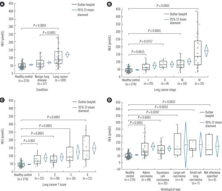
HE4 levels were significantly higher in patients with lung cancer (median 87.6 pmol/L, range 25.5–437.1 pmol/L; P <0.0001) and patients with benign lung diseases (60.4 pmol/L, range 4.9–177.5 pmol/L; P <0.0001) compared with healthy controls (44.6 pmol/L, range 20.0–381.9 pmol/L) (Fig. 1A).
The AUC for the discrimination of lung cancer patients from healthy controls was 0.84 (95% CI 0.78–0.89), significantly dif- ferent from the non-discriminant bisector (z test: P <0.0001).
The estimated sensitivity and specificity of HE4 level for lung cancer diagnosis at different cut-offs are shown in Table 1. The AUC value for the discrimination of lung cancer from benign lung disease was 0.70 (95% CI 0.62–0.78).
When comparing HE4 levels in lung cancer patients accord- ing to TNM stages, HE4 was elevated in TNM stage 1 and 2 (not statistically significant), as well as in stage 3 and 4 (statistically significant) compared with healthy controls. The median values by histological type are detailed in Fig. 1B.
In terms of T scores, serum HE4 levels were significantly
Table 1. Optimal cut-off values of HE4 for diagnosis between lung cancer and healthy control and benign lung disease (sensitivity, specific- ity, AUC, and 95% CI)
AUC 95% CI Cut-off (pmol/L) Sensitivity Specificity Youden’s index J SE P
Healthy control 0.84 0.78–0.89 41.1
57.9 74.4
0.908 0.806 0.633
0.449 0.770 0.908
0.353 0.576 0.541
0.026 <0.0001
Benign lung disease 0.71 0.62–0.79 46.2
70.0 134.3
0.890 0.660 0.260
0.333 0.684 0.930
0.223 0.362 0.207
0.042 <0.0001
Abbreviations: HE4, human epididymis protein 4; AUC, area under the curve; CI, confidence interval; SE, standard error of AUC.
Fig. 1. Distribution of human epididymis protein 4 (HE4) levels in different groups. (A) The serum HE4 levels of 100 lung cancer patients were significantly higher than those of 57 patients with benign lung diseases and 274 healthy controls (P <0.0001). (B) Serum HE4 levels were significantly higher in TNM stage 3 and 4 patients than in healthy controls (P <0.0001). (C) HE4 levels were significantly higher in pa- tients with all tumor scores (T1, T2, T3, and T4) than in healthy controls. (D) HE4 levels were significantly higher in patients with any histo- logical subtype of lung cancer than in healthy controls.
Abbreviation: CI, confidence interval.
C 450 400 350 300 250 200 150 100 50
0Healthy control
(n=274) 1
(n=22)
Lung cancer T score (n=28)2 3
(n=18) 4
(n=21)
HE4 (pmoI/L)
P <0.0001 P <0.0001 P <0.0001 P =0.002
Outlier boxplot 95% CI mean diamond 450
400 350 300 250 200 150 100 50
0 Healthy control
(n=274) Benign lung disease (n=57) Condition
Lung cancer (n=100)
HE4 (pmoI/L)
A
P <0.0001 P <0.0001
Outlier boxplot 95% CI mean diamond
B 450 400 350 300 250 200 150 100 50
0 Healthy control (n=274)
(n=20)I
Lung cancer stage (n=8)II III
(n=33) IV
(n=35)
HE4 (pmoI/L)
P <0.0001 P <0.0001
P =0.0615 P =0.0757
Outlier boxplot 95% CI mean diamond
450 400 350 300 250 200 150 100 50 0 –50 Healthy
control (n=274)
Adeno carcinoma
(n=48)
Squamous cell carcinoma
(n=35)
Large cell carcinoma (n=4)
Small cell lung carcinoma
(n=7)
Not otherwise specified
(n=6) Histological type
HE4 (pmoI/L)
P =0.0021 P =0.0033 P =0.0397 P <0.0001 P <0.0001 D
Outlier boxplot 95% CI mean diamond
higher in lung cancer patients with any T score than in healthy controls. The median values by T score are detailed in Fig. 1C.
Significant differences in HE4 levels were evident comparing lung cancer patients (all histological types) with healthy controls.
The median values by histological type are detailed in Fig. 1D.
We found that serum HE4 levels in patients with any histologi- cal type of lung cancer were significantly elevated than in healthy controls and patients with benign lung diseases. More- over, even patients with T scores of 1 could be distinguished from healthy controls.
HE4 is a member of the whey-acidic-protein (WAP) domain family, which shares 50 well-conserved amino acids. Two WAP family genes encode leukocyte protease inhibitors: secretory leukocyte protease inhibitor (SLPI) and elafin [11, 12]. SLPI and elafin are expressed in various carcinomas, including lung can- cer; thus, these genes and proteins may play roles in cancer development or progression [13, 14].
In this study, the serum HE4 levels in patients with T score 1 were significantly different from those in healthy controls, al- though the Stage 1 and 2 results were not statistically signifi- cant. This may be due to patients with low T scores having nodal involvement or distal metastasis. However, we hypothesize that serum HE4 can be used as an ancillary detection tool for small-size lung cancer tumors. Interestingly, the metastasis (M) scores showed a significant difference, while the node (N) scores did not, in contrast to the report of Liu et al [15]. Patients with any histological type of lung cancer exhibited higher serum HE4 levels than healthy controls. Small-cell lung cancer was as- sociated with the highest median HE4 level, although the differ- ences were not statistically significant. Nagy et al [16] reported that the highest HE4 levels were exhibited by patients with large-cell carcinomas but other histological types yielded similar values. However, a number of studies have found that non- small-cell lung cancer is associated with higher HE4 levels than small-cell lung cancer [15, 17, 18]. These discrepancies may be attributable to the fact that the small-cell lung cancer sub- groups were comprised of fewer patients. Although no signifi- cant difference was detected among histological types, addi- tional studies involving a greater number of small-cell lung can- cer cases are needed to enable a better understanding of the relationship between HE4 levels and histological subtypes of lung cancer.
Our study has certain limitations. First, the work was per- formed at a single center, which may limit the generalizability of our findings. Moreover, relatively few patients with large-cell car- cinoma, small-cell lung cancer, and early stage lung cancer
were included. Second, several factors, including sex, age, menopausal status, glomerular filtration rate, caffeine consump- tion, and smoking, which influence serum HE4 levels, were not considered [19]. Third, evaluation of other lung cancer detec- tion markers was not performed because they are not recom- mended for the diagnosis of early-stage lung cancer [3, 4].
We found that serum HE4 was overexpressed in patients with any histological type of lung cancer compared with those with benign lung diseases and healthy controls, even when the tu- mor size was small (T score 1). Thus, serum HE4 testing may have potential as an ancillary lung cancer detection tool, thus improving clinical outcomes by facilitating timely treatment.
Authors’ Disclosures of Potential Conflicts of Interest
No potential conflicts of interest relevant to this article are re- ported.
Acknowledgments
This work was supported by Abbott Diagnostics, Korea and the Soonchunhyang University Research Fund.
REFERENCES
1. Howlader N, Noone AM, Krapcho M, Garshell J, Miller D, Altekruse SF, et al. (Eds.), SEER Cancer Statistics Review, 1975-2012, National Can- cer Institute. Bethesda, MD. http://seer.cancer.gov/csr/1975_2012 (Up- dated on Apr 2015).
2. Manser R, Lethaby A, et al. eds. Screening for lung cancer. Cochrane database of systematic reviews 2013, Issue 6. Art. No.:CD001991.
DOI:10.1002/14651858.CD001991.pub3. John Wiley & Sons, Ltd, 2013:3-10.
3. Tas F, Aydiner A, Topuz E, Yasasever V, Karadeniz A, Saip P. Utility of the serum tumor markers: CYFRA 21.1, carcinoembryonic antigen (CEA), and squamous cell carcinoma antigen (SCC) in squamous cell lung cancer. J Exp Clin Cancer Res 2000;19:477-81.
4. Brower V. Biomarker studies abound for early detection of lung cancer.
J Natl Cancer Inst 2009;101:11-3.
5. Hellström I, Raycraft J, Hayden-Ledbetter M, Ledbetter JA, Schummer M, McIntosh M, et al. The HE4 (WFDC2) protein is a biomarker for ovarian carcinoma. Cancer Res 2003;63:3695-700.
6. Kong SY, Han MH, Yoo HJ, Hwang JH, Lim MC, Seo SS, et al. Serum HE4 level is an independent prognostic factor in epithelial ovarian can- cer. Ann Surg Oncol 2012;19:1707-12.
7. Galgano MT, Hampton GM, Frierson HF Jr. Comprehensive analysis of HE4 expression in normal and malignant human tissues. Mod Pathol 2006;19:847-53.
8. Iwahori K, Suzuki H, Kishi Y, Fujii Y, Uehara R, Okamoto N, et al. Serum HE4 as a diagnostic and prognostic marker for lung cancer. Tumour Biol 2012;33:1141-9.
9. Sobin LH, Gospodarowicz MK, et al. eds. TNM classification of malig- nant tumors. 7th ed. Oxford, UK: Wiley- Blackwell, 2009.
10. Galateau-Salle F, Churg A, Roggli V, Travis WD; World Health Organiza- tion Committee for Tumors of the Pleura. The 2015 World Health Orga- nization classification of tumors of the pleura: Advances since the 2004 classification. J Thorac Oncol 2016;11:142-54.
11. Thompson RC and Ohlsson K. Isolation, properties, and complete ami- no acid sequence of human secretory leukocyte protease inhibitor, a potent inhibitor of leukocyte elastase. Proc Natl Acad Sci U S A 1986;
83:6692-6.
12. Wiedow O, Schröder JM, Gregory H, Young JA, Christophers E. Elafin:
an elastase-specific inhibitor of human skin. Purification, characteriza- tion, and complete amino acid sequence. J Biol Chem 1990;265:
14791-5.
13. Devoogdt N, Revets H, Ghassabeh GH, De Baetselier P. Secretory leu- kocyte protease inhibitor in cancer development. Ann N Y Acad Sci 2004;1028:380-9.
14. Zhang M, Zou Z, Maass N, Sager R. Differential expression of elafin in human normal mammary epithelial cells and carcinomas is regulated at
the transcriptional level. Cancer Res 1995;55:2537-41.
15. Liu W, Yang J, Chi PD, Zheng X, Dai SQ, Chen H, et al. Evaluating the clinical significance of serum HE4 levels in lung cancer and pulmonary tuberculosis. Int J Tuberc Lung Dis 2013;17:1346-53.
16. Nagy B Jr, Krasznai ZT, Balla H, Csobán M, Antal-Szalmás P, Hernádi Z, et al. Elevated human epididymis protein 4 concentrations in chronic kidney disease. Ann Clin Biochem 2012;49:377-80.
17. Tang QF, Zhou ZW, Ji HB, Pan WH, Sun MZ. Value of serum marker HE4 in pulmonary carcinoma diagnosis. Int J Clin Exp Med 2015;8:
19014-21.
18. Escudero JM, Auge JM, Filella X, Torne A, Pahisa J, Molina R. Compari- son of serum human epididymis protein 4 with cancer antigen 125 as a tumor marker in patients with malignant and nonmalignant diseases.
Clin Chem 2011;57:1534-44.
19. Ferraro S, Braga F, Lanzoni M, Boracchi P, Biganzoli EM, Panteghini M.
Serum human epididymis protein 4 vs carbohydrate antigen 125 for ovarian cancer diagnosis: a systematic review. J Clin Pathol 2013;
66:273-81.