hepatocellular carcinoma with cirrhosis
Jian-Xun Chen, Heng-Quan Ran, Chang-Qin Sun
Department of Hepatopancreatobiliary Surgery, Panzhihua Central Hospital, Panzhihua, China
INTRODUCTION
Since the article about associating liver partition and portal vein ligation for staged hepatectomy (ALPPS) was published in March 2012 [1], this novel technique has been drawing the attention of surgeons. Although it had been demonstrated that ALPPS could induce extensive and rapid future liver remnant (FLR) hypertrophy [1,2], the morbidity form ALPPS was as high as 33% to 64% [3]. To decrease the morbidity rate and simplify the procedure of ALPPS, we performed a modified ALPPS, which used microwave ablation to replace the in situ splitting of the liver. We named this novel technique “AMAPS”
(associating microwave ablation and portal vein ligation for staged hepatectomy). This paper demonstrates that AMAPS was successfully applied in the treatment of huge hepatocellular carcinoma with cirrhosis.
CASE REPORT
A 43yearold man (weight, 60.0 kg; height, 171.0 cm;
body mass index, 20.5 kg/m2) was admitted to our hepa
topancreatobiliary surgery department, an asymptomatic 14.0
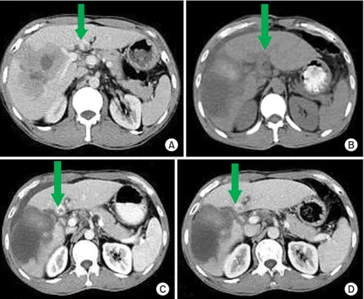
cm mass in the right liver was diagnosed by ultrasound. The patient was infected by chronic HBV 20 years proir. The αFP level was 1,000.00 ng/mL and the liver function was normal (Childpugh A). CT scan showed that the liver was cirrhotic with a huge lesion in the right liver, which was considered as hepatocellular carcinoma, and a small lesion in the left liver was a typical hemangioma (Fig. 1). The FLR volume was calculated to be 356.0 cm3 (29.1% of 1,223.3 cm3 of the stander liver volume) based on the preoperative CT scan. It is reported that an FLR >30% of the stander liver volume is considered as a safe scope for hepatoectomy [4]. However, our criteria is FLR >45% of the stander liver volume for patients with cirrhosis. Given the high risk of postoperative liver failure for the patient, a 2stage surgery should be adopted to increase the Associating liver partition and portal vein ligation for staged hepatectomy (ALPPS) could induce extensive and rapid future liver remnant hypertrophy. However, the morbidity for ALPPS is very high. This paper reports a modified ALPPS (associating microwave ablation and portal vein ligation for staged hepatectomy, AMAPS), which was successfully applied in the treat- ment of huge hepatocellular carcinoma with cirrhosis, and the procedure of operation was greatly simplified. Hence, AMAPS is feasible and safe in selected patients with primary hepatocellular carcinoma and cirrhosis.
[Ann Surg Treat Res 2016;90(5):287-291]
Key Words: Hepatectomy, Hepatocellular carcinoma, Liver cirrhosis
Reviewed January February March April May June July August September
Received December 8, 2015, Revised January 23, 2016, Accepted March 2, 2016
Corresponding Author: Heng-Quan Ran
Division of Hepatobiliary Pancreatic Surgery, Panzhihua Central Hospital,
Copyright ⓒ 2016, the Korean Surgical Society
cc Annals of Surgical Treatment and Research is an Open Access Journal. All articles are distributed under the terms of the Creative Commons Attribution Non- Commercial License (http://creativecommons.org/licenses/by-nc/4.0/) which
A B
C D
Fig. 1. The preoperative CT scan showed a huge hepatocellular carcinoma in the right liver (red arrow), and a typical heman- gioma in the left liver (green arrow). (A) The plain CT scan. (B) The hepatic arterial phase. (C) The portal venous phase. (D) The equilibrium phase.
A B
C D
Fig. 2. Surgical procedure. (A) The hepatic pedicle was dis- sected, the right hepatic artery (red arrow) and theright portal vein (blue arrow) were isolated in the first-stage operation respec- tively. (B) The right hepaticartery was tagged by the homemade rubber strip in the first-stage opera tion (green arrow). (C) The micro wave ablated the fu- ture transection plane with the ultra sound-guided in the first- stage operation. (D) The blood- less blunt liver resection in the second-stage operation.
sected carefully, and the right hepatic artery was isolated and tagged with the homemade rubber strip, which also reduced adhesion around the right hepatic artery by the large flat area.
Then, the right portal vein was freed and ligated (Fig. 2A, B).
After the falciform ligament was divided, the future transection plane was marked with ultrasound guiding. The microwave antenna, which was guided by ultrasound, was inserted into the liver parenchyma on the right future transection plane, setting a 4minute and 70 watts output cycle (Fig. 2C). This manner was repeated step by step every 2.5 cm to create an avascular groove along the future transection plane.
The duration of the firststage operation was 210 minutes with a blood loss of 100 mL with no need for blood transfusion or intensive care unit (ICU) admission. The postoperative FLR was reevaluated by CT scan weekly. The FLR was estimated at 430.6 cm3 (35.2% of the stander liver volume) after 1 week, and was increased to 500.3 cm3 (40.9% of the stander liver volume) after 2 weeks. On postoperative day 20, the volume of left hepatic was increased to 626.3 cm3 (51.2% of the stander liver
future transaction plane with an anterior approach (Fig. 2D).
The right biliary duct was indentified and sutured during the parenchymal transection. The median and right hepatic veins were divided and sutured last.
The duration of the secondstage operation was 100 mi
nutes with a blood loss of 200 mL, and there was no need for blood transfusion or ICU admission. The result of path ology demonstrated a hepatocellular carcinoma with poor differ
entiation, and the resection margin was tumorfree (R0). The patient recovered without any complications and was dis
charged on postoperative day 7. No tumor relapse or metastasis was detected (Fig. 4), and the αfetoprotein level was normal at three months after the surgery.
DISCUSSION
In 2012, Schnitzbauer et al. [1] report a 24% bile leakage and 12% 90day mortality for ALPPS. Because of the high occurrence rate of bile leakage, the safety of ALPPS has been
A B
Fig. 3. Comparison of the future liver remnant at different time after the first-stage operation.
(A) The preoperative CT image.
(B) The image after 1 week. (C) The image after 2 weeks. (D) The image after 20 days. All images
questioned by some surgeons [5]. Thus, some modified ALPPS have been performed, such as using tourniquet to ligate the future transection plane, which replaced the in situ splitting of the liver [6,7]. Although this method could help avoid the complication of bile leakage, it requires creating a hole between the right hepatic vein and the liver surface in the firststage operation, which increases the risk of hemorrhage, and requires more skillful surgeons. Compared with this method, our AMAPS not only avoided postoperative bile leakage, but also re
duced the risk of hemorrhage, as it does not require dissection of the hepatic vein in the firststage operation. Furthermore, as microwave ablation creates an avascular groove, it might greatly simplify the procedure and decrease surgical trauma in the secondstage operation, which finally led to a better recovery.
Another potential advantage of our method would be to better meet the “notouch” standard. By our method, we only microwave ablated the transection plane under ultrasound guiding, which did not require dissection of the second hepatic hilum or the right coronary ligament or the right triangular ligament. That could at most avoid tumortouch. It might improve the prognosis of the patients.
To our knowledge, this is the third case of microwave abla
tion applied in the ALPPS. The previous two cases were per
formed successfully without serious complications as well. [8,9].
However, unlike their cases, our patient had a comorbidity of cirrhosis, which could increase the risk of postoperative liver failure. Thus, we did not perform the secondstage operation until the FLR increased to 51.2% of the stander liver volume.
In the report of Cillo et al. [9], the FLR increased 35 cm3/day in a patient who was diagnosed with liver metastases and underwent microwave ablation assisting ALPPS, but the FLR hypertrophy was only 13.5 cm3/day in our patient. The major reason for this difference should likely be the comorbidity of liver cirrhosis. Thus, when AMAPS is applied in patients with cirrhosis, a longer time should be provided for liver regeneration.
In conclusion, AMAPS is feasible and simplified in selected patients with primary hepatocellular carcinoma and cirrhosis.
Further studies are needed to evaluate this modified ALPPS.
CONFLICTS OF INTEREST
No potential conflict of interest relevant to this article was reported.
Fig. 4. Follow-up computed to- mography scan 3 months later showed no recurrence or metas- tasis. The left liver lesion with high density was heman gioma (green arrow). (A) The plain CT scan. (B) The hepatic arterial phase. (C) The portal venous phase. (D) The equilibrium phase.
A B
C D
extended right hepatic resection in small
forsize settings. Ann Surg 2012;255:405
14.
2. Schadde E, Ardiles V, Slankamenac K, Tschuor C, Sergeant G, Amacker N, et al.
ALPPS offers a better chance of complete re section in patients with primarily un
resectable liver tumors compared with conventionalstaged hepatectomies: re
sults of a multicenter analysis. World J Surg 2014;38:15109.
3. Gall TM, Sodergren MH, Frampton AE, Fan R, Spalding DR, Habib NA, et al.
after major hepatectomy. Ann Surg Treat Res 2014;87:10811.
5. Kokudo N, Shindoh J. How can we safely climb the ALPPS? Updates Surg 2013;65:
1757.
6. Robles R, Parrilla P, LopezConesa A, Brusadin R, de la Pena J, Fuster M, et al.
Tourniquet modification of the associa
ting liver partition and portal ligation for staged hepatectomy procedure. Br J Surg 2014;101:112934.
7. Cai X, Peng S, Duan L, Wang Y, Yu H, Li Z. Completely laparoscopic ALPPS using
8. Gringeri E, Boetto R, D’Amico FE, Bassi D, Cillo U. Laparoscopic microwave abla
tion and portal vein ligation for staged hepatectomy (LAPS): a minimally invasive firststep approach. Ann Surg 2015;261:
e423.
9. Cillo U, Gringeri E, Feltracco P, Bassi D, D'Amico FE, Polacco M, et al. Totally lapar oscopic microwave ablation and portal vein ligation for staged hepatec
tomy: a new minimally invasive two
stage hepatectomy. Ann Surg Oncol 2015;
22:27878.