ISSN 2234-3806 • eISSN 2234-3814
https://doi.org/10.3343/alm.2020.40.2.142
Evaluation of the QuantaMatrix Multiplexed Assay Platform for Molecular Diagnosis of Multidrug- and Extensively Drug-Resistant Tuberculosis Using Clinical Strains Isolated in Myanmar
Yunhee Chang , B.S.1, Seoyong Kim , B.S.1, Yeun Kim , Ph.D.1, Phyu Win Ei , Ph.D.1, Dasom Hwang , B.S.1, Jongseok Lee , Ph.D.2, Chulhun L Chang , M.D., Ph.D.3, and Hyeyoung Lee , Ph.D.1
1Department of Biomedical Laboratory Science, College of Health Sciences, Yonsei University, Wonju, Korea; 2International Tuberculosis Research Center, Changwon, Korea; 3Department of Laboratory Medicine, Pusan National University Yangsan Hospital, Yangsan, Korea
Background: Although the incidence of tuberculosis (TB) is decreasing, cases of multi- drug-resistant (MDR) TB and extensively drug-resistant (XDR) TB continue to increase. As conventional phenotype drug susceptibility testing (pDST) takes six to eight weeks, molec- ular assays are widely used to determine drug resistance. we developed QuantaMatrix Multiplexed Assay Platform (QMAP) MDR/XDR assay (QuantaMatrix Inc., Seoul, Korea) that can simultaneously detect mutations related to both first- and second-line drug resis- tance (rifampin, isoniazid, ethambutol, fluoroquinolones, second-line injectable drugs, and streptomycin).
Methods: We used 190 clinical Mycobacterium tuberculosis (MTB) strains isolated from Myanmar, compared QMAP and pDST results, and determined concordance rates. Addi- tionally, we performed sequence analyses for discordant results.
Results: QMAP results were 87.9% (167/190) concordant with pDST results. In the 23 isolates with discordant results, the QMAP and DNA sequencing results completely matched.
Conclusions: The QMAP MDR/XDR assay can detect all known DNA mutations associ- ated with drug resistance for both MDR- and XDR-MTB strains. It can be used for molec- ular diagnosis of MDR- and XDR-TB to rapidly initiate appropriate anti-TB drug therapy.
Key Words: Mycobacterium tuberculosis, Multidrug-resistant tuberculosis, Extensively drug-resistant tuberculosis, QuantaMatrix Multiplexed Assay Platform
Received: May 28, 2019 Revision received: August 6, 2019 Accepted: October 11, 2019 Corresponding author:
Hyeyoung Lee, Ph.D.
Department of Biomedical Laboratory Science, College of Health Sciences, Yonsei University, 1 Yeonsedae-gil, Wonju 26493, Korea
Tel: +82-33-760-2740 Fax: +82-33-760-2561 E-mail: [email protected]
© Korean Society for Laboratory Medicine This is an Open Access article distributed under the terms of the Creative Commons Attribution Non-Commercial License (http://creativecom- mons.org/licenses/by-nc/4.0) which permits unrestricted non-commercial use, distribution, and reproduction in any medium, provided the original work is properly cited.
INTRODUCTION
Tuberculosis (TB), caused by Mycobacterium tuberculosis (MTB) infection, is the ninth highest cause of death worldwide.
There are an estimated 10 million cases of TB globally, with 1.6 million TB-associated deaths per year [1, 2]. Although the inci- dence of TB is decreasing by 2% every year, its prevalence and mortality rate remain high, necessitating comprehensive efforts
for eradication [2].
Multidrug-resistant (MDR) TB and extensively drug-resistant (XDR) TB are of particular concern as they are difficult to treat.
MDR-MTB exhibits resistance to two of the most important first- line drugs, rifampin (RIF) and isoniazid (INH), while XDR-MTB demonstrates resistance to RIF and INH, as well as to at least one fluoroquinolone (FQ) and at least one second-line injectable drug (SLID; kanamycin, amikacin, and capreomycin) [3]. Treat-
2017-03-16 https://crossmark-cdn.crossref.org/widget/v2.0/logos/CROSSMARK_Color_square.svg
ment success rates for MDR-TB and XDR-TB are low, at 54%
and 30%, respectively—and they are the main obstacles in TB eradication [1]. Therefore, rapid determination of the drug sus- ceptibility of the TB-causing bacteria is important to ensure ap- propriate treatment.
Drug susceptibility testing (DST), used to select appropriate drugs, is a culture-based method and requires approximately six to eight weeks for completion [4]. To overcome the limita- tions of conventional assays, DST based on molecular diagnos- tic assays has been developed. For example, GenoType MTB- DRplus (Hain Lifescience, Nehren, Germany) and GenoType MTBDRsl (Hain Lifescience) are employed for the rapid identifi- cation of gene mutations related to MDR- and XDR-MTB using a line probe assay, while GeneXpert MTB/RIF (Cepheid AB, Solna, Sweden) is used to determine RIF resistance by real-time PCR [5].
Recently, QuantaMatrix Inc. (Seoul, Korea) developed the QuantaMatrix Multiplexed Assay Platform (QMAP), which uti- lizes magnetic micro-particles and a reverse hybridization assay.
In QMAP, a probe for a specific gene is combined with a bar- coded magnetic micro-particle, which is a carboxyl-functional- ized magnetic disk with a 50-μm-thick silica-coated surface and a graphical barcode that allows >100-plex coding capacity in high-throughput analysis [6]. Each probe enables the capture of PCR products with a complementary sequence and then emits fluorescence. A 100-plex capacity in a single microwell system allows the testing of 100 types of pathogens in one microwell with one sample. Previously, we developed an assay for detect- ing MDR-MTB based on QMAP and evaluated the utility of the assay using strains isolated from TB patients in Korea [7].
We have now developed a more sophisticated QMAP MDR/
XDR assay by adding a probe to identify genetic mutations as- sociated with resistance to ethambutol (EMB), streptomycin (SM), FQ, and SLID in addition to RIF and INH. This molecular assay can simultaneously detect MDR- and XDR-MTB within six hrs. We evaluated the utility of this assay using clinical strains isolated from TB patients in Myanmar, which is among the 22 countries with the highest TB burden and is included in the global list of 27 countries with a high incidence of MDR-TB [8].
METHODS Clinical isolates
A total of 190 MTB strains isolated from sputum samples of pa- tients were collected from the National Tuberculosis Reference Laboratory (NTRL) in Yangon and the Upper Myanmar TB Lab-
oratory (UMTL) in Patheingyi, Myanmar, from 2015 to 2016.
Samples from patients suspected of having MDR-TB (113 from NTRL, and 77 from UMTL) were tested with the GeneXpert MTB/RIF assay, and positive samples were cultured in Ogawa egg slant medium to isolate MTB strains. This retrospective study was approved by the Ethics Review Committee of the De- partment of Medical Research in Yangon, Myanmar (Ethics/
DMR/2016/101).
DNA extraction from clinical isolates
Genomic DNA was extracted at the International Tuberculosis Research Center (ITRC, Changwon, Korea) using a simple boiling method with some modifications [9]. Briefly, the cultured colonies were suspended in 1 mL distilled water in an Eppendorf tube us- ing a loop and heated at 99°C for 20 minutes with vortexing at 5 minutes intervals. The tube was then centrifuged at 12,000 ×g, 23°C for 5 minutes and the supernatant was removed and stored at 4°C until used in the QMAP MDR/XDR assay.
QMAP MDR/XDR assay
Three oligonucleotide probes specific to the genus Mycobacte- rium and 65 drug resistance-related gene probes were synthe- sized to detect MDR- and XDR-MTB (Table 1). Each probe was combined with a carboxyl-functionalized magnetic microdisk (QuantaMatrix Inc.). To amplify 11 target areas simultaneously, primers specific to biotin-attached species-specific areas and drug resistance-related areas were prepared and used for multi- plex PCR. The PCR reactions consisted of 10 μL of AccuPower Multiplex PCR PreMix (Bioneer, Daejeon, Korea), 5 μL of the primer mixture, 1 μL of internal control, and 2 μL of molecular biology-grade water (GE Healthcare Life Sciences Korea, Seoul, Korea). PCR conditions were as follows: the mixture was dena- tured at 94°C for 5 minutes, followed by 45 cycles of 94°C for 20 seconds, 65°C for 1 minute, and 72°C for 5 minutes. The products were denatured at 25°C for 5 minutes by adding 10 μL of 2×denaturation solution (QuantaMatrix Inc.) to 10 μL of the biotinylated PCR products. The resulting solution was di- luted with 50 μL of hybridization solution and dispensed onto a glass MatriPlate (Brooks, Chelmsford, MA, USA). The dena- tured (single-stranded) PCR products were combined with the probe attached to the microdisk with INCUBATOR-micro mixer (FINEPCR, Gunpo, Korea) at 650 rpm and 35°C for 30 minutes.
The microdisks were washed three times with 100 μL of wash- ing buffer (QuantaMatrix Inc.) with shaking at 650 rpm, 25°C for 1 minute and then treated with staining buffer (QuantaMatrix Inc.) at 25°C and 650 rpm for 10 minutes. The microdisks were
washed three times with 100 μL of washing buffer (QuantaMa- trix Inc.) at 25°C for 1 minute, and the fluorescence intensity of each microdisk was automatically measured using the supplied software (QuantaMatrix Inc.).
Phenotypic DST
Phenotypic DST (pDST) for 12 first-line and second-line drugs was performed at ITRC using M-KIT plates (Korean Institute of Tuberculosis, Osong, Korea) with Löwenstein-Jensen medium, according to the manufacturer’s protocol. The strains were transported to ITRC under the conditions of the materials trans- fer agreement. The assay was conducted using the modified absolute concentration method. The critical concentrations for each drug were as follows: INH, 0.2; RIF, 40; SM, 10.0; EMB, 2.0; kanamycin, 30; capreomycin, 40; cycloserine, 30; para- aminosalicylic acid, 1.0; ofloxacin, 4.0; moxifloxacin, 1.0; ami- kacin, 30; and levofloxacin, 2.0 μg/mL.
Concordance rates and DNA sequence analysis
The results of QMAP and pDST were deemed concordant when the drug’s QMAP result indicated the same resistance as that in its pDST. The target DNA sequences of samples with discordant results were analyzed using ABI Prism 3730xl DNA Sequencer (ThermoFisher Scientific Korea, Seoul, Korea) in Genotech (Daejeon, Korea) and compared with sequences in the NCBI GenBank database. The corresponding samples were se-
quenced with designed primers for rpoB, katG, inhA, embB, gyrA, gyrB, eis, rrs, and rpsL.
RESULTS
pDST results of the clinical isolates and concordance between QMAP and pDST results
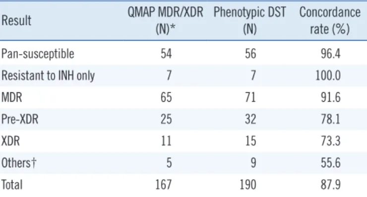
Table 2 shows the pDST results of all clinical isolates. Of the 190 isolates, 71 were MDR strains (37.4%), 56 were suscepti- ble to all tested drugs (29.5%), 32 were pre-XDR strains (16.8%), 15 were XDR strains (8.4%), and seven were resistant only to INH (3.7%).
The results for the 56 pan-susceptible isolates and seven iso- lates resistant only to INH showed a concordance rate of 96.4%
and 100%, respectively (Table 2). The results for MDR-MTB, pre-XDR-MTB, and XDR-MTB isolates showed concordance rates of 91.6%, 78.1%, and 68.8%, respectively. The results for the other nine isolates showed a concordance rate of 55.6%.
QMAP was able to detect 101 of 118 MDR- or XDR-MTB and pre-XDR-MTB isolates (85.6%) or 113 of 134 any-drug-resis- tant MTB isolates (84.3%).
Concordance rates for the susceptibility and resistance patterns of each drug
The QMAP and pDST concordance rates in each drug are shown in Table 3. Of the 70 RIF-susceptible and 120 RIF-resis- tant isolates, 95.7% and 96.7% showed concordant results us- ing QMAP assay, respectively. The results for all 61 INH-sus- ceptible isolates and 94.6% of INH-resistant isolates were con- cordant. Similarly, 96.7–100% of the EMB-, FQ-, SLID-, and SM-susceptible isolates were detected using QMAP. However, the detection rates of the resistant isolates were slightly lower with QMAP; 56.5% EMB-, 88.6% FQ-, 75.0% SLID-, and 90.6% SM-resistant strains were detected.
DNA sequencing of isolates with discordant results
The 23 isolates showing discordant QMAP and qDST results were subjected to sequence analysis of the respective gene tar- get region using the same primers (Table 4). Of the six MDR isolates identified by pDST, two showed no resistance; two, RIF resistance only; and two, INH resistance only in QMAP. Of the seven pre-XDR strains identified by pDST, six had neither FQ nor SLID resistance and one was susceptible to INH in QMAP.
Four XDR isolates identified by pDST showed pre-XDR geno- types in QMAP. For all these results, the sequence analysis re- sults completely matched QMAP results (Table 4).
Table 1. Target genes and regions of the probes used to detect my- cobacteria and their resistance to specific drugs
Purposes Drugs Target
genes Target regions Probes (N)
ID - rpoB Codons 302–420 3
DST RIF rpoB Codons 504–533 11*
INH katG Codons 315 4*
inhA 8–17 bp upstream promoter region 6*
EMB embB Codons 306 3
FQ gyrA Codons 88–94 10
gyrB Codons 538–540 5
SLID eis 8–14, 37 bp upstream promotor region 6 rrs Bases 1400–1402, 1445, 1484 7
rpsL Codons 4, 88 4
SM rpsL Bases 514–517 3
*Adapted from Wang, et al. [7]. Copyright by Korean Society for Laboratory Medicine.
Abbreviations: ID, identification; RIF, rifampin; INH, isoniazid; EMB, etham- butol; FQ, fluoroquinolones; SLID, second-line injectable drugs; SM, strepto- mycin; DST, drug susceptibility testing.
DISCUSSION
The utility of the QMAP assay was evaluated using MTB strains isolated from TB patients. We also compared the results of QMAP and pDST; the assays showed an overall concordance rate of 87.9%. The DNA sequencing results of the 23 isolates with discordant results matched QMAP results. Because of the inherent limitation of molecular DST methods, not all strains re- sistant to a specific drug can be detected as many drug resis- tance genes remain to be identified. The purpose of molecular DST is timely detection of resistant strains as many as possible.
In that context, our current assay could detect 87.9% of MDR- and XDR-MTB isolates accurately and rapidly.
Currently, several molecular DST assays, such as the AdvanS- ure MDR-TB GenoBlot assay kit (LG Chem, Seoul, Korea) and GenoType MTBDRplus, are clinically used. Both kits demon- strated good performance for MDR-MTB detection in clinical isolates, with concordance rates of 94.3% and 88.5–98.2%
[10-12], respectively. One study reported that the results of GenoType MTBDRsl, a kit for XDR-MTB detection, showed a concordance rate of 94.7% [13]. However, a pooled analysis re- ported lower sensitivity of 75–80%, accompanied by high speci- ficity of 91–100%, for detection of XDR-MTB isolates [14]. Our discordant pDST and QMAP results might have occurred for the following reasons. First, not all mutations associated with drug resistance are known [15]. Currently, only approximately 95%
of RIF resistance due to the rpoB gene, which contains the RIF resistance determining region (RRDR), is detectable [16]; in case of katG and inhA, which are genes related to INH resis- tance, the detection rates of INH resistance have been reported Table 2. Comparison of drug susceptibility patterns between QMAP
MDR/XDR assay and phenotypic DST results
Result QMAP MDR/XDR
(N)* Phenotypic DST
(N) Concordance rate (%)
Pan-susceptible 54 56 96.4
Resistant to INH only 7 7 100.0
MDR 65 71 91.6
Pre-XDR 25 32 78.1
XDR 11 15 73.3
Others† 5 9 55.6
Total 167 190 87.9
*Indicates the number of isolates showing the expected results in accor- dance with the phenotypic DST results; †Three isolates were resistant to only SM; three were resistant to INH and SM; one was resistant to RIF and SM;
one was resistant to RIF, EMB, and SM; and one was resistant to INH, EMB, fluoroquinolones, second-line injectable drugs, and SM.
Abbreviations: DST, drug susceptibility testing; QMAP, QuantaMatrix Multi- plexed Assay Platform; SM, streptomycin; RIF, rifampin; INH, isoniazid;
EMB, ethambutol; MDR, multidrug resistant; XDR, extensively drug resis- tant.
Table 3. Comparison of QMAP MDR/XDR assay and phenotypic DST results for each drug
Drug
QMAP assay
(Isolates, N) Phenotypic DST
(Isolates, N) Concordance rate (%)
S R S R S R
RIF 67 116 70 120 95.7 96.7
INH 61 122 61 129 100.0 94.6
EMB 82 67 82 108 100.0 56.5
FQ 141 39 146 44 96.7 88.6
SLID 170 15 170 20 100.0 75.0
SM 62 116 62 128 100.0 90.6
Abbreviations: S, susceptible; R, resistant; DST, drug susceptibility testing;
QMAP, QuantaMatrix Multiplexed Assay Platform; MDR, multidrug resistant;
XDR, extensively drug resistant; RIF, rifampin; INH, isoniazid; EMB, etham- butol; FQ, fluoroquinolones; SLID, second-line injectable drugs; SM, strepto- mycin.
Table 4. Sequence analysis of 23 isolates with discordant QMAP assay and phenotypic DST results
QMAP MDR/XDR Phenotypic DST Gene sequences for the discordant
results*
Isolates (N)
SM-R Pan-S rpsL 43 AAG → AGG
rpsL 88 AAG → AGG 2
Pan-S MDR rpoB, katG, inhA WT 2
RIF-R MDR katG, inhA WT 2
INH-R MDR ropB WT 2
MDR Pre-XDR (FQ-R) gyrA, gyrB WT 4
MDR Pre-XDR (SLID-R) eis, rrs, rpsL WT 2
RIF-, EMB-, FQ-R Pre-XDR (FQ-R) katG, inhA WT 1
Pre-XDR (SLID-R) XDR gyrA, gyrB WT 1
Pre-XDR (FQ-R) XDR eis, rrs, rpsL WT 3
Pan-S SM-R rpsL WT 1
RIF-R, SM-R RIF-, EMB-, SM-R embB WT 1
Pre-XDR (FQ-R) INH-, EMB-, SM-R rpoB 526 CAC → AAC gyrA 94 GAC → TAC 1 XDR INH-, EMB-, FQ-, SLID-,
SM-R rpoB 533 CTG → CAG
embB WT 1
*The target regions of each gene are described in Table 1.
Abbreviations: DST, drug susceptibility testing; QMAP, QuantaMatrix Multi- plexed Assay Platform; RIF, rifampin; INH, isoniazid; FQ, fluoroquinolones;
SM, streptomycin; SLID, second-line injectable drugs; Pan-S, pan-suscepti- ble; MDR, multidrug resistant; XDR, extensively drug resistant; pre-XDR, MDR with FQ resistance or MDR with resistance to SLID; WT, wild type;
EMB, ethambutol.
to be 70% and 10%, respectively [17]; and for the gyrA and gyrB regions associated with FQ resistance, the detection rates is only approximately 60% [18]. Second, the tested isolates pos- sibly exhibited heteroresistance. Heteroresistance is detected in 20–30% of TB patients and can be caused by a mixed infection of two different isolates or by acquisition of drug resistance dur- ing treatment [19-21]. Patients infected with heteroresistant iso- lates might convert to full resistance [22]. In such cases, ≤1%
of the DNA of resistant isolates could be detected by molecular methods [23]. Third, isolates with low-level resistance and bor- derline resistance exhibiting susceptibility in pDST could be de- tected by molecular methods [24]. For these reasons, although molecular assays might not replace the pDST, they could be more widely used for detecting drug resistance, as WHO has re- cently reported [25].
The following study limitations should be considered. First, the QMAP MDR/XDR assay results showed low concordance (56.5%) with pDST results for detecting EMB resistance. This low detection rate could be attributed to a lack of appropriate probes. EMB resistance occurs most frequently at codons 306, 406, and 497 in the embB gene [26, 27]; however, we de- signed probes related only to codon 306. Second, this study was conducted using only selected strains, including a high proportion of drug-resistant strains, and the performance evalu- ation was mainly focused on the detection of resistance. How- ever, in the real situation, there would be a larger proportion of susceptible strains, potentially leading to false-positive detection of resistance. Therefore, further studies using different and a greater number of probes and including an adequate number of susceptible strains are needed to improve the performance of QMAP MDR/XDR assay and confirm its specificity.
As not all types of mutations that cause drug resistance have been identified, using molecular assays in conjunction with pDST could enable the rapid and accurate determination of drug susceptibility of MTB isolates, thereby facilitating timely ini- tiation of appropriate anti-TB drug therapy [28]. In particular, the 100% concordance between DNA sequencing and QMAP results indicates that QMAP could detect all known, or at least, targeted DNA mutations associated with drug resistance. There- fore, the QMAP MDR/XDR assay can be used for molecular di- agnosis of MDR- and XDR-TB.
Acknowledgements
The Korea International Cooperation Agency (KOICA) has sup- ported TB eradication in Myanmar through activities such as
the establishment of the Advanced Molecular Research Center within the Department of Medical Research of Myanmar. The isolates were collected during such collaborative studies and were kindly provided to our research team.
Author Contributions
All authors participated in developing and evaluating the QMAP MDR/XDR assay in collaboration with QuantaMatrix Inc.
Conflicts of Interest
The QMAP MDR/XDR assay was developed and evaluated in collaboration with QuantaMatrix Inc. No other potential conflicts of interest relevant to this article were reported by the authors.
Research Funding
This study was supported by the National Research Foundation of Korea (NRF-2016S1A5B8925203).
ORCID
Yunhee Chang https://orcid.org/0000-0003-1857-1955 Seoyong Kim https://orcid.org/0000-0002-0071-3954 Yeun Kim https://orcid.org/0000-0002-5584-3318 Phyu Win Ei https://orcid.org/0000-0002-2486-3260 Dasom Hwang https://orcid.org/0000-0002-3426-8932 Jongseok Lee https://orcid.org/0000-0002-4340-9856 Chulhun L Chang https://orcid.org/0000-0001-9117-4919 Hyeyoung Lee https://orcid.org/0000-0003-1572-5250
REFERENCES
1. WHO. Global tuberculosis report 2017. https://www.who.int/tb/publica- tions/global_report/gtbr2017_main_text.pdf (Updated on Aug 2019).
2. WHO. Global tuberculosis report 2018. http://apps.who.int/iris/bit- stream/handle/10665/274453/9789241565646-eng.pdf (Updated on Aug 2019).
3. WHO. Definitions and reporting framework for tuberculosis–2013 revision.
https://apps.who.int/iris/bitstream/handle/10665/79199/9789241505345_
eng.pdf (Updated on Aug 2019).
4. Choi J, Yoo J, Kim KJ, Kim EG, Park KO, Kim H, et al. Rapid drug sus- ceptibility test of Mycobacterium tuberculosis using microscopic time- lapse imaging in an agarose matrix. Appl Microbiol Biotechnol 2016;100:
2355-65.
5. Pang Y, Dong H, Tan Y, Deng Y, Cai X, Jing H, et al. Rapid diagnosis of MDR and XDR tuberculosis with the MeltPro TB assay in China. Sci Rep 2016;6:25330.
6. Kim LN, Kim M, Jung K, Bae HJ, Jang J, Jung Y, et al. Shape-encoded silica microparticles for multiplexed bioassays. Chem Commun (Camb) 2015;51:12130-3.
7. Wang HY, Uh Y, Kim S, Cho E, Lee JS, Lee H. Detection of rifampicin- and isoniazid-resistant Mycobacterium tuberculosis using the Quanta- Matrix Multiplexed Assay Platform system. Ann Lab Med 2018;38:569- 77.
8. Han ET, Lee JS, Cheong JH, Chang CL, Nyunt MH, Aung WW, et al.
Current status of standard diagnostics and treatment for malaria, tuber- culosis, and hepatitis in Myanmar. Lab Med Online 2017;7:94-102.
9. Jensen MA, Webster JA, Straus N. Rapid identification of bacteria on the basis of polymerase chain reaction-amplified ribosomal DNA spacer polymorphisms. Appl Environ Microbiol 1993;59:945-52.
10. Kim J, Park YJ, Lee NY, Chang CL, Lee M, Shin JH. Evaluation of the AdvanSure MDR-TB GenoBlot assay for detection of rifampin and isoni- azid resistant Mycobacterium tuberculosis complex in respiratory speci- mens. Korean J Clin Microbiol 2012;15:117-24.
11. Maurya AK, Umrao J, Singh AK, Kant S, Kushwaha RA, Dhole TN.
Evaluation of GenoType MTBDRplus assay for rapid detection of drug susceptibility testing of multi-drug resistance tuberculosis in Northern India. Indian J Pathol Microbiol 2013;56:139-43.
12. Bedewi Omer Z, Mekonnen Y, Worku A, Zewde A, Medhin G, Moham- med T, et al. Evaluation of the GenoType MTBDRplus assay for detec- tion of rifampicin- and isoniazid-resistant Mycobacterium tuberculosis isolates in central Ethiopia. Int J Mycobacteriol 2016;5:475-81.
13. Tagliani E, Cabibbe AM, Miotto P, Borroni E, Toro JC, Mansjo M, et al.
Diagnostic performance of the new version (v2.0) of GenoType MTBDRsl assay for detection of resistance to fluoroquinolones and second-line in- jectable drugs: a multicenter study. J Clin Microbiol 2015;53:2961-9.
14. WHO. The use of molecular line probe assays for the detection of resis- tance to second-line anti-tuberculosis drugs: policy guidance. WHO HTM/TB/2016.07. http://www.who.int/iris/bitstream/10665/246131/1/
9789241510561-eng.pdf (Updated on Aug 2019).
15. Laurenzo D and Mousa SA. Mechanisms of drug resistance in Myco- bacterium tuberculosis and current status of rapid molecular diagnostic testing. Acta Trop 2011;119:5-10.
16. Lee AS, Lim IH, Tang LL, Wong SY. High frequency of mutations in the rpoB gene in rifampin-resistant clinical isolates of Mycobacterium tu- berculosis from Singapore. J Clin Microbiol 2005;43:2026-7.
17. Dookie N, Rambaran S, Padayatchi N, Mahomed S, Naidoo K. Evolu- tion of drug resistance in Mycobacterium tuberculosis: a review on the molecular determinants of resistance and implications for personalized care. J Antimicrob Chemother 2018;73:1138-51.
18. Chien JY, Chiu WY, Chien ST, Chiang CJ, Yu CJ, Hsueh PR. Mutations in gyrA and gyrB among fluoroquinolone- and multidrug-resistant My-
cobacterium tuberculosis isolates. Antimicrob Agents Chemother 2016;
60:2090-6.
19. Hofmann-Thiel S, van Ingen J, Feldmann K, Turaev L, Uzakova GT, Murmusaeva G, et al. Mechanisms of heteroresistance to isoniazid and rifampin of Mycobacterium tuberculosis in Tashkent, Uzbekistan. Eur Respir J 2009;33:368-74.
20. Chakravorty S, Aladegbami B, Thoms K, Lee JS, Lee EG, Rajan V, et al.
Rapid detection of fluoroquinolone-resistant and heteroresistant Myco- bacterium tuberculosis by use of sloppy molecular beacons and dual melting-temperature codes in a real-time PCR assay. J Clin Microbiol 2011;49:932-40.
21. Shin SS, Modongo C, Baik Y, Allender C, Lemmer D, Colman RE, et al.
Mixed Mycobacterium tuberculosis-strain infections are associated with poor treatment outcomes among patients with newly diagnosed tuber- culosis, independent of pretreatment heteroresistance. J Infect Dis 2018;218:1974-82.
22. Mekonnen D, Admassu A, Mulu W, Amor A, Benito A, Gelaye W, et al.
Multidrug-resistant and heteroresistant Mycobacterium tuberculosis and associated gene mutations in Ethiopia. Int J Infect Dis 2015;39:34- 8.
23. Liang B, Tan Y, Li Z, Tian X, Du C, Li H, et al. Highly sensitive detection of isoniazid heteroresistance in Mycobacterium tuberculosis by Deep- Melt assay. J Clin Microbiol 2018;56:e01239-17.
24. Ocheretina O, Escuyer VE, Mabou MM, Royal-Mardi G, Collins S, Vilb- run SC, et al. Correlation between genotypic and phenotypic testing for resistance to rifampin in Mycobacterium tuberculosis clinical isolates in Haiti: investigation of cases with discrepant susceptibility results. PLoS One 2014;9:e90569.
25. WHO. The use of next-generation sequencing technologies for the de- tection of mutations associated with drug resistance in Mycobacterium tuberculosis complex: technical guide. WHO CDS/TB/2018.19. https://
apps.who.int/iris/bitstream/handle/10665/274443/WHO-CDS-TB- 2018.19-eng.pdf (Updated on Aug 2019).
26. Khosravi AD, Sirous M, Abdi M, Ahmadkhosravi N. Characterization of the most common embCAB gene mutations associated with ethambu- tol resistance in Mycobacterium tuberculosis isolates from Iran. Infect Drug Resist 2019;12:579-84.
27. Sun Q, Xiao TY, Liu HC, Zhao XQ, Liu ZG, Li YN, et al. Mutations within embCAB are associated with variable level of ethambutol resistance in Mycobacterium tuberculosis isolates from China. Antimicrob Agents Chemother 2017;62:e01279-17.
28. Gkaravela L, Papadimitriou-Olivgeris M, Foka A, Kolonitsiou F, Spilio- poulou A, Charokopos N, et al. Combination of commercially available molecular assays and culture based methods in diagnosis of tuberculo- sis and drug resistant tuberculosis. Braz J Microbiol 2017;48:785-90.