http://dx.doi.org/10.4078/jrd.2014.21.3.132
132
<Received:April 22, 2014, Revised:May 26, 2014, Accepted:June 19, 2014>
Corresponding to:Seung-Geun Lee, Division of Rheumatology, Department of Internal Medicine, Pusan National University School of Medicine, Pusan National University Hospital, 179, Gudeok-ro, Seo-gu, Busan 602-739, Korea. E-mail:[email protected] pISSN: 2093-940X, eISSN: 2233-4718
Copyright ⓒ 2014 by The Korean College of Rheumatology
This is a Free Access article, which permits unrestricted non-commerical use, distribution, and reproduction in any medium, provided the original work is properly cited.
Association between Vitamin D Deficiency and Carotid Intima-media Thickness in Patients with Rheumatoid Arthritis
Jong-Man Park1, Seung-Geun Lee1, Eun-Kyoung Park1, Dae-Sung Lee1, Sung-Min Baek1, Kyung-Lim Hwang1, Joong-Keun Kim1, Ji-Heh Park1, Geun-Tae Kim2, Seon-Yoon Choi2
Department of Internal Medicine, Pusan National University School of Medicine, Pusan National University Hospital1, Department of Internal Medicine, Kosin University College of Medicine2, Busan, Korea
Objective. The present study determined if vitamin D defi- ciency is a potential risk factor for increased carotid in- tima-media thickness (CIMT) in patients with rheumatoid arthritis (RA).
Methods. This cross-sectional study analyzed 50 consecutive female RA patients without cardiovascular disease history at the Pusan National University Hospital between September and December of 2013. CIMT was measured us- ing a high-resolution ultrasonography. Serum 25-hydroxy vitamin D (25-OHD) levels were assessed by radioimmuno- assay, and vitamin D deficiency was defined as serum 25-OHD levels <20 ng/mL. Stepwise multivariable linear regression analyses were performed to evaluate the associa- tion between vitamin D deficiency and increased CIMT.
Results. The median 25-OHD level (inter-quartile range) was 14.0 (11.0∼20.7) ng/mL, and 74% of patients had vita- min D deficiency. The mean±standard deviation of CIMT was 0.58±0.08 mm. RA patients with vitamin D deficiency
had significantly higher CIMT than those without this fea- ture (0.59±0.07 vs 0.54±0.05, p=0.028). In univariable line- ar regression models, vitamin D deficiency (β(SE)=0.047 (0.021), p=0.028), older age (β(SE)=0.003 (7.2-4), p<0.001) and higher disease activity score 28-erythrocyte sed- imentation rate (β(SE)=0.021 (0.010), p=0.034) and Korean version of health assessment questionnaire score (β(SE)=0.051 (0.015), p=0.002) were significantly asso- ciated with increased CIMT. Vitamin D deficiency re- mained statistically significant in multivariable regression models after adjusting for confounders.
Conclusion. Vitamin D deficiency was associated with in- creased CIMT in female RA patients. Our finding suggests that hypovitaminosis D can be a risk factor for athero- sclerosis in RA patients.
Key Words Vitamin D, Rheumatoid arthritis, Atheroscle- rosis, Cardiovascular diseases
Introduction
Increasing evidence indicates that low serum 25-hydroxy vi- tamin D (25-OHD) is associated with a higher frequency of cardiometabolic outcomes including type 2 diabetes mellitus, hypertension, and cardiovascular diseases (CVDs) in the gen- eral population (1,2). The National Health and Nutritional Examination Surveys (NHANES 2000∼2004) revealed that the frequency of CVDs including coronary heart disease, heart failure and peripheral vascular disease are significantly higher
in adults with low 25-OHD levels (<20 ng/mL) than those with high 25-OHD levels (≥30 ng/mL) (3). In addition, lim- ited data suggest that vitamin D supplementation may reduce the risk of CVDs (4,5) and decrease mortality in elderly peo- ple (6). Experimental evidence that vitamin D regulates the rennin-angiotensin system (7), inhibits vascular smooth mus- cle cell proliferation (8) and improves endothelial function in- flammation (9) may support the epidemiological relationship between vitamin D deficiency and increased risks of CVDs.
Hence, studies demonstrating the contribution of vitamin D deficiency on the burdens of cardiovascular morbidity and mortality have prompted increased awareness.
CVDs are the major causes of mortality and morbidity in pa- tients with RA (10). RA was recently shown to be associated with a 1.48-fold increase in CVDs (11) and a 1.6-fold increase in CVD-related death (12), compared to the general population.
Epidemiological studies demonstrated that traditional risk fac- tors such as smoking, hypertension, dyslipidemia, diabetes, and obesity as well as the inflammatory burden of RA can cause premature atherosclerosis and eventually lead to CVD develop- ment. As mounting evidence indicating the immunoregulatory effect of vitamin D has prompted several studies investigating the association of hypovitaminosis D with the disease activity and outcomes of RA (13). However, little attention has been given to the association of vitamin D deficiency with athero- sclerosis or CVDs in patients with RA. Accordingly, the pres- ent study determined if vitamin D deficiency is a potential risk factor for carotid atherosclerosis assessed by carotid in- tima-meida thickness (CIMT) in patients with RA.
Materials and Methods Study design and subjects
This cross-sectional study included 50 consecutive female RA patients (aged 18∼75 years) from a single outpatient rheuma- tology clinic of the Pusan National University Hospital in Busan, South Korea between September 2013 and December 2013. Pusan National University Hospital is a tertiary referral center in South Korea and Busan is a harbor city with a tem- perate climate located in the southeastern part of South Korea at a latitude of 34o north. All RA patients met the American College of Rheumatology 1987 revised classification criteria for RA (14). Patients with previous CVDs, abnormal renal function (serum creatinine ≥1.2 mg/dL), and current use of vitamin D supplements were excluded. All subjects provided written informed consent in accordance with the Declaration of Helsinki prior to study participation. This study was ap- proved by the Research and Ethics Review Board of the Pusan National University Hospital, Busan, South Korea.
Assessments
General information was collected by an interview and re- view of medical records. Anthropometric parameters including height, weight, body mass index (BMI), waist and hip circum- ference and blood pressure were measured in all study subjects. BMI was calculated as body weight divided by the square of height in meters (kg/m2). Waist circumference was measured at the smallest circumference of the natural waist,
usually just above the belly button and hip circumference was measured at its widest part of the buttocks or hip. The waist-to-hip ratio (WHR) was subsequently calculated. Blood pressure was determined as the average of two measurements taken at an interval of 5 minutes using a TM-2655P apparatus (A&D Company Ltd., Tokyo, Japan). Hypertension was de- fined as blood pressure ≥140/90 mmHg or a requirement of antihypertensive medication.
Fasting blood samples of all participants were taken between 8:00 AM and 10:00 AM to determine concentration of total cholesterol (TC), triglycerides (TGs), low density lipoprotein cholesterol (LDL-C), high density lipoprotein cholesterol (HDL-C), fasting glucose, fasting insulin, erythrocyte sed- imentation rate (ESR), C-reactive protein (CRP) and 25-OHD.
The concentrations of TC, TGs, and HDL-C were analyzed using an enzymatic colorimetric reagent (Roche Diagnostics, Zurich, Switzerland) and a P800 Module (Roche Diagnostics).
LDL-C value was calculated using the Friedewald formula.
Fasting glucose and insulin were assessed by the glucose oxi- dase method (Synchron LX-20, Beckman Coulter Inc., Fullerton, CA, USA) and radioimmunoassay (Diagnostic Product Co., Los Angeles, CA, USA), respectively. CRP was measured with a particle-enhanced immunoturbidimetric assay (Tina-quant C-reactive protein assay, Roche Diagnostics) us- ing a P800 Module (Roche Diagnostics). Serum 25-OHD lev- els were measured by a radioimmunoassay kit (DIAsource, Belgium) using a γ-counter (1470 Wizard, PerkinElmer, Turku, Finland). Vitamin D deficiency was defined as a 25-OHD levels less than 20 ng/mL. Insulin resistance was evaluated by homeostatic model assessment-insulin resistance (HOMA-IR), which was calculated with the formula defined by Matthews et al. (15) as follows:
HOMA−IR=[fasting serum insulin (μIU/mL)×fasting se- rum glucose (mg/dL)×0.055/22.5]
The following additional data were collected: disease dura- tion, medication records, swollen joint count (SJC), tender joint count (TJC), general health, physical function, immunoglobulin M-rheumatoid factor (RF), anti-cyclic citrullinated peptide anti- body (anti-CCP; U/mL) and previous history of type 2 diabetes mellitus and dyslipidemia. For RA patients treated with gluco- corticoids (GCs), the cumulative dose (in prednisone equiv- alent) was calculated by multiplying the current daily dose by the number of days for which patients had received GCs since they were first prescribed. General health was assessed using a visual analogue scale (VAS) ranging form 0-100 and phys- ical function was assessed using the Korean Version of health
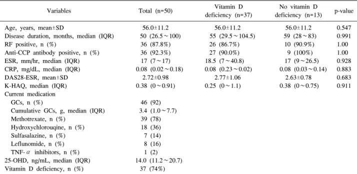
Table 1. Baseline demographics of 50 female patients with rheumatoid arthritis
Variables Total (n=50) Vitamin D
deficiency (n=37)
No vitamin D
deficiency (n=13) p-value Age, years, mean±SD
Disease duration, months, median (IQR) RF positive, n (%)
Anti-CCP antibody positive, n (%) ESR, mm/hr, median (IQR) CRP, mg/dL, median (IQR) DAS28-ESR, mean±SD K-HAQ, median (IQR) Current medication GCs, n (%)
Cumulative GCs, g, median (IQR) Methotrexate, n (%)
Hydroxychlorouqine, n (%) Sulfasalazine, n (%) Leflunomide, n (%) TNF-α inhibitors, n (%) 25-OHD, ng/mL, median (IQR) Vitamin D deficiency, n (%)
56.0±11.2 50 (26.5∼100) 36 (87.8%) 36 (92.3%) 17 (7∼17) 0.08 (0.02∼0.18)
2.72±0.98 0.38 (0∼0.91)
46 (92) 3.4 (1.0∼7.7)
39 (78) 18 (36) 7 (14) 8 (16) 1 (2) 14.0 (11.2∼20.7)
37 (74%)
56.0±11.2 55 (29.5∼104.5) 26 (86.7%) 27 (90.0%) 18.5 (7∼40.8) 0.08 (0.23∼0.02)
2.77±1.06 0.25 (0∼1.1)
56.0±11.2 59 (28∼83) 10 (90.9%)
9 (100%) 17 (9∼26.5) 0.08 (0.03∼0.14)
2.63±0.78 0.38 (0∼0.75)
0.547 0.991 1.00 1.00 0.928 0.883 0.683 0.911
IQR: interquartile range, RF: rheumatoid factor, Anti-CCP antibody: Anti-citrullinated protein antibody, ESR: erythrocyte sedimentation rate, CRP: C-reactive protein, DAS28: disease activity score 28, GCs: glucocorticoids, K-HAQ: Korean Version of health assessment questionnaire, TNF-α: tumor necrosis factor-alpha, 25-OHD: 25-hydroxy vitamin D.
assessment questionnaire (K-HAQ) (16). Immunoglobulin M-RF was assessed by particle- enhanced immunoturbido- metric assay (range 0∼14 IU/ml) and anti-CCP was measured using chemiluminescent microparticle immunoassay (range 0∼
5 U/mL). Disease activity score (DAS) 28-ESR was calculated using the following formula (17):
DAS28−ESRscore=[0.56×√((TJC28))]+[0.28×((STC28))]+
(0.70×in ESR)+(0.0014×VAS)
On the same day of blood sampling, CIMT was measured using high-resolution ultrasonography (Philips HD 15, Bothwell, WA, USA) with a 7.5 to 12.5-MHz linear array transducer. The far walls on both sides of the common carotid artery, carotid bulb and internal carotid artery were visualized at the lateral and anterior-oblique angles. CIMT measurements were performed automatically using QLAB’s CIMT quantifica- tion software (Philips Healthcare, DA Best, The Netherlands), which can enhance the consistency and reliability of measure- ment (18). The mean of maximal CIMT from all carotid seg- ments was collected from each study subjects. A CIMT ≥0.6 mm was considered as a maker of subclinical atherosclerosis (19,20). Carotid plaque was assessed in the common carotid artery, carotid bulb and internal carotid artery and was defined as a distinct protrusion of ≥50% from the adjacent wall into the vessel lumen.
Statistical analysis
No formal sample size calculation was conducted. Continuous variables are expressed as mean±standard deviation or median (interquartile range) and categorical variables as the number of cases with percentages. The Kolmogorov-Smirnov test was al- so applied to assess the normal distribution of each continuous variable. For group comparisons, the two-tailed Student’s t test or the Mann-Whitney U test was used to compare continuous variables, and the chi-squared test or Fisher’s exact test was performed to compare categorical variables. Correlation be- tween continuous variables was evaluated by the Spearman correlation test. The primary goal of our study is to investigate the relationship between vitamin D deficiency and CIMT.
Thus, the stepwise multivariate linear regression models that included demographic variables such disease duration and vari- ables with p<0.20 in the univariate regression analyses were used. Values of p<0.05 were considered to indicate statistical significance. All statistical analyses were performed using STATA version 11.1 for Windows (StataCorp LP, College Station, TX, USA) and SPSS software version 18.0 (SPSS Inc., Chicago, IL, USA).
Results
A total of 50 female patients with RA were enrolled in this study. Their baseline characteristics and biomarker levels are summarized in Table 1. Their mean±SD age was 56.0±11.2
Table 3. Linear regression models for carotid intima media thickness in study subjects
Univariable analysis Model 1* Model 2†
β (SE) p-value β (SE) p-value β (SE) p-value
Age, years
Vitamin D deficiency BMI, kg/m2
Disease duration, months HDL-C, mg/dL
DAS28-ESR K-HAQ
0.003 (7.2-4) 0.047 (0.021) 0.005 (0.003) 1.6-4 (1.6-4)
−0.001 (5.2-4) 0.021 (0.010) 0.051 (0.015)
<0.001 0.028 0.106 0.318 0.054 0.034 0.002
0.003 (6.9-4) 0.044 (0.017) 0.005 (0.002)
<0.001 0.014 0.032
0.003 (6.6-4) 0.041 (0.016) 0.006 (0.002)
0.044 (0.015)
<0.001 0.017 0.016
0.005 Adjusted R2*=0.421 Adjusted R2†=0.550 BMI: body mass index, HDL-C: high density lipoprotein cholesterol, DAS28: disease activity score 28, ESR: erythrocyte sedimentation rate, K-HAQ: Korean Version of health assessment questionnaire. *Model 1 includes age, vitamin D deficiency, BMI, disease duration, HLD-C and DAS28-ESR. †Model 2 includes age, vitamin D deficiency, BMI, disease duration, HLD-C and K-HAQ.
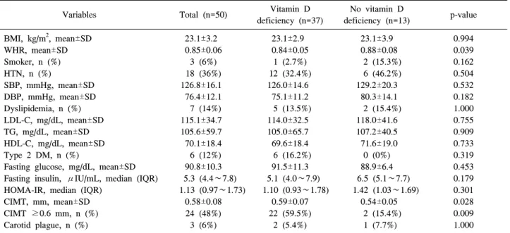
Table 2. Cardiovascular risk factors in study subjects
Variables Total (n=50) Vitamin D
deficiency (n=37)
No vitamin D
deficiency (n=13) p-value BMI, kg/m2, mean±SD
WHR, mean±SD Smoker, n (%) HTN, n (%)
SBP, mmHg, mean±SD DBP, mmHg, mean±SD Dyslipidemia, n (%) LDL-C, mg/dL, mean±SD TG, mg/dL, mean±SD HDL-C, mg/dL, mean±SD Type 2 DM, n (%)
Fasting glucose, mg/dL, mean±SD Fasting insulin, μIU/mL, median (IQR) HOMA-IR, median (IQR)
CIMT, mm, mean±SD CIMT ≥0.6 mm, n (%) Carotid plague, n (%)
23.1±3.2 0.85±0.06
3 (6%) 18 (36%) 126.8±16.1 76.4±12.1 7 (14%) 115.1±34.7 105.6±59.7 70.1±18.4 6 (12%) 90.8±10.3 5.3 (4.4∼7.8) 1.13 (0.97∼1.73)
0.58±0.08 24 (48%) 3 (6%)
23.1±2.9 0.84±0.05
1 (2.7%) 12 (32.4%) 126.0±14.6
75.1±11.2 5 (13.5%) 114.0±32.5 105.0±65.7 69.6±18.4 6 (16.2%) 91.5±11.3 5.1 (4.0∼7.9) 1.10 (0.93∼1.78)
0.59±0.07 22 (59.5%)
2 (5.4%)
23.1±3.9 0.88±0.08
2 (15.3%) 6 (46.2%) 129.2±20.3
80.3±14.1 2 (15.4%) 118.0±41.6 107.2±40.5 71.6±19.0 0 (0%) 88.9±6.4 6.5 (5.1∼7.7) 1.42 (1.03∼1.69)
0.54±0.05 2 (15.4%) 1 (7.7%)
0.994 0.039 0.162 0.504 0.532 0.182 1.000 0.755 0.909 0.733 0.319 0.453 0.179 0.301 0.028 0.009 1.000
BMI: body mass index, WHR: waist-to-hip ratio, HTN: hypertension, SBP: systolic blood pressure, DBP: diastolic blood pressure, LDL-C:
low density lipoprotein cholesterol, TG: triglycerides, HDL-C: high density lipoprotein cholesterol, DM: diabetes mellitus, IQR:
interquartile range HOMA-IR: homeostatic model assessment-insulin resistance, CIMT: carotid intima-media thickness.
years and the median (interquartile range, IQR) disease dura- tion was 50 (26.5∼100) months. The mean±SD DAS28-ESR score was 2.72±0.98. Twenty-four (48%) patients with RA had remission with a DAS28-ESR score less than 2.6. All of the patients with RA were treated with at least one disease modifying anti-rheumatic drugs (DMARDs), GCs or tumor necrosis factor-alpha (TNF-α) inhibitors. 46 (92%) patients were taking GCs and median (IQR) cumulative dose of GCs was 3.4 (1∼7.7) g. The median (IQR) 25-OHD was 14.0 (11.2∼20.7) ng/mL and 37 (74%) patients with RA had vita- min D deficiency. There were no differences in age, disease duration, ESR, CRP, DAS28-ESR and K-HAQ scores and the
proportion of RF and anti-CCP antibody positivity according the presence or absence of vitamin D deficiency.
Table 2 shows the baseline cardiovascular risk factors of the study subjects. The mean CIMT value was 0.58±0.08 mm and 48% had a CIMT >0.6 mm. Three patients (6%) showed evi- dence of carotid plaque. The proportion of RA patients with HTN, dyslipidemia and type II diabetes mellitus were 36%, 14% and 12%, respectively. Of note, RA patients with vitamin D deficiency had significantly higher CIMT than those with- out this feature (0.59±0.07 vs 0.54±0.05, p=0.028). In addi- tion, the proportion of subclinical carotid atherosclerosis (CIMT ≥0.6 mm) in RA patients with vitamin D deficiency
was significantly higher than in those without this feature (59.5% vs 15.4%, p=0.009).
The results of linear regression analysis for CIMT are shown in Table 3. Univariable analyses showed that older age, vita- min D deficiency and DAS28-ESR scores and K-HAQ scores were significantly associated with increased CIMT. There was a trend between higher HDL-C and increased CIMT, but it did not reach the statistical significance. Because DAS28-ESR and K-HAQ scores were significantly correlated (correlation coefficient=0.387, p=0.007), these variables were included in separate multivariable linear regression models to prevent multicollinearity. The association between vitamin D defi- ciency and increased CIMT remained statistically significant in multivariable regression models after adjusting for age, BMI, disease duration, HDL-C, and DAS28-ESR and K-HAQ scores (Table 3). In addition, low serum 25-OHD levels were also significantly associated with increased CIMT in the mul- tivariable regression models (data not shown).
Discussion
Little is known about the epidemiological association be- tween hypovitaminosis D and atherosclerosis in patients with RA. In this preliminary study, vitamin D deficiency was sig- nificantly associated with increased CIMT in patients with RA even after adjusting for traditional cardiovascular risk factors, disease activity and functional capacity. These results suggest that vitamin D deficiency may be a risk factor for athero- sclerosis and CVDs in patients with RA.
To our knowledge, 2 previous studies have evaluated the as- sociation between vitamin D deficiency and cardiometabolic risk in patients with RA. Haque et al. (21) reported that serum 25-OHD was significantly associated with HDL-C and in- versely associated with HOMA-IR in 179 RA patients. More recently, Goshayeshi et al. (22) reported that vitamin D defi- ciency was independently associated with metabolic syndrome in 120 RA patients. Similar to the present study, these studies found that vitamin D is linked to cardiometabolic inter- mediates in RA patients. However, these previous studies did not evaluate CIMT, which is a surrogate marker of atheros- clerosis. Increased CIMT increases the risks of myocardial in- farction, stroke and peripheral artery disease; therefore it is considered as an important tool for predicting future CVDs (23). Thus, the present study provides more comprehensive in- formation regarding the role of vitamin D deficiency in car- diovascular risk in patients with RA. The results of the present and previous studies collectively suggest that vitamin D defi- ciency increases the risks of CVDs in patients with RA.
Vitamin D is a steroid hormone involved in calcium and phos-
phate homeostasis as well as bone metabolism. Moreover, 25-OHD is a marker of “vitamin D status”; it is converted to active 1,25-OH2D by 1-α-hydroxylase. After binding vitamin D receptor (VDR), 1,25-OH2D exerts its biological action.
VDR has a broad tissue distribution including immune cells, endothelium, vascular smooth muscle cells and cardiomyocytes.
Accordingly, except for bone metabolism, a great deal of atten- tion has recently been given to the role of vitamin D in the immune and cardiovascular systems. Experimental studies showed that vitamin D regulates the renin angiotensin system (7), suppresses vascular smooth muscle cell proliferation (8), improves endothelial function (9) and inhibits myocardial hy- pertrophy (24). In addition, considering that inflammation is a predisposing factor for atherosclerosis, the anti-inflammatory property of vitamin D may reduce the risk of CVDs.
Concordantly, epidemiological studies suggests an association between vitamin D deficiency and CVDs. Vitamin D deficiency is reported to be associated with a increased risk of myocardial infarction (25), stroke (26), peripheral artery diseases (27) as well as cardiovascular mortality (28) in the general population.
In addition, hypovitaminosis D is linked to cardiovascular risk factors including hypertension, insulin resistance and type 2 diabetes mellitus as well as surrogate markers for athero- sclerosis such as increased CIMT and coronary artery calcium score (CACS) (1, 29∼31). However, the clinical implications of vitamin D deficiency in the atherosclerosis or CVDs of RA patients have not been extensively studied to date. Thus, the present results provide insight into the roles of vitamin D defi- ciency in CVDs in patients with rheumatic diseases.
Inflammatory rheumatic diseases including RA and systemic erythematous lupus has long been known to increase car- diovascular risks (32). Cardiovascular morbidity and mortality are significantly higher among patients with rheumatic diseases than the general population. The main cause of the CVDs is premature atherosclerosis which is attributable to traditional cardiovascular risk factors including type 2 diabetes mellitus and hypertension as well as the inflammatory burden of rheu- matic diseases. Therefore, the surveillance and prevention of atherosclerosis in patients with rheumatic diseases are im- portant issues in clinical practice. Clinicians should pay great attention to modify traditional risk factors and control disease activity to reduce comorbidty of CVDs in RA patients.
Considering our results, regular monitoring of serum 25-OHD levels and appropriate maintenance of sufficient levels of vita- min D may be needed in the management of patients with RA.
CIMT has become the most commonly used marker of sub- clinical atherosclerosis in patients with rheumatic diseases in- cluding RA (23). A CIMT ≥0.6 mm is considered as a marker
of atherosclerosis (19), whereas a CIMT >0.9 mm or the pres- ence of carotid plaque is associated with subclinical organ dam- age (33). In RA patients, increased CIMT and carotid plaque are predictive of CVDs (34). A recent meta-analysis shows that CIMT is significantly greater in RA patients than the general population and that age, disease duration and pre-existing athe- rogenic risk factors contributed to increase CIMT (35).
Concordant with these previous findings, older age, high BMI and low functional capacity were associated with increased CIMT in the present study. However, disease duration and tra- ditional cardiovascular risk factors including LDL-C, TG and HOMA-IR did not show the significant association with CIMT in our study, possibly because of small sample size or the char- acteristics of study subjects.
The epidemiological relationship between vitamin D defi- ciency and atherosclerosis in the present study should be in- terpreted cautiously, owing to potential reverse causality.
Among the various factors that influence vitamin D status in the human body, sun exposure is the primary determinants of serum 25-OHD. Young or physically active subjects tend to have sufficient vitamin D levels, whereas those who are eld- erly or sedentary owing to chronic illness are prone to vitamin D deficiency. Therefore, serum 25-OHD may represent gen- eral health status rather than a causative factor of CVDs. In addition, the degree of systemic inflammation is inversely as- sociated with the circulating levels of vitamin D (36). Thus, serum 25-OHD reflects the acute phase response, similar to ESR or CRP, and may not increase the risks of CVDs or atherosclerosis. Nevertheless, further investigation is required to better understand the association between vitamin D defi- ciency and CVDs in patients with RA.
Some studies report an association between CVDs and vita- min D deficiency in patients with other rheumatic diseases be- sides RA. For example, in a study of patients with Behcet’s diseases, CIMT was not associated with hypovitaminosis D but vitamin D supplementation improved CIMT (0.56 vs 0.42 mm, p=0.02) (37). Meanwhile, in patients with systemic lupus erythematosus, vitamin D deficiency was not associated with subclinical atherosclerosis assessed by CIMT and CACS (38).
The discrepancy in the effect of vitamin D deficiency on CVDs in patients with rheumatic diseases may be attributable to the differences in the methods for assaying serum 25-OHD levels and risk factors for vitamin D deficiency including sun exposure time, latitude and dietary habit; however, these fac- tors were not fully adjusted for in the present or previous studies. In addition, vitamin D metabolism may vary among rheumatic diseases. Therefore, further studies should compare the effects of vitamin D deficiency on CVDs or athero-
sclerosis among various rheumatic diseases.
This study has some limitations that warrant further discussion. First, the explanatory power of our model is lim- ited by the small sample size. In addition, because only 3 RA patients had carotid plaques, we could not investigate the as- sociation between carotid plaque and vitamin D deficiency.
Second, 48% of patients with RA in the present study had remission, with DAS-ESR scores less than 2.6 and there were few active RA patients. Thus, the present subjects may not represent of the entire RA population. Third, this study was a single center study, which could have led to a selection bias.
Conclusion
Vitamin D deficiency was significantly associated with in- creased CIMT in female patients with RA after adjusting for confounding factors in our study. Thus, the results suggest that hypovitaminosis D is a risk factor for subclinical athero- sclerosis in patients with RA. Randomized controlled trials demonstrating that vitamin D supplementation reduces the risks of atherosclerosis or future CVDs in patients with RA are required to corroborate the present findings.
Acknowledgements
The authors declare no conflict of interest. We specially thank the late Professor Sung-Il Kim who was devoted him- self to education, research and patient care in Division of Rheumatology, Department of Internal Medicine, Pusan National University School of Medicine (1963∼2011). This work was supported by clinical research grant form Pusan National University Hospital 2014.
References
1. Pittas AG, Lau J, Hu FB, Dawson-Hughes B. The role of vitamin D and calcium in type 2 diabetes. A systematic review and meta-analysis. J Clin Endocrinol Metab 2007;92:2017-29.
2. McGreevy C, Williams D. New insights about vitamin D and cardiovascular disease: a narrative review. Ann Intern Med 2011;155:820-6.
3. Kim DH, Sabour S, Sagar UN, Adams S, Whellan DJ.
Prevalence of hypovitaminosis D in cardiovascular dis- eases (from the National Health and Nutrition Examina- tion Survey 2001 to 2004). Am J Cardiol 2008;102:
1540-4.
4. Mao PJ, Zhang C, Tang L, Xian YQ, Li YS, Wang WD, et al. Effect of calcium or vitamin D supplementation on vascular outcomes: A meta-analysis of randomized con- trolled trials. Int J Cardiol 2013;169:106-11.
5. Wang L, Manson JE, Song Y, Sesso HD. Systematic re- view: Vitamin D and calcium supplementation in pre- vention of cardiovascular events. Ann Intern Med
2010;152:315-23.
6. Bjelakovic G, Gluud LL, Nikolova D, Whitfield K, Wetterslev J, Simonetti RG, et al. Vitamin D supple- mentation for prevention of mortality in adults. Cochrane Database Syst Rev 2011;(7):CD007470.
7. Li YC, Kong J, Wei M, Chen ZF, Liu SQ, Cao LP.
1,25-Dihydroxyvitamin D(3) is a negative endocrine regu- lator of the renin-angiotensin system. J Clin Invest 2002;110:229-38.
8. Carthy EP, Yamashita W, Hsu A, Ooi BS. 1,25-Dihydro- xyvitamin D3 and rat vascular smooth muscle cell growth. Hypertension 1989;13:954-9.
9. Borges AC, Feres T, Vianna LM, Paiva TB. Effect of cholecalciferol treatment on the relaxant responses of spontaneously hypertensive rat arteries to acetylcholine.
Hypertension 1999;34:897-901.
10. Wolfe F, Mitchell DM, Sibley JT, Fries JF, Bloch DA, Williams CA, et al. The mortality of rheumatoid arthritis.
Arthritis Rheum 1994;37:481-94.
11. Avina-Zubieta JA, Thomas J, Sadatsafavi M, Lehman AJ, Lacaille D. Risk of incident cardiovascular events in pa- tients with rheumatoid arthritis: a meta-analysis of ob- servational studies. Ann Rheum Dis 2012;71:1524-9.
12. Meune C, Touzé E, Trinquart L, Allanore Y. Trends in cardiovascular mortality in patients with rheumatoid ar- thritis over 50 years: a systematic review and meta-analy- sis of cohort studies. Rheumatology (Oxford) 2009;48:1309-13.
13. Gatenby P, Lucas R, Swaminathan A. Vitamin D defi- ciency and risk for rheumatic diseases: an update. Curr Opin Rheumatol 2013;25:184-91.
14. Arnett FC, Edworthy SM, Bloch DA, McShane DJ, Fries JF, Cooper NS, et al. The American Rheumatism Association 1987 revised criteria for the classification of rheumatoid arthritis. Arthritis Rheum 1988;31:315-24.
15. Matthews DR, Hosker JP, Rudenski AS, Naylor BA, Treacher DF, Turner RC. Homeostasis model assessment:
insulin resistance and beta-cell function from fasting plas- ma glucose and insulin concentrations in man.
Diabetologia 1985;28:412-9.
16. Bae SC, Cook EF, Kim SY. Psychometric evaluation of a Korean Health Assessment Questionnaire for clinical research. J Rheumatol 1998;25:1975-9.
17. Prevoo ML, van 't Hof MA, Kuper HH, van Leeuwen MA, van de Putte LB, van Riel PL. Modified disease ac- tivity scores that include twenty-eight-joint counts.
Development and validation in a prospective longitudinal study of patients with rheumatoid arthritis. Arthritis Rheum 1995;38:44-8.
18. Kang Y, Park HJ, Kang MI, Lee HS, Lee SW, Lee SK, et al. Adipokines, inflammation, insulin resistance, and carotid atherosclerosis in patients with rheumatoid arthritis. Arthritis Res Ther 2013;15:R194.
19. Veller MG, Fisher CM, Nicolaides AN, Renton S, Geroulakos G, Stafford NJ, et al. Measurement of the ul- trasonic intima-media complex thickness in normal subjects. J Vasc Surg 1993;17:719-25.
20. Targońska-Stepniak B, Drelich-Zbroja A, Majdan M. The relationship between carotid intima-media thickness and the activity of rheumatoid arthritis. J Clin Rheumatol 2011;17:249-55.
21. Haque UJ, Bathon JM, Giles JT. Association of vitamin D with cardiometabolic risk factors in rheumatoid arthritis.
Arthritis Care Res (Hoboken) 2012;64:1497-504.
22. Goshayeshi L, Saber H, Sahebari M, Rezaieyazdi Z, Rafatpanah H, Esmaily H, et al. Association between met- abolic syndrome, BMI, and serum vitamin D concen- trations in rheumatoid arthritis. Clin Rheumatol 2012;31:1197-203.
23. Kerekes G, Soltész P, Nurmohamed MT, Gonzalez-Gay MA, Turiel M, Végh E, et al. Validated methods for as- sessment of subclinical atherosclerosis in rheumatology.
Nat Rev Rheumatol 2012;8:224-34.
24. Simpson RU, Hershey SH, Nibbelink KA. Characteriza- tion of heart size and blood pressure in the vitamin D re- ceptor knockout mouse. J Steroid Biochem Mol Biol 2007;103:521-4.
25. Giovannucci E, Liu Y, Hollis BW, Rimm EB.
25-hydroxyvitamin D and risk of myocardial infarction in men: a prospective study. Arch Intern Med 2008;168:
1174-80.
26. Pilz S, Dobnig H, Fischer JE, Wellnitz B, Seelhorst U, Boehm BO, et al. Low vitamin d levels predict stroke in patients referred to coronary angiography. Stroke 2008;39:2611-3.
27. Melamed ML, Muntner P, Michos ED, Uribarri J, Weber C, Sharma J, et al. Serum 25-hydroxyvitamin D levels and the prevalence of peripheral arterial disease: results from NHANES 2001 to 2004. Arterioscler Thromb Vasc Biol 2008;28:1179-85.
28. Dobnig H, Pilz S, Scharnagl H, Renner W, Seelhorst U, Wellnitz B, et al. Independent association of low serum 25-hydroxyvitamin d and 1,25-dihydroxyvitamin d levels with all-cause and cardiovascular mortality. Arch Intern Med 2008;168:1340-9.
29. Pittas AG, Chung M, Trikalinos T, Mitri J, Brendel M, Patel K, et al. Systematic review: Vitamin D and car- diometabolic outcomes. Ann Intern Med 2010;152:307-14.
30. Reis JP, von Mühlen D, Michos ED, Miller ER 3rd, Appel LJ, Araneta MR, et al. Serum vitamin D, para- thyroid hormone levels, and carotid atherosclerosis.
Atherosclerosis 2009;207:585-90.
31. de Boer IH, Kestenbaum B, Shoben AB, Michos ED, Sarnak MJ, Siscovick DS. 25-hydroxyvitamin D levels in- versely associate with risk for developing coronary artery calcification. J Am Soc Nephrol 2009;20:1805-12.
32. Symmons DP, Gabriel SE. Epidemiology of CVD in rheumatic disease, with a focus on RA and SLE. Nat Rev Rheumatol 2011;7:399-408.
33. Mancia G, De Backer G, Dominiczak A, Cifkova R, Fagard R, Germano G, et al; Management of Arterial Hypertension of the European Society of Hypertension;
European Society of Cardiology. 2007 Guidelines for the Management of Arterial Hypertension: The Task Force
for the Management of Arterial Hypertension of the European Society of Hypertension (ESH) and of the European Society of Cardiology (ESC). J Hypertens 2007;25:1105-87.
34. González-Gay MA, González-Juanatey C, Llorca J.
Carotid ultrasound in the cardiovascular risk stratification of patients with rheumatoid arthritis: when and for whom?
Ann Rheum Dis 2012;71:796-8.
35. Tyrrell PN, Beyene J, Feldman BM, McCrindle BW, Silverman ED, Bradley TJ. Rheumatic disease and carotid intima-media thickness: a systematic review and meta-analysis. Arterioscler Thromb Vasc Biol 2010;30:
1014-26.
36. Welsh P, Peters MJ, Sattar N. Is vitamin D in rheumatoid arthritis a magic bullet or a mirage? The need to improve the evidence base prior to calls for supplementation.
Arthritis Rheum 2011;63:1763-9.
37. Can M, Gunes M, Haliloglu OA, Haklar G, Inanç N, Yavuz DG, et al. Effect of vitamin D deficiency and re- placement on endothelial functions in Behçet's disease.
Clin Exp Rheumatol 2012;30(3 Suppl 72):S57-61.
38. Kiani AN, Fang H, Magder LS, Petri M. Vitamin D defi- ciency does not predict progression of coronary artery cal- cium, carotid intima-media thickness or high-sensitivity C-reactive protein in systemic lupus erythematosus.
Rheumatology (Oxford) 2013;52:2071-6.