Received:
Revised:
Accepted:
May 15, 2015 July 29, 2015 July 29, 2015
Corresponding Author: Kwang Jae Lee, Department of Internal Medicine, Daedong Hospital, 187, Chungnyeol-daero, Dongnae-gu, Busan 47737, Korea
Tel: +82-51-550-9963, Fax: +82-51-553-7575, E-mail: [email protected]
This is an Open Access article distributed under the terms of the creative Commons Attribution Non-Commercial License (http://creativecommons.org/licenses/by-nc/3.0) which permits unrestricted non-commercial use, distribution, and reproduction in any medium, provided the original work is properly cited.
pISSN 2287-2892 • eISSN 2288-2561
JLA
New Diagnostic Criterion of Ankle Brachial Index for Peripheral Arterial Disease
Jin Doo Kang, Chang Woo Yeo, Hye Won Lee, Sung Woon Yoon, A Ra Jo, Byung Soo Kim, Kwang Jae Lee
Department of Internal Medicine, Dae Dong Hospital, Busan, Korea
말초 동맥 질환 진단을 위한 발목 상완 지수의 새로운 기준
강진두, 여창우, 이혜원, 윤성운, 조아라, 김병수, 이광재 대동병원 내과
Background: The ankle brachial index (ABI) is a simple, inexpensive diagnostic test for peripheral arterial disease (PAD).
However the diagnostic criterion of 0.9 has shown variable accuracy for identification of stenosis. We investigated more specific and sensitive diagnostic criterion of ABI for the diagnosis of PAD.
Methods: Among 5,379 patients who performed ABI test, 398 patients with abnormal ABI results or PAD symptoms underwent computed tomography angiography to confirm PAD. Each ABI results were compared with its sensitivity, specificity, positive and negative predictive values. ROC analysis and cross-tabulation analysis were performed to yield proper ABI criterion.
Results: ABI of 0.9 showed very high level of sensitivity (92.2%) and very low specificity(59.3%). ABI of 0.84 showed high level of specificity (81.4%), sensitivity (82.2%) and diagnostic correspondent rate (0.607).
Conclusion: The ABI of 0.84 could be more accurate and useful diagnostic Criterion for identifying PAD.
Key Words: Ankle brachial index, Peripheral arterial disease, Sensitivity, Specificity, CT angiography
서 론
말초 동맥 질환(peripheral arterial disease: PAD)은 협착, 혈전, 동맥류, 그 중에서도 주로 죽상경화증에 의해 혈관의 내경이 좁아져 그 구조와 기능에 이상이 생기는 질환을 이르는 것으로 협착의 정도와 위치에 따라서 무증상에서부터 파행, 통증, 궤양, 괴저까지 다양한 임상양상을 보인다. 심혈관 질환과 연관성이 깊은 것으로 알려져 있으며, PAD가 있을 경우 심혈관 질환으로 사망할 위험도가 2.5배에서 6배까지 증가하는 것으로 보고되어
조기 진단이 요구된다.1
PAD는 최근 고령, 비만, 흡연, 당뇨, 고혈압, 고지혈증, 만성신 부전과 같은 혈관 질환을 일으키는 위험인자가 증가하면서 그 유병률 또한 증가하고 있으며 전 인구의 4.5%, 당뇨병 환자군에서 7%를 차지할 정도로 그 빈도가 높은 질환이다.2,3
PAD는 증상이 없는 경우가 1/3 정도를 차지 할 정도로 무증상 비율이 높아서, 적절한 시기에 발견하여 처치하지 않을 경우 허혈 로 인한 괴저, 하지 절단과 같은 극단적인 결과를 초래하게 되므로 적절한 선별검사의 필요성이 높다.
최근 영상학적 진단기술의 발전으로 컴퓨터단층촬영(computed tomography), 자기공명영상(magnetic resonance imaging), 컬러 도플러 초음파 등의 다양한 종류의 영상 기법이 PAD를 진단하는데 사용되고 있다. 고식적 혈관조영술(conventional angiography)의 경우 확진과 치료를 동시에 시행할 수 있는 검사 방법으로 PAD 확진에 표준지침으로 사용되어 왔으나, 침습 적인 검사로 현재는 컴퓨터단층촬영 혈관조영술, 초음파 등의 비침습적인 영상학적 진단방법으로 대체되어 가고 있다.4,5 하지만 이러한 영상학적 검사의 경우 검사 비용이 비싸고 장비보유 여부에 따라 일차 진료에서 선별검사로 쉽게 사용하는 데에는 제한적이라 는 단점이 있다.
진단을 위한 검사 중에서 발목 상완 지수(ankle brachial index, ABI)를 측정하는 것은 전자 혈압계를 비롯한 혈압을 확인할 수 있는 비교적 간단한 장비만 있으면 일차 진료실에서 쉽고 간편하게 시행할 수 있어 PAD를 진단하는데 있어서 가장 적절한 선별검사 방법이다.6
현재까지 PAD를 진단할 때 ABI는 일반적으로 0.9라는 기준치 를 사용하고 있으나 임상에서 이 수치를 바탕으로 확진을 위한 추가적인 영상학적 검사를 시행했을 때 병변이 확인되지 않는 경우가 상당수 있었다.6,7,8
적절한 ABI의 기준치에 대해서는 연구에 따라 많은 이견이 있어 왔으며 ABI 0.9를 진단 기준으로 했을 때 특이도의 경우 64%-99%, 민감도의 경우 68%-95%까지 다양하게 보고되고 있다.7,8 본 연구는 PAD의 진단에 있어서 ABI의 가장 높은 특이도와 민감도를 가지는 cut-off value를 찾아 선별검사에서 낮은 특이도 로 인하여 필요가 없는 고가의 확진검사를 시행하는 것을 줄이면서 임상에서 가장 유용한 진단 기준치를 새롭게 제시하고자 한다.
대상 및 방법 1. 연구 대상
2010년부터 2014년까지 임상시험 실시기관인 대동병원에 내원 또는 입원하여 ABI 검사를 시행한 만 19세에서 80세의 5,379명의 환자 중 컴퓨터단층촬영 혈관조영술을 시행한 398명 을 대상으로 의무기록을 통한 후향적 조사를 하였다.
2. 연구 방법
본 연구는 대동병원 임상연구 심의위원회의 승인을 획득하여
시행하였다.
임상시험 실시기관인 대동병원에서 ABI 검사를 시행하여 측정 된 수치가 0.9 이하 또는 임상증세에 따라 PAD 진단을 위해 컴퓨터단층촬영 혈관조영술(Computed tomographic Angio- graphy)을 시행한 환자의 ABI와 PAD 유무를 확인하여 PAD 예측에 적절한 ABI를 구하였다.
PAD의 진단은 임상시험 실시기관의 영상의학과 전문의 1인이 ABI 결과를 모르는 상태로 목측으로 컴퓨터단층촬영 혈관조영술 상 총장골동맥(Common iliac artery), 바깥장골동맥(External iliac artery), 총대퇴동맥(Common femoral artery), 표재성대 퇴동맥(Superficial femoral artery), 전후경골동맥(Anterior and Posterior tibial artery), 비골동맥(Peroneal artery)의 폐색 또는 50% 이상 협착이 있다고 판독한 경우로 하였다. 컴퓨터단층 촬영 혈관조영술은 다중검출(multidetector) 기술을 사용한 4채 널 CT (GE Light speed Plus, USA)를 통해 혈관 영상을 얻었으며, 촬영한 혈관 영상을 3D로 재구성하여 진단에 참조하였다.
ABI는 VP2000 (Colin Medical Technology company, Komaki, Japan) 기계를 사용하여 측정하였다. 바로 누운(Supine) 자세에서 10분 이상 안정을 취한 후 사지에 커프를 감아 양측 상완 동맥(Brachial artery)과 후경골 동맥의 수축기 혈압을 측정 하여 후경골 동맥압을 양측 상완 동맥압 중에서 높은 쪽 압력으로 나눈 값으로 그 비를 계산하여 구하였다.
3. 통계 분석
통계분석은 SPSS 11.0 software (SPSS version 11.0 windows, Inc Chicago, USA)를 사용하였다. PAD 진단에 적절 한 ABI 확인을 위해 ROC 분석(Receiver operation charac- teristic analysis)을 이용하였으며, ABI 0.8부터 0.9까지 0.01 증가에 따른 각 ABI의 민감도, 특이도, 양성예측도, 음성예측도 및 진단일치도를 교차 분석을 통하여 산출하였다. 통계적 유의성 은 p값이 0.05 미만인 경우로 하였다.
결 과
1. 대상 환자의 임상적 특성
2010년부터 2014년까지 임상시험 실시기관인 대동병원에 내원 또는 입원하여 ABI 검사를 시행한 만 19세에서 80세의 5,379명의 환자 중 컴퓨터단층촬영 혈관조영술을 시행한 환자는
Table 1. Characteristics of the patients
Total PAD Non PAD
Total No.
(Male/Female)
398 (302/96)
217 (171/46)
181 (131/50)
Mean Age (years) 65.0±10.8* 68.7±9.1* 60.6±11.0*
Mean BMI 23.0±3.3 22.6±3.0 23.6±3.5
Hypertension, N (%) 274(68.8) 156(71.9) 118(65.2)
Diabetes, N (%) 268(67.3) 147(67.7) 121(66.9)
Hyperlipidemia, N (%) 178(44.7) 84(38.7) 68(51.9)
Current Smoker, N (%) 156(39.2) 88(40.6) 45(37.6)
Mean±Standard deviation.
*p<0.001 PAD versus Non PAD
PAD; peripheral arterial disease, BMI (kg/m2); body mass index
0.0 0.2 0.4 0.6 0.8 1.0 1 - specificity
1.0 0.8
0.6
0.4
0.2 0.0
Receiver Operating Characteristic (ROC) curves
(AUC=0.893)
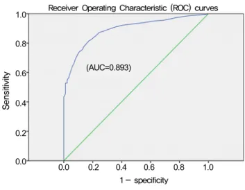
Fig. 1. Receiver Operating Characteristic (ROC) curves for ABI for diagnosing peripheral vascular disease. The cutoff value of ABI was 0.84
398명이었다. 이 중 남성은 302명(75.9%), 여성은 96명 (24.1%)으로 남성이 많았다. 대상 환자들의 평균연령은 65세였 다.
전체 환자 중 컴퓨터단층촬영 혈관조영술상 한 개 이상의 혈관 에서 50% 이상의 의미있는 협착 또는 폐색을 보여 PAD로 확진된 경우는 217명이었으며 이 중 남성은 171명, 여성은 46명으로 남성이 많았다. PAD로 확진된 환자들의 평균 연령은 68.7세로 PAD가 없는 환자군의 평균연령인 60.6세보다 연령이 의미있게 높았다(p<0.001). PAD와 체질량 지수, 고혈압, 당뇨, 고지혈증, 흡연과의 상관관계는 없었다(p>0.1)(Table 1).
2. ABI에 따른 민감도, 특이도, 양성예측도, 음성예측도, 진단일치도 및 ROC 분석
ABI와 컴퓨터단층촬영 혈관조영술상 혈관의 협착 및 폐색의 상관관계를 ROC 분석을 통해 알아본 결과, ABI 0.905의 경우 민감도 92.2%, 특이도 59.3%로 확인되어 ABI 0.9일 때 민감도 91% 이상, 특이도 64% 이하로 높은 민감도와 매우 낮은 특이도 를 보여주었다. ABI 0.835의 경우 민감도 81.7%, 특이도 82.6%, ABI 0.845의 경우 민감도 82.2%, 특이도 81.4%를 보여 ABI 0.84인 경우 민감도 81%이상, 특이도 81%이상으로 적절한 선별진단 기준값임을 보여주었다(Fig. 1).
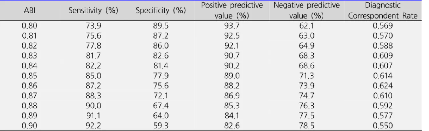
ABI 0.8부터 0.9까지 0.01 상승에 따른 민감도, 특이도, 양성예 측도, 음성예측도 및 진단일치도를 산출한 결과, ABI 0.9일 때 민감도는 92.7%, 특이도 59.3%, 양성예측도 82.6%, 음성예측 도 78.5%로 민감도는 높았지만 매우 낮은 특이도를 보여주었다.
진단일치도 또한 0.550으로 매우 낮았다. ABI 0.84일 때 민감도
82.2%, 특이도 81.4%, 양성예측도 90.2%, 음성예측도 68.6%, 진단일치도 0.607을 보였고 ABI 0.86인 경우 민감도 87.2%, 특이도 75.6%, 양성예측도 88.2%, 음성예측도 73.9%, 진단일치도 0.624를 보였다. ABI 0.86이 ABI 0.84에 비해 진단일치도는 높았으나 특이도가 75.6%로 ABI 0.84일 때 81.4%보다 낮아 ABI 0.84가 적절한 선별진단 기준임을 보여 주었다(Table 2).
고 찰
PAD는 그 유병률이 증가하고 있어 위험인자를 가진 사람을 대상으로 선별 검사의 필요성이 강조되고 있다. 미국심장협회
Table 2. Sensitivity, Specificity, Positive predictive value, Negative predictive value, Diagnostic Correspondent Rate of ABI 0.8 to 0.9
ABI Sensitivity (%) Specificity (%) Positive predictive
value (%) Negative predictive
value (%) Diagnostic Correspondent Rate
0.80 73.9 89.5 93.7 62.1 0.569
0.81 75.6 87.2 92.5 63.0 0.570
0.82 77.8 86.0 92.1 64.9 0.588
0.83 81.7 82.6 90.7 68.3 0.609
0.84 82.2 81.4 90.2 68.6 0.607
0.85 85.0 77.9 89.0 71.3 0.614
0.86 87.2 75.6 88.2 73.9 0.624
0.87 88.3 72.1 86.9 74.7 0.610
0.88 90.0 67.4 85.3 76.3 0.592
0.89 91.1 64.0 84.1 77.5 0.577
0.90 92.2 59.3 82.6 78.5 0.550
(AHA, American Heart Association)에서는 70세 이상의 노인, 흡연을 하거나 당뇨가 있는 50-69세, 그리고 50세 이하의 인구에 서는 당뇨환자 중에서 흡연, 고지혈증, 고혈압, 그리고 고호모시스 틴혈증과 같은 죽상경화증의 위험인자가 있는 경우 선별 검사를 시행하도록 권고하고 있다. 그 외 PAD를 시사하는 증상이 있는 경우 즉, 휴식 시에 허혈성 통증이 있거나, 신체검진에서 족부맥박 이상 소견을 보이는 경우, 관상동맥질환, 경부, 신장 동맥 이상이 있는 환자 역시 선별검사를 하도록 권고하고 있다.6
간편하고 쉽게 시행할 수 있는 ABI 측정이 현재 선별검사로 널리 시행되고 있다. PAD를 진단하기 위한 적절한 ABI 수치에 대해서는 연구마다 다른 수치를 제시하고 있으며 많은 경우 0.9를 진단 기준으로 하고 있다. 그러나 보편적으로 사용되고 있는 0.9라 는 수치를 임상에서 적용할 경우 확진을 위해 컴퓨터단층촬영 혈관조영술을 시행했을 때 PAD가 확인되지 않은 경우가 많았다.
본 연구는 필요 없는 고가의 침습적 검사 시행을 줄일 수 있는 보다 적절한 ABI의 기준을 알아보고자 하였다.
PAD의 확진법으로 고식적 혈관조영술(conventional angio- graphy)이 이용되나 본 연구에서 확진검사로 시행한 4채널 컴퓨 터단층촬영 혈관조영술은 폐색 또는 50% 이상 협착을 보이는 경우를 PAD로 진단했을 때 민감도 95%, 특이도 96%로 보고되 어, 매우 높은 민감도와 특이도로 다른 영상학적 검사방법과 비교 하여도 확진 검사로 이용하기에 무리가 없을 것으로 사료된 다.4,5,18,19,20
연령의 경우 PAD의 위험 인자로 연령이 증가할수록 그 유병률 이 증가하는 것으로 보고되고 있으며 본 연구에서도 역시 연령이 증가함에 따라 PAD의 진단율이 높게 나타나, 나이와 PAD는
상관관계가 있음을 보여주었다(p<0.001).3,6,9 그러나 비만, 흡 연, 당뇨, 고혈압, 고지혈증은 PAD의 위험 인자로 보고되고 있으나 본 연구에서는 PAD와의 상관관계는 없었다(p>0.1).
Ouriel 등은 ABI 0.9를 기준으로 하였을 때 민감도 90%, 특이도 98%의 결과를 보였다고 보고하였다.16 Guo 등은 민감도 76%, 특이도 90%의 결과를 보였다고 보고하였다.17 그러나 이전의 연구와 다르게 본 연구에서는 ABI 0.9인 경우 ROC 분석상 91% 이상의 높은 민감도 및 64% 이하의 매우 낮은 특이도를 보였고 교차 분석 상에서도 59.3%의 매우 낮은 특이도를 보이면 서 82.6%의 상당히 높은 음성예측도 및 0.550로 낮은 진단일치 도를 보여 실제 임상에서의 경험과 일치하게 위 양성률이 높은 결과를 보여주었다.
ROC 분석상 ABI 0.835일 때 민감도 81.7%, 특이도 82.6%, ABI 0.845일 때 민감도 82.2%, 특이도 81.4%를 보여 그 중간 값인 ABI 0.84가 적절한 선별진단 기준임을 보여주었다. 교차 분석상 ABI 0.84일 때 민감도 82.2%, 특이도 81.4%, 양성예측 도 90.2%, 음성예측도 68.6%, 진단일치도 0.607을 보였다.
ABI 0.86일 때 민감도 87.2%, 특이도 75.6%, 양성예측도 88.2%, 음성예측도 73.9%, 진단일치도 0.624를 보여 ABI 0.86이 진단일치도는 높으나 특이도가 떨어져 ABI 0.84가 적절한 선별진단 기준으로 판단된다. 두 가지 분석 결과를 토대로 ABI 0.84가 ABI 0.9일 때에 비해 민감도가 감소하고 위 음성률이 증가하나 특이도 및 진단일치도가 향상되고 위 양성률이 감소되는 결과를 보여 본 연구의 목적인 필요 없는 고가의 침습적 검사 시행을 줄일 수 있는 보다 적절한 PAD 선별진단 기준으로 사료된 다. 추가로 임상에서 사용시에 보다 편리한 기준으로 ABI 0.85도
고려해 볼 수 있을 것으로 사료되며 이 경우 ROC 분석상 82%
이상의 민감도 및 77% 이상의 특이도를 보였고, 교차 분석상 민감도 85.0%, 특이도 77.9%, 양성예측도 89.0%, 음성예측도 71.3%, 진단일치도 0.614를 보였다.
본 연구는 단일기관에서 ABI를 시행한 5,379명의 환자 중 고식적 혈관조영술이 아닌 컴퓨터단층촬영 혈관조영술을 확진 검사로 시행한 398명의 비교적 적은 수의 내국인 환자만을 대상으 로 결과를 산출한 후향적 연구로 향후 더 진단가치가 높고 정확한 진단기준을 위해서는 보다 많은 다양한 인종군을 대상으로 한 연구가 필요할 것으로 생각된다.
참고문헌
1. Attinger CE, Beckman J, Criqui M, Froelich JB, Giurini JM, Hamdan A, et al. American Diabetes Association. Peri- pheral arterial disease in people with diabetes. Diabetes Care 2003;26:3333-3341.
2. Gregg EW, Sorlie P, Paulose-Ram R, Gu Q, Eberhardt MS, Wolz M, et al. 1999-2000 national health and nutrition examination survey. Prevalence of lower- extre- mity disease in the US adult population ≥40 years of age with and without diabetes: 1999-2000 national health and nutrition examination survey. Diabetes Care 2004;
27:1591-1597.
3. Peach G, Griffin M, Jones KG, Thompson MM, Hinchliffe RJ. Diagnosis and management of peripheral arterial disease. BMJ 2012;345 aug14 1:e5208.
4. Heijenbrok-Kal MH, Kock MC, Hunink MG. Lower extre- mity arterial disease: multidetector CT angiography meta-analysis. Radiology 2007;245:433-439.
5. Met R, Bipat S, Legemate DA, Reekers JA, Koelemay MJ.
Diagnostic performance of computed tomography angiography in peripheral arterial disease: a systematic review and meta-analysis. JAMA 2009;301:415-424.
6. Hirsch AT, Haskal ZJ, Hertzer NR, Bakal CW, Creager MA, Halperin JL, et al. ACC/AHA Guidelines for the Manage- ment of Patients with Peripheral Arterial Disease (Lower Extremity, Renal, Mesenteric, and Abdominal Aortic).
Circulation 2006;113:1474-1547.
7. Khan TH, Farooqui FA, Niazi K. Critical review of the ankle brachial index. Curr Cardiol Rev 2008;4:101-106.
8. Dachun Xu, Jue Li, Liling Zou, Yawei Xu, Dayi Hu, Pagoto
SL, et al. Sensitivity and specificity of the ankle--brachial index to diagnose peripheral artery disease: a structured review. Vasc Med 2010;15:361-369.
9. Hiatt WR. Medical treatment of peripheral arterial disease and claudication. N Engl J Med 2001;344:1608- 1621.
10. Schaper NC, Andros G, Apelqvist J, Bakker K, Lammer J, Lepantalo M, et al. Diagnosis and treatment of peripheral arterial disease in diabetic patients with a foot ulcer. A progress report of the International Working Group on the Diabetic Foot. Diabetes Metab Res Rev 2012;28 Suppl 1:218-224.
11. Criqui MH. Peripheral arterial disease--epidemiological aspects. Vasc Med 2001;6 Suppl:3-7.
12. Feigelson HS, Criqui MH, Fronek A, Langer RD, Molgaard CA. Screening for peripheral arterial disease: the sensi- tivity, specificity, and predictive value of noninvasive tests in a defined population. Am J Epidemiol 1994;140:526- 534.
13. Hirsch AT, Criqui MH, Treat-Jacobson D, Regensteiner JG, Creager MA, Olin JW, et al. Peripheral arterial disease detection, awareness, and treatment in primary care.
JAMA 2001;286:1317-1324.
14. Selvin E, Erlinger TP. Prevalence of and risk factors for peripheral arterial disease in the United States: results from the National Health and Nutrition Examination Survey, 1999-2000. Circulation 2004;110:738-743.
15. Taylor-Piliae RE, Fair JM, Varady AN, Hlatky MA, Norton LC, Iribarren C, et al. Ankle brachial index screening in asymptomatic older adults. Am Heart J 2011;161:979- 985.
16. Ouriel K, McDonnell AE, Metz CE, Zarins CK. Critical evaluation of stress testing in the diagnosis of peripheral vascular disease. Surgery 1982;91:686-693.
17. Guo X, Li J, Pang W, Zhao M, Luo Y, Sun Y, et al.
Sensitivity and specificity of ankle-brachial index for detecting angiographic stenosis of peripheral arteries.
Circ J 2008;72:605-610.
18. Kock MC, Adriaensen ME, Pattynama PM, van Sambeek MR, van Urk H, Stijnen T, et al. DSA versus multi-detector row CT angiography in peripheral arterial disease:
randomized controlled trial. Radiology 2005;237:727- 737.
19. Schernthaner R, Stadler A, Lomoschitz F, Weber M,
Fleischmann D, Lammer J, et al. Multidetector CT angiography in the assessment of peripheral arterial occlusive disease: accuracy in detecting the severity, number, and length of stenoses. Eur Radiol 2008;
18:665-671.
20. Ouwendijk R, de Vries M, Pattynama PM, van Sambeek MR, de Haan MW, Stijnen T, et al. Imaging peripheral arterial disease: a randomized controlled trial comparing contrast-enhanced MR angiography and multi-detector row CT angiography. Radiology 2005;236:1094-1103.