부속비장은 선천적으로 비장의 주조직과 떨어져 있는 조직 학적으로 정상 비장조직으로 수 밀리미터에서 수 센티미터로 크기가 다양하며 비교적 흔하나 대개 임상적인 문제를 일으 키지는 않는다. 그러나 드물게 염전이나 경색으로 인해 급성 복증을 유발 할 수 있다 ( 1 - 3 ) .
증례 보고
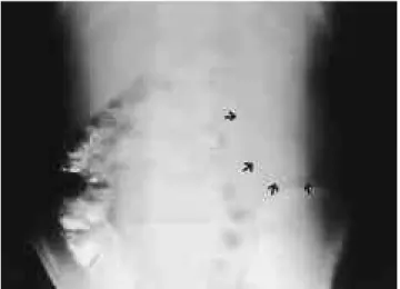
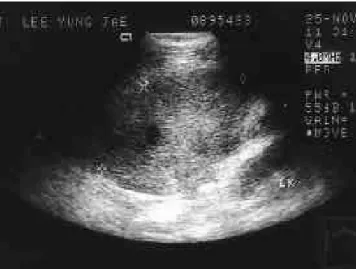
1 1세의 남아가 3일 전부터 시작된 좌상복부 동통과 발열을 주소로 내원하였다. 과거력이나 진찰 소견및 검사 소견에서 특이 소견은 없었다. 단순 복부 사진에서 좌상복부에 비교적 경계가 분명한 연부조직 음영의 종괴가 관찰되었으며 ( F i g . 1), 상복부 초음파 검사에서 좌상복부에 약 9 cm×8 cm 크기 의 고에코의 종괴가 발견되어 (Fig. 2) 복부 CT 를 시행하였 다. 조영전 C T에서 좌측 신장 앞에 약 10 cm×9 cm×6 cm 크 기의 평활하면서도 뚜렷한 경계를 가진 고음영의 종괴가 관찰 되었으며, 종괴 후방에 약 5 cm×3 cm×2 cm 크기의 미약한 저음영 부위가 있었다 (Fig. 3A). 조영후 CT 에서 종괴는 테 두리 조영증강을 보이는 저음영으로 관찰되었으며, 조영전 C T에서 저음영을 보였던 병변은 정상 비장보다 약간 낮은 정 도의 조영증강을 보였다 (Fig. 3B). 또한, 부속비장의 영양혈관 (feeding vessels)으로 여겨지는 조영증강되는 관상의 구조물이 종괴 후방에 관찰되었으며 (Fig. 3B) 주변 장간막에 염증성 변 화와 많은 양의 복수가 있었다. 간, 비장, 췌장, 신장 등 주변 장기는 정상이었고 약간의 복수가 간주위에서 관찰되었다.
진단과 치료를 목적으로 수술을 시행하였다. 수술 소견상 좌상복부에 매끈한 표면의 적갈색 종괴가 대망에 둘러 쌓여 있었고, 4-5 cm 의 혈관경이 꼬여있었다. 적출물은 13 cm×8
c m×6 cm 크기와 270 gm의 무게였으며 그 절단면은 광범위한 심한 울혈성 변화를 보였다 (Fig. 4). 현미경 소견상, 대부분 허혈성 괴사를 보였으며 정상 비장 조직이 부분적으로 관찰되 었다. 병리학적 진단은 부속비장의 염전으로 인한 경색이었다.
고 찰
부속비장은 분리되어있던 비장 조직의 융합이 이루어지지 않 아 발생하는 것으로 부검 시 10-30% 정도에서 발견되며, 수 m m에서 수 cm 까지 그 크기가 다양하다 (1-3). 흔하게는 비문 부 또는 인대를 따라서 위치하나, 약 2 0 %정도에서는 복강 또는 후복강 특히 췌미문부에서 발견할 수 있으며, 주로 좌측 신문부 상방에 위치한다. 비 전형적인 위치에 있는 경우 종양으로 오인 될 수 있으며 가끔 비장 절제술 후에 부속비장이 현저하게 커 대한방사선의학회지 2 0 00;42: 8 05- 8 0 7
─ 8 0 5 ─
경색을 동반한 부속비장의 경 염전 : CT 소견 1예 보고1
윤정경・이준식・김미은・편해욱・이일기・이종길・김희진・김익수2
부속비장의 염전은 다양한 임상 증상을 보이는 드문 질환이다. 저자들은 최근 1 1세 남아 에서 혈관경의 염전으로 부속비장의 경색을 초래하고 이로 인해 급성 복증을 주소로 내원한 증례를 경험하였기에 문헌고찰과 함께 보고하고자 한다. CT 상 좌상복부에 주변 장간막에 염증 변화를 동반하고 내부에 조영증강되는 고형성분을 가진 저음영 종괴로 보였으며, 수술 후 경색을 동반한 부속비장의 경 염전으로 확인되었다.
1대구 파티마병원 진단방사선과
2대구 파티마병원 해부병리과
이 논문은 2 0 0 0년 2월 3일 접수하여 2 0 0 0년 3월 2 7일에 채택되었음.
Fig. 1. The simple abdomen shows a relatively well-defined large soft tissue density mass lesion (arrows) at the left upper a b d o m e n .
져 좌상복부 종괴로 나타나기도 하고, 특히 자가 면역성 용혈성 빈혈, 특발성 혈소판 감소성 자반증, 유전성 구상 적혈구증 환 자에서 비장절출술 후 재발의 원인이 되기도 한다 (1-3). 대개 부속비장은 임상적인 문제를 일으키지는 않지만 드물게 경색이 나 출혈, 파열이 있는 경우 급성 복증을 초래하고 응급 수술이 필요하다. 특히 혈관경이 길고 유동적이라면, 부속비장은 복강 내 어디에나 위치할 수 있으며 혈관경도 쉽게 감돈 된다 ( 4 ) .
이 증례의 경우 혈액학적 질환 혹은 간질환 없이 부속비장이 커진 경우로서 그 원인에 관해서는 명확하지는 않지만 혈관경 의 염전으로 인해 심한 정맥 울혈로 커졌을 것으로 추측 된다.
A l e x a n d e r등이 처음 보고한 이래로, 23예의 부속비장의 염전
이 문헌에 보고 되어있으며 환자들은 발열과 비특이적인 급 성 복증을 주소로 내원 했고 수술로 진단 되었다 (3-4). 환자 연령은 영아에서 장년에 이르기까지 다양하며 소아에서 절반 이상 보고 되어있다 (4). 부속비장의 염전은 보통 염증성 종 괴의 양상을 보이므로, 복강 내 염증성 종괴와 연관된 급성 복증의 감별 진단에 고려되어야 한다.
부속비장의 경 염전으로 인한 종괴는 다양한 방사선학적 검사를 통하여 발견하고 최근에는 US 와 CT 를 병행하여 시 행한다. 그러나 이 두 검사는 위치와 크기, 모양, 주변과의 관 계를 보는데 적합하나 종양이나, 비대증, 염증과 같은 그 외 원인을 규명하는 데는 덜 유용하며 종괴의 양상과 병리의 추 윤정경 외 : 경색을 동반한 부속비장의 경 염전
─ 8 0 6 ─ Fig. 2. Transverse sonogram of the left upper abdomen shows a heterogeneous hyperechoic mass lesion.
Fig. 4. Photograph of resected specimen shows a well-defined mass measuring 13cm×8 c m×6cm and 270 gm. The external surface is smooth and reddish brown. Vascular pedicle is not- ed (arrows).
Fig. 3. A. Precontrast CT shows a slightly high-attenuating mass (arrows) with low-attenuating portion within the lesion (arrow- h e a d s ) .
B. Contrast-enhanced CT shows low-attenuating mass with a thick high-attenuating pseudocapsule (arrows) in the mesentery just anterior to the lower pole of the left kidney. Note long and mobile vascular pedicle arising from the splenic hilum in the posterior to the mass (arrowhead). Normally enhancing spleen is noted (open arrows).
A B
정에 있어서는 MRI 가 유용한 것으로 되어있다 (5). 혈관 조 영술과 Tc - 99m Sulfur colloid를 이용한 신티그라피를 사용할 수 있으나 (2-4) 혈관(afferent blood vessels) 이 완전히 폐색된 경우 부속비장의 발견에 도움이 되지 않기 때문에 정확한 진 단이 어려울 수 있다 ( 4 ) .
이 증례의 경우 C T상 테두리 조영증강을 보이는 저음영의 장간막 종괴로, 종괴의 후방에 비장과 비슷한 정도의 조영증 강을 보이는 병변과 조영 증강되는 관상의 구조물이 관찰 되 었으며, 후향적으로 고찰을 했을 때 진단적 실마리가 될 수 있을 것으로 생각된다.
결론적으로, 염증성 변화를 동반한 복강 내 종괴로 나타나 는 급성 복증의 감별 진단에 부속비장의 급성 염전도 한 원인 으로 고려되어야 한다.
참 고 문 헌
1 . 최진영,김은경,정재준등. 비장의 비외상성 양성병변. 대한방사선 의학회지 1 9 9 9 ; 4 0 : 7 3 7 - 7 4 4
2 . Dachman AH. Anomalies and congenital disorders of the spleen. In Gore RM, Levine MS, Laufer I. Textbook of gastrointestinal radiology.
1st ed. Philadelphia: WB Saunders, 1994:2238-2250
3 . Valls C, Mones L, Guma A, Lopez- Calonge E. Torsion of a wander- ing accessory spleen: CT findings. Abdom Imaging 1 9 9 8 ; 2 3 : 1 9 4 - 1 9 5 4 . Seo T, Ito T, Watanabe Y, Umeda T. Torsion of an accessory spleen
presenting as an acute abdomen with an inflammatory mass: US, CT, and MRI findings. Pediatr Radiol 1 9 9 4 ; 2 4 : 5 3 2 - 5 3 4
5 . Partain CL, Price RR, Patton JA, Kulkarni MV, James AE Jr.
Magnetic resonance imaging. 2nd ed. Philadelphia: Saunders, 1988:
1 3 3 대한방사선의학회지 2 0 00;42: 8 05- 8 0 7
─ 8 0 7 ─
J Korean Radiol Soc 2000;42:8 05- 8 0 7
Address reprint requests to : Jung-Kyung Yun, M.D., Department of Diagnostic Radiology, Fatima Hospital, Taegu, 302-1, Sinam-dong, Dong-ku, Taegu 700-600, Korea.
Tel. 82-53-940-7167 Fax. 82-53-954-7417
To rsion of the Accessory Spleen with Infarction :
CT Fe a t u res in a Case Re p o r t
1J u n g - Kyung Yun, M.D., Jun-Sik Lee, M.D., Mee-Eun Kim, M.D., Hae-Wook Pyun, M.D., Il-Gi Lee, M.D., Jong-Gil Lee, M.D., Hee-Jin Kim, M.D., Ik - Su Kim, M.D.2
1Department of Diagnostic Radiology, Fatima Hospital, Taegu
2Department of Anatomical Pathology, Fatima Hospital, Taegu
Torsion of the accessory spleen is a rare entity that can have variable clinical presentations. We report case involving an 11-year-old boy with severe abdominal pain and a mass that was found to be due to infarction of the accessory spleen, which was twisted on its pedicle. CT revealed a low-attenuating mass with peripheral in- flammatory changes in the left upper abdomen. The mass was pathologically confirmed as torsion of the ac- cessory spleen with infarction.
Index words : Spleen, abnormalities Spleen, infarction Spleen, CT
─ 808 ─