자기공명영상(magnetic resonance imaging: MRI)은 골반 부 해부에 관한 정확한 정보를 제공하며 부인과적 종양을 지 닌 환자의 진단 및 치료에 있어 점차로 중요한 역할을 담당 하고 있는 진단 매체이다. 종양의 정확한 진단을 위해 MRI 소견과 실제 병리학적 소견과의 비교연구는 필수적이나 이를 인체에서 실행하기에는 많은 제한점이 있어 실험동물을 이용 하게 된다(1-3). 최근에 적용되고 있는 자궁종양에 대한 치 료로서 자궁혈관색전술 등이 사용되고 있으나, 현재까지 실험
동물을 통한 부인과적 종양에 관한 연구는 없었다. 이에 저자 들은 실험적으로 가토의 자궁에 VX-2 암종을 유발할 수 있 는지의 여부를 시도하고, 이 암종의 자기공명영상과 조직학적 인 소견과의 비교를 통해 앞으로의 자궁암종에 대한 실험적 인 모델을 마련하고자 하였다.
대상과 방법
체중 3-3.5 kg내외의 암컷 가토(New Zealand White
가토의 자궁내에 실험적으로 유발한 VX-2 암종:
자기공명영상 소견과 병리조직학적인 소견
1강병철・김윤경・이민선・이근영・한수연
목적: 실험적으로 가토의 자궁에 VX-2 암종을 유발할 수 있는지를 시도하고, 이 암종의 자 기공명영상과 조직학적 소견의 비교를 통하여 앞으로의 자궁 암종에 대한 실험적인 모델을 마련하고자 하였다.
대상과 방법: VX-2 종양세포현탁액 1cc를 담은 2개의 주사기와 18-guage의 바늘을 이용하
여 13마리의 가토의 각각의 자궁각의 자궁벽에 주입한 후 2주(group A, n=5) 또는, 2주와 4주(group B, n=8)에 자기공명영상을 얻었고, 그 다음날 토끼를 희생하여 자기공명영상과 조 직학적인 연관성을 연구하였다. 자기공명영상은 extremity coil을 이용하여 T2-강조영상을 얻었고, 종양과 자궁벽의 신호강도를 측정하고 신호대 잡음비(signal to noise ratio, SNR)를 구하였으며, 종양의 크기와 모양을 측정하고 이들을 병리조직과 비교하였다.
결과: Group A, B의 모든 예(n=13)에서 2주에 얻은 자기공명영상에서는 T2-강조영상에서 고신호강도를 보이는 자궁벽 비후를 관찰할 수 있었다. 자궁벽의 두께는 3-10 mm(평균 6.5 mm)로 측정되었다. 또한 13예중 3예(23%)에서 복막결절(peritoneal nodules)로 관찰되었으 며, 크기는 1.5-1.8×3.0 cm 이었으며, 3예(23%)에서는 자궁종양이 관찰되었고, 그 크기는 0.6-1.5×1.3-1.5 cm 이었다. Group B 즉, 4주 후에 얻은 자기공명영상에서는 모든 예(100%) 에서 자궁벽 비후가 관찰되었으며 두께는 5-14 mm(평균 7.6 mm)였다. 자궁종양은 8예중 6예(75%)에서 보였으며 크기는 1.3-3.0×1.5-6.0 cm 이었다. 자기공명영상에서 발견된 종 양은 모든 예에서 T2-강조영상에서 주변의 정상적인 토끼의 장벽의 SNR을 비교하였을 때, 약간 높은 SNR였고, 내부와 테두리의 신호강도의 차이가 거의 없었다. 토끼를 희생한 후 조 직학적인 검사를 하였을 때, 모든 예(n=13)에서 자궁종양조직의 내부는 괴사되어 살아있는 세포가 적은 반면, 테두리에는 살아있는 세포(viable tumor cell)가 많이 발견되었다.
결론: 토끼의 자궁에 VX-2 암종세포를 이용하여 자궁종양을 유발하여 동물모델을 만들 수 있고 그 종양은 내부에 괴사가 발생하였으며, 실험적으로 유발된 토끼의 자궁종양의 자기공 명영상은 병리학적으로 연관성을 나타내고 있다.
탁액 1 cc를 주입하였다. 바늘구멍으로 VX-2 종양현탁액이 빠져나오는 것을 막기위해 6-0 Dexon과 Glue를 이용하여 바 늘구멍을 폐쇄하였다.
자기공명영상은 1.5-T GE Signa(GE Medical Systems, Milwaukee, U.S.A.)를 사용하였고, extremity coil을 이용하여 T2-강조영상(TR/TE : 11200.8-14400.5/101.5 msec, FA:
90, FOV: 280 mm, section thickness: 5 mm)을 얻었다. 토 끼를 두 그룹으로 나누어 2주(group A, n=5) 후와 2주와 4 주(group B, n=8) 후에 자기공명영상을 얻었다. 종양이 가 장 잘 나타나는 영상을 선택하여 종양의 크기, 모양 등을 계 측하였으며, 종양 및 정상자궁벽에서 신호강도를 측정한 후 신호대 잡음비(signal-to-noise ratio; SNR)를 측정하였다.
종양과 자궁벽의 중심부에서 1×1×1 mm의 ROI(region of interest)를 정하여서 신호강도를 측정하였다. 신호강도의 비 교를 위하여 2마리의 토끼의 자기공명영상을 얻고 자궁각의 모양 및 자궁각 벽의 신호강도를 측정하고 이의 SNR을 비교 의 기준으로 정하였다. 정상 토끼 2마리의 자궁각의 자궁벽 10군데에서 그 두께를 측정하여 자궁벽 두께의 기준을 마련 하였다.
자기공명영상을 얻은 다음날 토끼를 희생하여 자기공명영 상과 조직학적인 연관성을 연구하였다.
결 과
정상 토끼 2마리의 자궁벽의 두께는 0.8-1.2 mm(평균 1.0 mm)로 측정되었다. Group A와 B 모두 2주에 얻은 자기공 명영상에서는 T2-강조영상에서 고신호강도를 보이는 관상으 로 비후된 자궁벽을 관찰할 수 있었다. 자궁벽의 두께는 3- 10 mm(평균 4.5 mm)로 측정되었다. 또한 13예중 3예(23%) 에서 복막결절(peritoneal nodules)로 관찰되었으며, 크기는 1.5-1.8×3.0 cm 이었다. 3예(23%)에서는 자궁종양이 관찰
되었고, 그 크기는 0.6-1.5×1.3-1.5 cm 이었다(Table 1).
4주 후에 얻은 자기공명영상에서 8예 모두(100%)에서 관상 의 자궁벽 비후가 관찰되었으며(Fig. 1A) 두께는 5-14
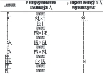
Table 1. Wall Mass (or Peritoneal Nodules) and Wall Thickeness of Uterine Horn, 2 Weeks after Inoculation (Group A)
Rabbit Mass or peritoneal Wall thickeness (mm) nodules (cm) of uterine horn
01 none 4
02 1.5×3 10
03 2×3 5
04 none 4
05 1.8×3.0 3
06 none 3
07* 0.6×1.3 5
08 none 5
09* 1.5×1.5 3
10 none 3
11* 1×1.3 7
12 none 4
13 none 3
* Rabbits demonstrated uterine masses.
Table 2. Wall Mass (or Peritoneal Nodules) and Wall Thickeness of Uterine Horn, 4 Weeks after Inoculation (Group B)
Rabbit Wall mass (cm) Peritoneal Wall thickness (mm) nodules (cm) of uterine horn
6 3×4.5 0.6×0.6 5
7 3×5 none 7
8 2×3 2×3 7
9 3×3×6 none 14
10 none 1×1 8
11 1.3×1.5 none 10
12 2×5 2×2 5
13 none 1.5×3 5
Fig. 1. Two different types of the uter- ine VX-II tumors experimentally devel- oped in rabbits.
A. T2-weighted MRI showed thickened rabbit uterine horn (arrow).
B. T2-weighted MRI demonstrated a mass on rabbit uterine horns (arrow).
mm(평균 7.6 mm)였다(Table 2). 자궁종양은 6/8(75%)에 서 보였으며, 크기는 1.3-3.0×1.5-6.0 cm 이었다(Table 2, Fig. 1B).
자기공명영상에서 발견된 종양은 모두 T2-강조영상에서 약 간 높은 SNR을 보였고, 내부와 테두리의 신호강도의 차이가 거의 없었다. VX-2 암종의 주입 4주 후에 두꺼워진 자궁각 과 자궁종양의 SNR을 측정한 결과, 정상토끼의 자궁각의 벽
의 SNR은 130-200이었는데 반해 VX-2 암종을 주사한 토 끼의 비후된 자궁각의 SNR은 230-392(평균 318±38)이었 으며 종양의 SNR는 291-463(360±52)로 높게 나타났다 (Table 3).
토끼를 희생한 후 얻은 병리조직에서 자궁각의 부종이 모 든 경우에서 관찰되었으며 모든 자궁종양의 내부는 괴사되어 살아있는 세포가 적은 반면, 종양의 테두리에서 많은 살아있 는 세포를 발견할 수 있었다(Fig. 2).
고 찰
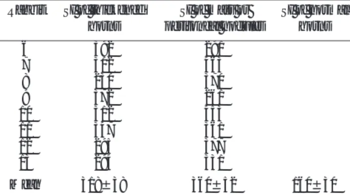
VX-2 종양은 초기에는 과혈관성의 종괴에서 점차 성장하 면서 종양의 주변부위에 동정맥단락 등의 비정상 혈관을 유 발하고 내부에서는 괴사에 의한 무혈관지역이 나타난다. VX- 2 암종의 혈관분포는 초기에는 종양의 주위 조직에서 기시하 는 굵은 혈관이 종양을 둘러싸며 이로부터 분지하는 가늘고 구불구불하며 불규칙한 혈관들이 내부로 주행하며 중심부로 갈수록 주변부에 비하여 저혈관성 혹은 무혈관성의 소견을 보 인다. 혈관중 일부는 서로 불규칙하게 문합하여 동굴 모양 (sinusoidal)을 보일 수도 있다. 종양의 크기가 커지면 내부괴 사가 진행되어 주위를 둘러싸는 피막구조물이 확실해지면서 Table 3. Signal Intensity of Thickened Uterine Horns and Masses
Compared with the Signal Intensity of Normal Uterine Horns of Rabbit Uterus, 4 Weeks after Inoculation
Rabbit SI of thickened SI of mass or SI of normal horns peritoneal nodules horns
6 392 291
7 302 463
8 230 370
9 372 261
10 312 453
11 347 361
12 295 377
13 294 331
Mean 318±38 360±52 160±30
SI: Signal Intensity
A B
Fig. 2. Microsocpic examinations of VX-II carcinomas of rabbit uterus.
A. Microscopic examination of the wall of uterine horns of rab- bit shows fibrotic tissues and cancerous cells (arrows) (H & E stain, ×400).
B. Microscopic examination of the center of the rabbit uterine tumors shows fibrotic tissues and some sparse cancerous cells with massive aptosis (arrows) (H & E stain, ×400).
C. Microscopic examination of the periphery of rabbit uterine tumor shows fibrotic tissues (white arrows) and viable cancer- ous cells (black arrows) (H & E stain, ×400).
이 피막양 구조와 그 주위에 구불구불하고 불규칙한 내경을 가진 신생혈관으로 형성된 과혈관성 부위가 형성되고 혈관의 분포도 더욱 치밀해진다. 종양내부는 거의 관류되지 않는 무 혈관성 괴사의 소견을 보인다(4). 실험동물에 쉽게 이식시킬 수 있고 성장속도가 빠르며 그 양상이 인체의 종양과 유사하 여 동물실험에 많이 이용되고 있다. 또한 급속히 성장하는 특 성으로 인해 내부괴사를 잘 동반한다(5-8). 실험동물을 이용 한 VX-2 암종의 자기공명영상 소견에 관한 연구는 장딴지 근육(9), 간(10), 신장(11), 대퇴골(12) 등에서 유발시킨 종 양에 관한 것들이 있었으나 자궁에 유발시킨 경우는 지금까 지 없었다. 저자들은 13마리의 가토의 자궁에 VX-2 암종을 주입한 결과 13마리 모든 예에서 정도의 차이는 있으나, 자 궁벽 비후를 관찰하였으며(Table 1, 2), 이중 6마리(46%)에 서 종괴가 생성되었고 이는 조직병리소견에서 관찰된 자궁각 의 부종 및 종괴형성과 일치하는 것으로 자기공명영상이 자 궁에 유발시킨 VX-2 암종의 발견에 매우 적합하다고 할 수 있겠다.
T2-강조영상에서 자궁종양의 중심부와 주변부 간의 신호 강도 차이가 관찰되지 않은 것은 자궁종양의 내부가 괴사가 심하지만, 초기에는 괴사된 부분에도 살아있는 종양세포와 섬 유조직이 함께 관찰되기 때문으로 사료된다.
또한, 일부의 보고에서 VX-2 암종은 크기가 작은 초기에 는 균질한 양상을 보이다가 성장함에 따라 내부에 괴사조직 을 형성하고 주변부로 중등도의 혈관 분포를 가지는 살아있 는 세포의 변연을 이루며, 종양 주위로 풍부한 혈관분포를 함 유한 섬유화된 피막을 형성한다고 알려져 있다(5, 7, 13, 14).
기존의 연구에서는 이러한 종양세포와 종양을 둘러싸는 피막 부위가 조영증강되는 부분이라고 보고하였으며(5, 7, 13, 14), 장기가 다르므로 직접적인 비교는 어려우나 종양 내부의 괴 사와 주변부의 살아있는 종양세포와 섬유조직으로 만약 조영 제를 사용하여 영상을 얻는다면 유사한 결과를 얻으리라고 추 정할 수 있다.
VX-2 종양의 신호강도는 다양하게 보고 되었는데, Sundaram 등(15)의 보고에 의하면 대부분의 종양은 T2-강 조영상에서 고신호강도를 보이나 섬유조직이 많거나 저세포 충실성종양인 경우는 저신호강도로 나타난다고 하였으며, 최 (9) 등의 보고에 의하면, 괴사부위에서는 여러 형태의 신호강 도를 나타낼 수 있는데 초기형태에서는 저신호강도, 중기형태 괴사는 동등 혹은 고신호강도로, 후기 액화성 괴사시기에는 높은 고신호강도로 나타난다고 하여 괴사의 신호강도가 시간 적 변화에 따라 T2-강조영상에서 저신호강도에서 고신호강 도로 이행한다고 보고했다. 본 연구에서는 T2-강조영상에서 자궁종양의 내부와 테두리의 신호강도의 차이가 거의 없이 약 간의 고신호강도로 보였는데 이는 시기적으로 이른 시기에 자 기공명영상을 실시하여 얻은 결과로 사료되며 이는 T2 강조
영상에 비하여 Gd-DTPA를 이용한 조영증강영상이 내부의 괴사를 설명해 줄 수 있는 방법으로 사료되며 앞으로 조영증 강 자기공명영상을 이용한 토끼 자궁의 VX-2 암종의 내부 에 대한 연구가 필요할 것으로 사료된다.
요약하면, 저자들은 가토의 자궁에 VX-2 암종세포를 주입 하여 자궁암종을 유발하여 동물모델을 만들 수 있었으며, 암 종의 자기공명영상 소견은 자궁벽 비후와 종양으로 나타났다.
또한 이러한 소견은 병리조직학적 소견과 잘 부합되었다.
참 고 문 헌
1. Worthington JL, Balfe DM, Lee JK, Gersell DJ, Heiken JP, Ling D, et al. Uterine neoplasms: MR imaging. Radiology 1986;159:725-730 2. Sahdev A, Sohaib SA, Jacobs I, Shepherd JH, Oram DH, Reznek RH. MR imaging of uterine sarcomas. AJR Am J Roentgenol 2001:
177:1307-1311
3. Moon KL Jr, Davis PL, Kaufman L, Crooks LE, Sheldon PE, Miller T, et al. Nuclear magnetic resonance imaging of a fibrosarcoma tu- mor implanted in the rat. Radiology 1983;148:177-181
4. 김지혜. 실험적으로 유발한 VX2 종양의 자기공명영상, 미세혈관 조 영술, 및 병리조직소견의 비교연구. 서울대 석사학위논문 1994 5. Hough A Jr, Seyberth H, Oates J, Hartmann W. Changes in bone
and bone marrow of rabbits bearing the VX-2 carcinoma. A com- parison of local and distant effects. Am J pathol 1977;87:537-552 6. Burgener FA. Peripheral hepatic artery embolization in rabbits
with VX-2 carcinomas of the liver. Cancer 1980;46:56-63
7. Young DM, Fioravanti JL, Prieur DJ, Ward JM. Hypercalcemic VX-2 carcinoma in rabbits: a clinicopathologic study. Lab Invest 1976;35:30-46
8. Kobayashi N, Allen N, Clendenon NR, Ko LW. An improved rat brain-tumor model. J Neurosurg 1980;53:808-815
9. 최병인, 이동호, 서정욱, 한준구, 김성엽, 한문희, 등. 가토 VX-2 car- cinoma의 종양부위와 괴사부위의 자기공명영상소견에 관한 연구.
대한방사선의학회지 1990;26:230-241
10. 최병인, 임효근, 주성욱, 신용문, 김재성, 김주완, 등. 가토 간의 VX- 2 암종: 초음파검사, 전산화단층촬영술 및 자기공명영상의 발견율 에 대한 비교연구. 대한방사선의학회지 1994;30:521-529
11. Yancey JM, Ackerman N, Kaude JV, Googe RE, Fitzsimmons JR, Scott KN, et al. Gadolinium-DTPA enhancement of VX-2 carcino- ma of the rabbit kidney on T1 weighted magnetic resonance im- ages. Acta Radiol 1987;28:470-482
12. 강흥식, 정성훈, 김철우, 김성문, 임정기, 한만청. 가지공명영상을 이용한 악성 근골격계 종양의 병기결정: 가토 VX-2 carcinoma를 이 용한 실험적 연구. 대한방사선의학회지 1993;29:507-515
13. Young DM, Ward JM, Prieur DJ. Hypercalcemia of malignancy.
Animal model: VX-2 carcinoma of rabbits. Am J Pathol 1978;93:
619-622
14. Revel D, Brasch RC, Paajancen H, Rosenau W, Grodd W, Engelstad B, et al. Gd-DTPA contrast enhancement and tissue dif- ferentiation in MR imaging of experimental breast carcinoma.
Radiology 1986;158:319-323
15. Sundaram M, McGuire MH, Herbold DR, Beshany SE, Fletcher JW. High signal intensity soft tissue masses on T1 weighted puls- ing sequences. Skeletal Radiol 1987;16:30-36
J Korean Radiol Soc 2004;51:649-653
Address reprint requests to : Byung Chul Kang, M.D., Department of Radiology, Mokdong Hospital, Ewha Womans University, 911-1, Mokdong, Yangcheon-gu, Seoul 158-710, Korea.
Tel. 82-2-2650-5092 Fax. 82-2-2650-5302 E-mail: [email protected]
Development of VX-II Carcinoma Model in Rabbit Uterus:
Evaluation with MR Imaging and Histopathology
1Byung Chul Kang, M.D., Yoon Kyung Kim, M.D., Min Sun Lee, M.D., Geun Young Lee, M.D., Soo Yeon Hahn, M.D.
1Department of Radiology, Mokdong Hospital, Ewha Womans University
Purpose: To develop a VX-II carcinoma model in the rabbit uterus and to describe the MR imaging findings of an experimentally induced VX-II uterine carcinoma along with the histopathologic findings.
Materials and Methods: 13 New Zealand rabbits were included in this study. Two pieces of tissue suspension (1 mm3×2) of VX-II carcinoma were loaded in an 18 gauge disposable needle and the tissue embedded in the wall of each horn of the rabbits’uterus. We obtained the MR images at 2 weeks in group A (n=5), or at 2 and 4 weeks in group B (n=8). T2-weighted images were obtained using an extremity coil. On MR imaging, we mea- sured the signal intensity of the tumor and the uterine wall. We also measured the size and shape of the tumor and we compared this with the histopathologic results.
Results: On MR images obtained 2 weeks after inoculation, all the rabbit uteruses (group A and B, n=13) show a thick tubular wall, and the uteruses demonstrated a high signal intensity on T2-weighted images. The thickened uterine walls were measured as 3-10 mm (mean: 6.5 mm). Peritoneal nodules were observed in 3/13 rabbits (23%), the nodules measured 1.5-1.8×3.0 cm; uterine masses were observed in 3/13 rabbits (23%), and they measured 0.6-1.5×1.3-1.5 cm. On MR images obtained 4 weeks after the inoculations (group B, n=8), rabbit uteruses that had VX-II carcinoma show thick tubular wall in all cases (n=8, 100%) and round uterine masses (n=6, 75%). The thickened uterine walls measured 5-14 mm (mean 7.6 mm) and uter- ine masses measured 1.3-3.0×1.5-6.0 cm. All the rabbit uteruses having VX-II carcinoma showed high sig- nal-to-noise ratios along the thickened uterine walls or masses on T2WI. On the histopathologic exam after sac- rificing the rabbit, a few viable tumor cells were found because of necrosis in the inner portion of the uterine tumors, and abundant viable tumor cells were found at the periphery of the uterine tumors.
Conclusion: We can develop an animal model with uterine tumor in rabbit uterus using VX-II carcinoma.
Experimentally induced VX-II carcinomas in rabbit uterus demonstrated central necrosis, and MR imagings of experimentally induced uterine VX-II carcinomas were well correlated with the histopathology.
Index words :Uterine neoplasms, MR Magnetic resonance (MR)