submit.radiology.or.kr J Korean Soc Radiol 2011;65(2):167-170
167 INTRODUCTION
Synovial sarcoma is the fourth most common type of soft tissue sarcoma (1). It most commonly arises in the extremities and other locations, but in rare cases can arise in the extra- peritoneal space (2). Most extraperitoneal synovial sarcomas that were reported arose in the abdominal retroperitoneum (3). Here, we report a case of primary synovial sarcoma arising in the rectovesical space. We present the radiological findings of this disease and discuss the relevant literature review.
CASE REPORT
A 48-year-old man presented with constipation and urinary symptoms such as frequency and urgency for a month. On a physical examination, abdominal distension was noted and a mass-like lesion was palpable anteriorly by digital rectal ex- amination. Laboratory findings were normal. Computed to- mography (CT) revealed a large soft tissue mass measuring 10 cm in a diameter in the pelvic cavity. The mass had a well-de-
fined, well-encapsulated and slightly lobulated margin. It was well demarcated from the surrounding structures and com- pressed the bladder anteriorly and rectum posteriorly. The precontrast image showed the mass had a homogenously low- er attenuation than that of skeletal muscle. No calcification was noted in the mass (Fig. 1A). Arterial phase CT images af- ter contrast administration showed that the mass was a het- erogeneous enhancing lesion containing tubular enhancing structures, which were presumed to be blood vessels and mul- tiple septa. Further, on 10 minute late CT images after con- trast administration, the mass revealed strong enhancement (Fig. 1B, C). Magnetic resonance imaging (MRI) showed that the mass abutted to the bladder and rectum, and compressed the seminal vesicle inferiorly. Its signal intensity on both T1- weighted image (T1WI) and T2-weighted image (T2WI) was higher than that of skeletal muscles. Further, on the T2WI, the mass was found to be a tubular structure with a signal void and multiple intratumoral septa. After contrast adminis- tration, the mass showed heterogeneous enhancement with a poorly enhancing portion within it, similar to that seen on the
Case Report
pISSN 1738-2637
J Korean Soc Radiol 2011;65(2):167-170
Received March 24, 2011; Accepted June 20, 2011 Corresponding author: Bum Sang Cho, MD
Department of Radiology, Chungbuk National University Hospital, 410 Seongbong-ro, Heungdeok-gu, Cheongju 361-711, Korea.
Tel. 82-43-269-6490 Fax. 82-43-269-6479 E-mail: [email protected]
Copyrights © 2011 The Korean Society of Radiology
Synovial sarcoma is an uncommon soft tissue malignancy usually arising in the ex- tremities of young adults. Synovial sarcomas at unusual anatomic locations have been reported; however, to the best of our knowledge, there are no reports on primary synovial sarcoma in the rectovesical space. Here, we describe the radiologic findings of primary synovial sarcoma in the rectovesical space and review relevant literature.
Index terms Synovial Sarcoma Rectovesical Space Computed Tomography Magnetic Resonance Imaging
Synovial Sarcoma in the Rectovesical Space: A Case Report
1 직장방광극에서 발생한 윤활막육종: 1예1Min Chul Kil, MD
1, Bum Sang Cho, MD
1, Gi Seok Han, MD
1, Kil Sun Park, MD
1, Sung Jin Kim, MD
1, Sang-Hoon Cha, MD
1, Seung Young Lee, MD
1, Min Ho Kang, MD
1, Ok-Jun Lee, MD
2Departments of 1Radiology, 2Pathology, Chungbuk National University Hospital, Cheongju, Korea
Synovial Sarcoma in the Rectovesical Space
submit.radiology.or.kr
J Korean Soc Radiol 2011;65(2):167-170
168
On a histological examination, we found that the tumor was comprised of short-spindle cells with rich blood vessels and foci of geographic necrosis, and had myxoid changes.
Moreover, the tumor cells were uniform and relatively small in size with inconspicuous nucleoli, contained sparse cyto- CT scan (Fig. 1D-G). Hence, the mass was considered to be a
soft tissue sarcoma arising in the extraperitoneal region.
At operation, the tumor was found to be located in the recto- vesical space adhered to the bladder and the rectum. It was ex- cised using mass excision, partial cystectomy, and cystoplasty.
COLOR
A
D
G
B
E
H
C
F
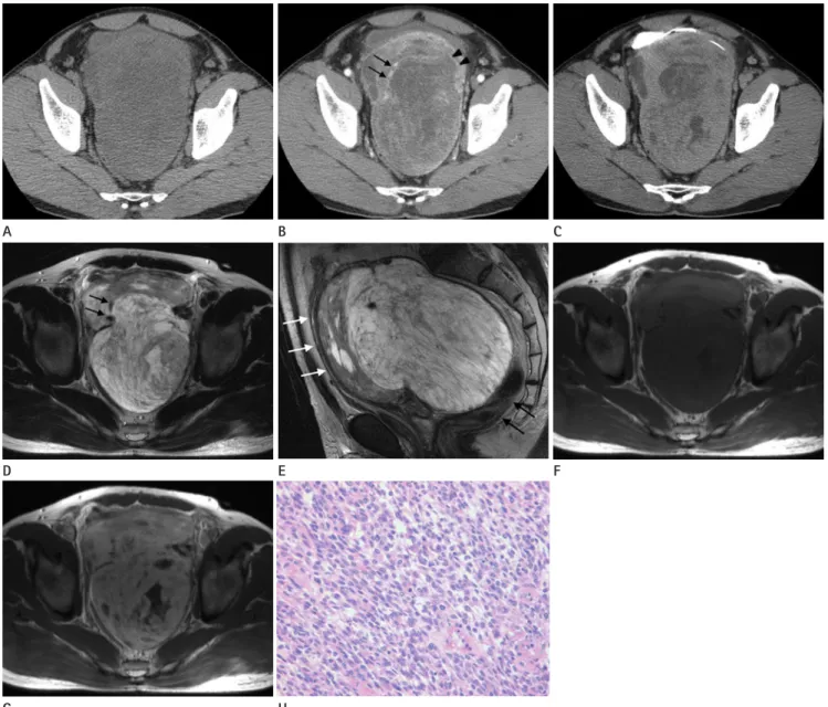
Fig. 1. The synovial sarcoma in the rectovesical space of a 48-year-old man.
A. Precontrast-phase CT scan shows a well-defined homogenous soft tissue mass without any calcification in the entire pelvic cavity.
B. Arterial phase CT scan shows an intratumoral tubular structure (arrows) indicating an enhanced vascular structure. Multiple enhancing septa (arrowheads) and poorly enhanced lesions are also seen in the mass.
C. 10 minute late CT scan after IV contrast administration shows strong enhancement in the mass.
D. T2-weighted axial MR image shows signal heterogeneity and multiple septa in the mass. An intratumoral serpentine tubular structure with a signal void (arrows) is seen.
E. T2-weighted sagittal MR image shows the mass surrounded anteriorly by the urinary bladder (white arrows) and posteriorly by the rectosig- moid colon (black arrows).
F. Precontrast T1-weighted axial MR image shows that the mass has a slightly higher signal intensity than that of the skeletal muscle.
G. Postcontrast T1-weighted axial MR image shows that the mass contains a strongly enhancing solid portion and poorly enhancing portion.
H. The tumor has uniform and relatively small ovoid or short-spindle cells with occasional mitosis (Hematoxylin-eosin staining, × 400).
Min Chul Kil, et al
submit.radiology.or.kr J Korean Soc Radiol 2011;65(2):167-170
169
able from other mesenchymal tumors (1, 7, 9). The most common finding on CT is the heterogeneous attenuation of the mass, which is usually similar to or slightly lower than that of skeletal muscles (1). In addition, areas of low attenua- tion representing necrosis or hemorrhage are common, al- though smaller lesions may be mostly homogeneous (1, 2).
CT is a useful imaging technique for identifying calcifica- tions, especially those that are subtle or located in regions with a complex anatomy (1, 2). After administration of intra- venous contrast agents, synovial sarcomas may show irregular peripheral enhancement; poor central area enhancement, re- flecting necrotic, cystic, and hemorrhagic regions; and nodu- lar enhancement (1). In our case, CT scans showed heteroge- neous attenuation that was slightly lower than that of skeletal muscle on the precontrast image and poorly enhancing por- tions within the tumor and heterogeneous delayed enhance- ment on the postcontrast image. These features are similar to those of synovial sarcomas arising in other locations. Howev- er, it did not show calcification. The enhancement pattern of synovial sarcoma has not been reported to date. In this case, delayed enhancement of the mass was seen. We assumed this finding was caused by increased vascularity, cellularity, and myxoid change such as focal nodular hyperplasia of the liver.
Further, a serpentine vascular channel was observed in the contrast-enhanced CT (Fig. 1B). This finding has not been re- ported thus far on CT, although Murphey et al. (1) identified the vascular channel on MRI. They reported the presence of the serpentine vascular channel in approximately one-third of synovial sarcomas, and stated that the use of these radiologic findings could limit the differential diagnoses to alveolar soft tissue sarcoma, metastatic renal cell carcinoma, hemangio- pericytoma, hemangioendothelioma, rhabdomyosarcoma, ex- traskeletal Ewing’s sarcoma, and synovial sarcoma. Nishimura et al. (10) found that the flow void on MRI was common in the case of hemangiopericytoma, arteriovenous hemangioma, and alveolar soft part sarcoma.
Previous reports on the MRI findings of synovial sarcoma mentioned the triple sign, bowl of grapes appearance, inter- vening septa, neurovascular encasement, and calcification, but no serpentine vascular channel (1, 7, 9). In contrast, the synovial sarcoma in our case showed a vascular channel, tri- ple sign, and intervening septa, but no neurovascular encase- plasm, and had distinct cell borders. Mitosis was occasionally
observed. On immunohistochemical examination using for- malin-fixed and paraffin-embedded tissue blocks, the tumor cells were found to be positive for vimentin, CD99, CD10 (fo- cal), and Bcl-2 but negative for cytokeratin, EMA, CD34, CD56, CD117, desmin, smooth muscle actin, S100, and HMB45. On the basis of these histological and immunohisto- chemical features, the tumor was diagnosed as a monophasic synovial sarcoma showing myxoid change (Fig. 1H).
DISCUSSION
The rectovesical space is the region between the bladder and the rectum. It is surrounded superiorly by the peritone- um; inferiorly by the fascia overlying the posterior part of the urogenital diaphragm; laterally by the sheath arising from the parietal pelvic fascia that contains vessels, nerves, and lym- phatics going to the prostate in the male and to the vagina, uterus, and ovaries in the female; and posteriorly by the ‘rectal stalks’. This space is divided into the retrovesical and prerectal compartments by the rectovesical (Denovillier’s) fascia in the male, and by the vagina and cervix uteri in the female (4). Thus far, only 2 reports have described primary tumors (lymphoma and dermoid cyst) arising in the rectovesical space (5, 6).
Synovial sarcoma is an uncommon malignant mesenchy- mal tumor that accounts for 2.5-10.5% of all primary soft tis- sue malignancies worldwide (1). Although this tumor is seen across all age groups and at any anatomical location, most sy- novial sarcomas (80-95%) occur in the extremities, especially in the lower limbs around the knees (1, 3). Furthermore, in more than 90% of cases, synovial sarcomas are located in the para-articular regions in the limbs (1-3, 7). This tumor has been founded to arise in the extraperitoneal region, the most uncommon place of occurrence, occurring in only 0.3% of cas- es (1, 2), for the most part arising in the abdominal retroperi- toneum (3, 8). Less than 10 cases of synovial sarcomas in the pelvic extraperitoneal region have been reported thus far, and these reports focus only on the pathologic findings (3). To the best of our knowledge, this is the first report on a synovial sarcoma arising in the rectovesical space.
Synovial sarcomas, irrespective of their location, have no specific imaging features, and this makes them indistinguish-
Synovial Sarcoma in the Rectovesical Space
submit.radiology.or.kr
J Korean Soc Radiol 2011;65(2):167-170
170
pathologic correlation. Radiographics 2006;26:1543-1565 2. Ulusan S, Kizilkilic O, Yildirim T, Hurcan C, Bal N, Nursal TZ.
Radiological findings of primary retroperitoneal synovial sarcoma. Br J Radiol 2005;78:166-169
3. Chatzipantelis P, Kafiri G. Retroperitoneal synovial sarco- ma: a clinicopathological study of 6 cases. J BUON 2008;
13:211-216
4. Janke WH, Block MA. Chronic retroperitoneal pelvic ab- scesses. Arch Surg 1965;90:380-384
5. Tosaka A, Yamazaki A, Hirokawa M, Matsushita K, Asakura S. [A bulky mass non-Hodgkin’s lymphoma with dysuria in the rectovesical space]. Hinyokika Kiyo 1990;36:701-705 6. Wilson RG. Dermoid cyst of the rectovesical space: report
of a case. Dis Colon Rectum 1973;16:530-531
7. Morton MJ, Berquist TH, McLeod RA, Unni KK, Sim FH. MR imaging of synovial sarcoma. AJR Am J Roentgenol 1991;
156:337-340
8. Tateishi U, Hasegawa T, Beppu Y, Satake M, Moriyama N.
Synovial sarcoma of the soft tissues: prognostic signifi- cance of imaging features. J Comput Assist Tomogr 2004;
28:140-148
9. Jones BC, Sundaram M, Kransdorf MJ. Synovial sarcoma:
MR imaging findings in 34 patients. AJR Am J Roentgenol 1993;161:827-830
10. Nishimura H, Zhang Y, Ohkuma K, Uchida M, Hayabuchi N, Sun S. MR imaging of soft-tissue masses of the extraperi- toneal spaces. Radiographics 2001;21:1141-1154
ment, calcification, or bowl of grapes appearance. The combi- nation of a large cystic area and hemorrhagic foci often leads to the latter feature (1). Jones et al. (9) reported marked signal heterogeneity of the mass on the T2WI as the triple sign.
Pathologically, this finding reflects a mixture of solid elements (intermediate signal intensity), hemorrhagic or necrotic ele- ments (high signal intensity), and calcified or fibrous ele- ments (low signal intensity) (1, 9). However, the triple sign is also observed in other soft tissue neoplasms, particularly ma- lignant fibrous histiocytoma; therefore, this finding by itself is not specific (1). Moreover, T2WIs show the intervening sep- tum in approximately 60% of synovial sarcomas (1, 8), and this was also observed in our case.
In conclusion, the radiological findings of synovial sarcoma arising in the rectovesical space are similar to those of tumors at other locations. Synovial sarcomas in the rectovesical space are difficult to distinguish from other retroperitoneal soft tis- sue masses. However, if a soft tissue mass in the rectovesical space has intervening septa, vascular channels, and signal heterogeneity, synovial sarcoma should be included in the dif- ferential diagnosis.
REFERENCES
1. Murphey MD, Gibson MS, Jennings BT, Crespo-Rodríguez AM, Fanburg-Smith J, Gajewski DA. From the archives of the AFIP: Imaging of synovial sarcoma with radiologic-
직장방광극에서 발생한 윤활막육종: 1예1
길민철
1· 조범상
1· 한기석
1· 박길선
1· 김성진
1· 차상훈
1· 이승영
1· 강민호
1· 이옥준
2윤활막육종은 주로 젊은 성인의 사지에서 드물게 발생하는 악성 연부 조직 종양이다. 윤활막육종이 드물게 발생하는 부 위에 대한 여러 보고가 있으나, 이제까지 직장방광극에서 발생한 윤활막육종에 대한 보고는 없었다. 이에 저자들은 직장 방광극에서 발생한 윤활막육종을 보고하고자 한다.
충북대학교병원 1영상의학과, 2병리과