Printed in the Republic of Korea DOI 10.5012/jkcs.2011.55.3.436
사배위 N2O2 Schiff 염기 리간드와 그 희토류 금속착물의 합성 및 특성
Vinod A. Shelke, Sarika M. Jadhav, Sunil G. Shankarwar, Achut S. Munde†, and Trimbak K. Chondhekar*
Department of Chemistry, Dr. Babasaheb Ambedkar Marathwada University, Aurangabad-431 004, Maharashtra, India
†Department of Chemistry, Milind College of Science, Aurangabad (접수 2010. 11. 30; 수정 2011. 4. 1; 게재확정 2011. 4. 18)
Synthesis and Characterization of Tetradentate N2O2 Schiff Base Ligand and its Rare Earth Metal Complexes
Vinod A. Shelke, Sarika M. Jadhav, Sunil G. Shankar war, Achut S. Munde†, and Trimbak K. Chondhekar*
Department of Chemistry, Dr. Babasaheb Ambedkar Marathwada University, Aurangabad-431 004, Maharashtra, India.
*E-mail: [email protected]
†Department of Chemistry, Milind College of Science, Aurangabad (Received November 30, 2010; Revised April 1, 2011; Accepted April 18, 2011)
요 약. o-phenylenediamine, 3-acetyl-6-methyl-(2H)pyran,2,4(3H)-dione (dehydroacetic acid 또는 DHA) 및 2, 4-dihydroxy benzaldehyde에서 비롯된 4-hydroxy-3-(1-{2-(2,4-dihydroxy-benzylidene)-amino phenylimino}–ethyl)-6-methyl-pyran-2-one (H2L)의 La(III), Ce(III), Pr(III), Nd(III), Sm(III) 및 Gd(III) 고체착물을 합성하여 원소분석, 전도도법, 자기수자율, UV-vis, FT-IR, 1H-NMR, X-선 회절, 열분석으로 특성을 규명하였으며, 항미생물 활성도를 조사하였다. FT-IR 스펙트럼 결과에 의 하면 이 리간드는 중심금속에 대해 ONNO 주개원자를 통해 이염기성 사배위 리간드로 행동함을 알 수 있다. 원소분석 결 과로부터 이들 착물의 화학량론비는 1:1 (금속: 리간드)임을 알 수 있다. 물리 및 화학적 실험결과로부터 La(III), Ce(III), Pr(III), Nd(III), Sm(III) 및 Gd(III) 착물은 일그러진 팔면체의 기하구조를 가짐을 알 수 있다. X-선 회절 데이터로부터 La(III) 및 Ce(III) 착물은 단사정계, 그리고 Pr(III) 및 Nd(III) 착물은 사방정계를 가짐을 알 수 있다. 이들 착물의 열적 행 동(TG/DTA)을 연구하였으며, 속도론적 파라메타를 Horowitz-Metzger 및 Coats-Redfern 방법으로 결정하였다. 리간드와 그 착물의 항박테리아 활성도를 Staphylococcus aureus, Escherichia coli 및 Bacillus Sp.에 대해, 그리고 살균활성도를 Aspergillus Niger, Trichoderma 및 Fusarium oxysporum에 대해 각각 조사하였다.
주제어: 디하이드로 아세트산, 비대칭 사배위 Schiff 염기, 희토류 금속착물, 분말 X-선 회절, 생물학적 활성도
ABSTRACT. The solid complexes of La(III), Ce(III), Pr(III), Nd(III), Sm(III) and Gd(III) with 4-hydroxy-3-(1-{2-(2,4-dihy- droxy-benzylidene)-amino phenylimino}-ethyl)-6-methyl-pyran-2-one (H2L) derived from o-phenylenediamine, 3-acetyl-6- methyl-(2H)pyran,2,4(3H)-dione (dehydroacetic acid or DHA) and 2, 4-dihydroxy benzaldehyde have been synthesized and characterized by elemental analysis, conductometry, magnetic susceptibility, UV-visible, FT-IR, 1H-NMR, X-ray dif- fraction, thermal analysis study, and screened for antimicrobial activity. The FT-IR spectral data suggest that the ligand behaves as a dibasic tetradentate ligand with ONNO donor atoms sequence towards central metal ion. From the microan- alytical data, the stoichiometry of the complexes has been found to be 1:1 (metal: ligand). The physico-chemical data sug- gests distorted octahedral geometry for La(III), Ce(III), Pr(III), Nd(III), Sm(III) and Gd(III) complexes. The X-ray diffraction data suggests monoclinic crystal system for La(III) and Ce(III) and orthorombic crystal system for Pr(III) and Nd(III) complexes. Thermal behavior (TG/DTA) of the complexes was studied and kinetic parameters were determined by Horowitz-Metzger and Coats-Redfern methods. The ligand and its metal complexes were screened for antibacterial activity against Staphylococcus aureus, Escherichia coli, Bacillus Sp. and fungicidal activity against Aspergillus Niger, Trichoderma and Fusarium oxysporum.
Keywords: Dehydroacetic acid, Unsymmetrical tetradentate Schiff base, Rare Earth metal complexes, Powder X-ray diffrac- tion. Biological activity
INTRODUCTION
Tetradentate Schiff bases with N2O2 donor atoms are well known to coordinate with various metal ions and
have attracted a great deal of interest in recent years due to their rich coordination chemistry.1-5 Schiff bases of o-phe- nylenediamine reported to have variety of applications including biological,6 clinical7 and analytical8 fields. Many
symmetrical tetradentate bis-type Schiff bases of 1, 2- diamines with o-hydroxy ketones/aldehydes have been prepared and studied intensively. However, much less attention has been focused on unsymmetrical tetradentate Schiff bases derived from 1, 2-diamines and different aldehydes. In particular, those derived from aromatic 1, 2 diamines have been under-investigated.9 It is worthwhile to mention here that the unsymmetrical Schiff bases of this type are difficult to obtain and are not easily isolated.10
Coordination chemistry of lanthanides has become of increasing significance in last few years due to the wide variety of applications of lanthanide complexes in super- amolecular photochemistry and in medicine.11,12 Coordi- nation compounds of the lanthanides are frequently used as catalysts, which is demonstrated by the work of Shiba- ski et al., Kobayashi and Voropai and others.13-15 One of the oxygen heterocyclic compounds, 3-acetyl-6-methyl- 2H-pyran 2,4(3H)-dione (DHA) was reported to be an excellent chelating agent and possesses promising fungi- cidal, bactericidal, herbicidal and insecticidal activities.16-19 It is also a versatile starting material for the synthesis of a wide variety of heterocyclic ring systems.20 The structural and interesting biological properties of DHA appeals to inorganic chemists working in the field of coordination chemistry. Schiff bases and their metal complexes exhibit a wide range of biological activities and various structural features. In view of the enormous importance of DHA and its metal complexes it is thought worthwhile to synthesise the Schiff base of DHA and its metal complexes.
A search of literature revels that no work has been done on the rare earth metal complexes of the asymmetrical Schiff bases derived from aromatic 1, 2-diamine, dehy- droacetic acid and 2, 4-dihydroxy benzaldehyde. In con-
tinuation of our earlier work on synthesis of bioactive transition metal complexes,21 in this communication we report the synthesis of asymmetrical tetradentate Schiff base formed by the condensation of o-phenylenediamine, dehydroacetic acid and 2, 4-dihydroxy benzaldehyde (Scheme 1). The solid complexes of La(III), Ce(III), Pr(III), Nd(III), Sm(III) and Gd(III) with this ligand have been prepared and characterized by different physico-chemical techniques.
EXPERIMENTAL
Dehydroacetic acid purchased from Merck was used as supplied. o-phenylene diamine and 2, 4-dihydroxy ben- zaldehyde of AR grade were used for synthesis of ligand.
AR grade metal nitrates were used for the complex prep- aration. The carbon, hydrogen and nitrogen contents were determined on Perkin Elmer (2400) CHNS analyzer. FTIR spectra were recorded on Jasco FTIR-4100 spectrometer using KBr pellets. 1H-NMR spectra of ligand was obtained in CDCl3 using TMS as an internal standard. The TG/
DTA and XRD were recorded on Perkin Elmer TA/SDT- 2960 and Philips 3701 respectively. The UV-visible spec- tra of the complexes were recorded on JascoUV-530 spec- trometer. Magnetic susceptibility measurements of the metal chelates were determined on a Guoy balance at room temperature using Hg[Co(SCN)4] as calibrant. Molar conductance of complexes was measured on Elico CM 180 conductivity meter using 10-3 M solution in DMF.
General procedure for the synthesis of ligand Step I: The ligand was prepared by modification of reported method.21 In a 50 mL solution of 0.001 mol (0.168 g) of DHA, 0.001 mol (0.108 g) of o-phenylenediamine was refluxed in super dry ethanol for about 3 h. Then it was cooled to room temperature. On cooling, the white coloured intermediate solid compound, mono-Schiff base was obtained with 80% yield.
Step II: 0.001 mol of intermediate (0.258 g) was then refluxed with 0.001 mol of 2, 4-dihydroxy benzaldehyde (0.138 g) in super dry ethanol for 6 h. The precipitate thus formed was filtered, dried in vacuum over CaCl2 and recrystallised in ethanol (yield: 73%).
General procedure for the synthesis of metal com- plexes
To a hot solution of ligand (0.01 mol) in chloroform, methanolic solution (25 mL) of metal nitrate (0.01 mol) was added under constant stirring. The pH of the reaction Scheme 1. Synthesis of Ligand.
mixture was adjusted to 7-8 by adding 10% alcoholic ammonia solution and refluxed for about 10-11 hrs. The precipitated solid metal complex was filtered off in hot condition and washed with hot methanol, petroleum ether and dried over calcium chloride in vacuum desiccator (Yield=50%).
Antimicrobial Activity
The antibacterial activity of free ligand, its metal com- plexes, the metal salts and control (DMF solvent) was tested in vitro against gram + ve bacteria (Staphylococcus) and gram -ve bacteria (E. coli) by paper disc method.22 Sterile (10 mm) diameter Whatmann No. 42 paper discs were soaked in different concentrations of the ligand/
complexes (250 ppm and 500 ppm) in DMF, dried and then placed on the lawn culture of nutrient agar plates. The plates were then incubated for 24 h at 37 ºC and the inhi- bition zone around each disc was measured. The results obtained were compared with known antibiotics, Cipro- floxin. Three replicates were taken and average values are given in (Table 4 and 5).
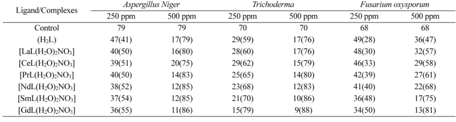
The free ligand, its metal complexes, metal salts and control were screened for antifungal activity against the fungi Aspergillus niger and Trichoderma at 250 ppm and 500 ppm levels respectively by mycelia dry weight method.23 The culture of fungi was purified by single spore isolation technique. The glucose nitrate (GN) medium was used for the growth of fungi. The mycelial biomass was then dried along with filter paper in an oven at 655 ºC to constant weight, cooled and finally weighed. The mycelial dry weight (MDW) was obtained by subtracting the weight of mycelium free filter paper from final dry weight.23 Three replicates of each treatment were repeated in all experi- ments. The MDW was corrected each time by subtracting the dry weight obtained from incubated flask under sim- ilar experimental conditions. The yields of MDW in mg are presented in (Table 5). The percentage error was found to be 0.01. The percent decrease in mycelia dry weight of
the test compound in each case was calculated and tab- ulated in terms of average percentage inhibition. The results indicate that the ligand and its metal complexes arrested the growth of fungi.
RESULTS AND DISCUSSION
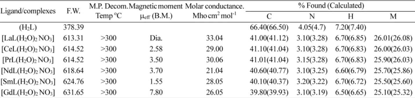
Physical characteristics, micro analytical, molar con- ductance data of ligand and metal complexes are given in Table 1. The analytical data of complexes revels 1:1 molar ratio (metal: ligand) and corresponds well with the general formula [ML(H2O)2. NO3.] [where M=La(III), Ce(III), Pr(III), Nd(III), Sm(III) and Gd(III)]. The magnetic sus- ceptibilities of La (III), Ce (III), Pr (III), Nd (III), Sm (III) and Gd (III) complexes at room temperature are consis- tent with distorted octahedral structure with two water molecules and one nitrate molecule coordinated to metal ion. The presence of two coordinated water molecules was also confirmed by TG-DTA. The metal chelate solu- tions in DMF show low conductance which suggest their non-electrolytic nature.
1H-NMR spectra of ligand
The 1H-NMR spectra of free ligand in CDCl3 at room temperature shows the following signals. δ 2.10 (s, 3H, -CH3), 2.55 (s, 3H, N=C-CH3), 5.20 (s, 1H, phenolic OH), 5.80 (s, 1H, Ar-H), 6.10-7.20 (m, 8H, Ar-H), 9.20 (s, 1H, N=C- H), 9.50 (s, 1H, enolic OH of DHA moiety).
Mass Spectra of the ligand
Mass spectral data confirmed the structure of the ligand HL as indicated by the peaks corresponding to their molec- ular masses.
Magnetic measurements
The effective magnetic moments (Table 1) of all the complexes indicate their paramagnetic nature except La(III), which is diamagnetic. The values obtained are
Table 1. Physical characterization, analytical and molar conductance data of ligand and its metal complexes Ligand/complexes F.W. M.P. Decom.
Temp oC
Magnetic moment µeff (B.M.)
Molar conductance.
Mhocm2 mol-1
% Found (Calculated)
C N H M
(H2L) 378.39 66.40(66.50) 4.05(4.7) 7.20(7.40)
[LaL(H2O)2 NO3] 613.31 >300 Dia. 33.04 41.00(41.12) 3.10(3.28) 6.70(6.85) 26.01(26.08) [CeL(H2O)2 NO3] 614.52 >300 2.58 29.00 41.10(41.04) 3.10(3.28) 6.70(6.83) 26.00(26.03) [PrL(H2O)2 NO3] 614.52 >300 3.50 30.06 41.01(41.04) 3.15(3.28) 6.70(6.83) 25.90(26.03) [NdL(H2O)2 NO3] 618.64 >300 3.70 21.04 40.60(40.77) 3.10(3.25) 6.60(6.79) 25.70(25.86) [SmL(H2O)2 NO3] 624.76 >300 1.55 28.05 40.10(40.37) 3.20(3.22) 6.70(6.72) 25.50(25.60) [GdL(H2O)2 NO3] 631.65 >300 7.80 26.05 39.80(39.93) 3.10(3.19) 6.50(6.65) 25.10(25.32)
similar to the Van Vleck and Frank24 and Hund’s values except in case of Sm(III) where slightly higher value was obtained. This is due to low J-J separation, which leads to thermal population of higher energy levels. The values obtained are similar to those of typical lanthanide ions25 and indicate the non involvement of 4f electrons in bond- ing due to their very effective shielding by the 5s2 5p6 octet. Gd(III) ion has 4f 7 electronic configuration with 8S7/2
single-ion ground state.
FTIR spectra
The FTIR spectrum of free ligand shows characteristic bands at 3060-3312, 1705, 1663, 1360 and 1224 cm-1 assign- able to intramolecular hydrogen bonded (υ OH), lactone carbonyl (υ C=O), azomethine (υ C=N), aryl azomethine (υ C-N) and phenolic (υ C-O) stretching modes respec- tively.21,26 The absence of a weak broad band in the 3060- 3312 cm-1 region, noted in the spectra of the metal com- plexes, indicates deprotonation of the intramolecular hydrogen bonded OH group on complexation and subse- quent coordination of phenolic oxygen to the metal ion.
This is further supported by upward shift in phenolic (υ C- O)27 to the extent of 30-50 cm-1. On complexation, the (υ C=N) band is shifted to lower wave number with respect to free ligand, denoting that the nitrogen of the azome- thine group is coordinated to the metal ion. This is sup- ported by upward shift in (υ C-N)to the extent of 10-50 cm.-1 28 The FTIR spectra of metal chelates showed new bands in 425-550 cm-1 range assignable to (M-N)29-32 and 400-450 cm-1 range assignable to (M-O) modes.33 The FTIR spectra showed a strong band in 3150-3600 cm-1 region, suggesting the presence of coordinated water in these metal complexes. Infrared spectra of the nitrato com- plexes reveled two additional strong bands around 1491 and 1279 cm-1, which were absent in the free ligand.34 The presence of coordinated water is further confirmed by the appearance of non-ligand band in 830-840 cm-1 region, assignable to the rocking mode of water.34 The presence of coordinated water is also established and supported by TG/DT analysis of these complexes. Hence it is clear that the coordination takes place via phenolic oxygen and azomethine nitrogen of ligand molecule.
Electronic Spectra
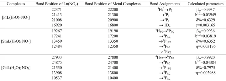
The electronic spectra of the aqueous solutions of the nitrates of Pr, Nd and Sm are compared with the corre- sponding complexes. The data are summarized in Table 4 The data indicates that the energy of f-f transitions in the complexes is slightly reduced compared to the corre-
sponding aquo ions either because of the slight covalent interaction of the 4f orbitals with vacant ligand orbitals, leading to some delocalization with consequent reduction in interelectronic repulsion33 or by increased nuclear shield- ing of the orbitals due to a slight covalent ligand-metal electron drift. The bonding parameters (b1/2), the cova- lency parameter (δ) and nephelauxetic ratio (β) have been calculated using literature procedures.35-37 The value of (1-β) being less than unityfor the complexes, the small and positive values of the bonding parameter b1/2 and Sinha’s parameter δ% suggests the possibility of a partial covalent nature of the metal-ligand bond.38,39 Based on electronic spectral studies, a coordination number of seven around the metal ion has been suggested, as reported for Nd(CF3COCHCOMe).H2O by Karraker40 and for [Ln(Hapfh)2Cl]Cl2 (Hapfh=acetylpyridine-2-furoylhy- drazone) by Singh and Singh.41
Thermal analysis
The simultaneous TG/DT analysis of metal complexes was studied from ambient temperature to 1000oC in nitro- gen atmosphere using α-Al2O3 as reference. The La(III), and Sm(III) complexes were chosen for thermal study. On the TG curve of La(III) complex, the first step shows a steep slope between 140-230oC with a mass loss of 5.7%
(calculated 5.86%), indicating the removal of two mole- cules of coordinated water. An endothermic peak in the range 140-200oC (∆Tmax=170oC) on the DTA curve corresponds to the dehydration step. The anhydrous com- plex first show slow decomposition from 200-300oC, with 10.00% mass loss, (calcd. 10.10%) and a broad exotherm (∆Tmax = 275oC) in DTA may be attributed to removal of coordinated nitrate part of the complex.42 The second step decomposition is from 400 to 600oC with 22.70%
mass loss (calcd 23.00%) which corresponds to decom- position of coordinated part of ligand. A broad endotherm in DTA is observed for this step. The mass of the final res- idue corresponds to stable oxide, 52.00% (calcd. 52.82%).
The TG curve of Sm(III) complex shows first mass loss 5.78% (calcd. 5.76%) in the range 100-225oC and an endothermic peak in this region ∆Tmax=195oC, which indicates removal of two coordinate water molecules. The anhydrous complex show slow decomposition from 225- 320oC with 10.20% (calcd. 9.92%) mass loss. A broad exotherm ∆Tmax = 299oC in DTA may be attributed to the removal of coordinated nitrate part of the complex.
The second step decomposition from 400 to 650oC with 24.20% mass loss (calcd 23.50%) corresponds to decom- position of coordinated part of ligand. A broad endotherm
in DTA is observed for this step. The mass of final residue corresponds to stable Sm2O3, 54.20% (calcd. 55.81%).
Powder X-ray diffraction
The X-ray diffraction of metal complexes was scanned in the range 5-80o at wave length 1.543Å. The diffracto- gram and associated data depict the 2θ value for each peak, relative intensity and inter-planar spacing (d-values).The diffractogram of La(III) complex had fourteen reflactions with maxima at 2θ = 41.57 corresponding to d value 2.17Å.
The diffractogram of Ce(III) complex shows twelve reflactions with maxima at 2θ = 40.92o corresponding to d value 2.20. The diffractogram of Pr(III) complex had twelve reflactions with maxima at 2θ = 23.09 correspond- ing to d value 3.08. The diffractogram of Nd(III) complex had nine reflactions with maxima at 2θ = 70.43 corre- sponding to d value 1.33. The x-ray diffraction pattern of these complexes with respect to major peaks having rel- ative intensity greater than 10% have been indexed by using computer programme (Powder X-ray data analysis system, Cheng Dong, Institute of Physics, Chinese Acad- emy of Science) The above indexing method also yields Miller indices (hkl), unit cell parameters and unit cell vol- umes.
The unit cell of La (III) complex yielded values of lat- tice constants, a = 12.18Å, b = 4.40Å, c = 5.58Å and unit cell volume V = 299.4321Å3. In concurrence with these cell parameters, the condition such as a≠b≠c and α = γ = 90o≠β. required for sample to be monoclinic were tested and found to be satisfactory. Hence it can be concluded that La(III) complex has monoclinic crystal system. The unit cell of Ce(III) complex yielded values of lattice con- stants, a =16.49Å, b = 5.34Å, c = 5.06Å and unit cell vol- ume V = 746.1900Å3. Pr(III) complex yielded values of lattice constants, a = 14.91Å, b = 7.82Å, c = 7.68Å and unit cell volume V = 897.5200Å. Nd(III) complex yielded values of lattices constants a = 10.59Å, b = 6.86Å, c = 4.35Å and unit cell volume V = 316.6349Å3. Gd(III) complex yielded values of lattices constants a = 17.26Å, b = 7.62Å, c = 4.10Å and unit cell volume V = 539.8008Å3. In concurrence with these cell parameters, the condition such as a≠b≠c and α
= β = γ = 90 required for crystal to be orthorombic were tested and found to be satisfactory. Hence it can be con- cluded that Ce(III), Pr(III), Nd(III) and Gd(III) complexes has orthorombic crystal system. The experimental density values of the complexes were determined by using spe- cific gravity method (Shoemaker and Garland, 1989) and found to be 1.24, 3.32, 1.65 and 4.71 gcm-3 for La(III), Ce(III), Pr(III) and Nd(III) complexes respectively. By
using experimental density values, molecular weight of complexes, Avogadro’s number, volume of the unit cell, the number of molecules per unit cell were calculated by using equation ρ = nM/NV and was found to be one for La(III) and four for Ce(III), Pr(III) and Nd(III) complexes.
With these values, theoretical densities were computed and found to be 1.23, 3.31, 1.64 and 4.70 gcm-3 for respec- tive complexes. Comparison of experimental and theo- retical density values showed good agreement within the limits of experimental error.43
Kinetic data
The kinetic and thermodynamic parameters viz order of reaction (n), energy of activation (Ea), frequency factor (log A), entropy of activation (∆S#) and free energy change (∆G#) together with correlation coefficient (r) for non-iso- thermal decomposition of metal complexes have been determined by Coats-Redfern integral and Horowitz- Metzer method30 and the data is given in Table 2. The results showed that the values obtained by two methods are comparable. The calculated free energy of activation is relatively low indicating the autocatalytic effect of metal ions on thermal decomposition of the complexes.31,32 ∆S# values were negative, which indicate a more ordered activated Fig. 1. X-ray diffractograms of La(III), Ce(III), Pr(III), Nd(III) and Gd(III) complexes.
state that may be possible through the chemisorption of oxygen and other decomposition products. The more ordered nature may be due to the polarization of bonds in activated state which might happen through charge trans- fer electronic transitions.
Antimicrobial activity
The Schiff base ligand H2L and its metal complexes were evaluated for antimicrobial activity against bacteria such as Staphylococcus aureus, Escherichia coli and Bacil- lus Sp by paper disc method.21 The compounds were
tested at the concentration 0.25 mgmL-1 and 0.5 mgmL-1 in DMF and compared with known antibiotics viz. Cipro- floxin (Table 4). For fungicidal activity, compounds were screened in vitro against Aspergillus Niger, Trichoderma and Fusarium oxysporum by mycelia dry method with glucose nitrate media.23 The compound were tested at the concentration 250 and 500 ppm in DMF and compared with control (Table 5). From Tables 4 and 5, it is clear that the inhibition by metal chelates is higher than that of ligand.
The results showed that the free ligand as well as all the metal complexes exhibit antimicrobial activity against Table 2. Electronic spectral data (cm-1) and related bonding parameters of lanthanide (III) complexes.
Complexes Band Position of Ln(NO3) Band Position of Metal Complexes Band Assignments Calculated parameters
[PrL(H2O)2 NO3]
22371 21413 21008 16920
22200 21300 20900 16800
3H43→P2
→3P1
→3P1
→1D2
βav=0.9937 b1/2=0.03949
δ%=0.6329 γ=0.003165 [SmL(H2O)2 NO3]
19267 17241 13459 12484
19190 17200 13350 12350
4H5/2→6P7/2
→4P9/2
→4P13/2
→4F9/2
→ 4F9/2
βav=0.9936 b1/2=0.03819
δ%=0.6352 η=0.003176
[GdL(H2O)2 NO3]
27933 24875 21550 13908 10537
27800 24700 21400 13800 10400
4H5/2→6P7/2
→4P9/2
→4P13/2
→4F9/2
→4F9/2
βav=0.9920 b1/2=0.04384
δ%=0.7975 η=0.003988
Table 3. The kinetic parameters of metal complexes calculated by the methods Horowitz-Metzger (HM) and Coats-Redfern (CM) methods Complex Step n Method Ea (kJmol-1) Z (S-1) ∆S# (JK-1mol-1) ∆G# (kJmol-1) Corelation coefficient (r)
La(III)
I 0.5 HM
CR
18.86 26.22
6.5105 6.8108
-234.72 -238.18
35.03 42.63
0.9995 0.9957
II 1.3 HM
CR
63.01 45.23
6.3102 5.4102
-222.18 -228.45
95.69 78.29
0.9985 0.9992
Sm(III)
I 0.9 HM
CR
16.35 22.09
3.1108 3.2108
-240.96 -245.24
32.95 38.99
0.9975 0.9999
II 1.3 HM
CR
31.50 26.53
9.910-3 1.3105
-257.61 -258.24
69.39 64.57
0.9995 0.9997
Table 4. Antibacterial activity of ligand and its metal complexes.
inhibition zone (mm)
E. coli Staphylococcus Bacillus sp.
Ligand/Complexes 250 ppm 500 ppm 250 ppm 500 ppm 250 ppm 500 ppm
Ciprofloxin 29 32 31 35 25 27
(H2L) 11 14 11 15 10 13
[LaL(H2O)2NO3] 11 15 13 16 11 14
[CeL(H2O)2NO3] 12 16 13 17 12 15
[PrL(H2O)2NO3] 12 18 13 16 11 17
[NdL(H2O)2NO3] 12 18 14 17 11 16
[SmL(H2O)2 NO3] 13 19 14 17 11 18
[GdL(H2O)2NO3] 13 19 15 18 12 18
both the strains. The comparative study of the ligand and its metal complexes indicates that the metal complexes exhibit higher antimicrobial activity than free ligand under identical experimental conditions. Antimicrobial inhibi- tions were compared with that of Ciprofloxin as a stan- dard, showing that the activity of all the tested compounds is less than the standard. The metal salts used for the syn- thesis of complexes exhibit negligibly small antimicro- bial activity.22,23 The remarkable enhancement of activity of ligand after chelation can be explained on the basis of Overtones concept and Tweedy’s concept. According to these concepts, chelation tends to make a ligand more potential bacterial agent. The increased activity upon che- lation is attributed to the positive charge of metal partially shared with donor atoms present on ligand and possible π-electron delocalization over the whole chelate ring.44 This, in turn, increases the lipophilic character of the metal chelate and favors its permeation through the lipid layers of the bacterial membranes resulting in interfer- ence with normal process. Inhibition was found to increase with increasing concentration of metal complex.45
CONCLUSION
La(III), Ce(III), Pr(III), Nd(III), Sm(III) and Gd(III) complexes with N2O2 donor Schiff base ligand derived from o-phenylenediamine, 3-Acetyl-6-methyl-pyran-2, 4- dione (dehydroacetic acid) and 2, 4-dihydroxy benzalde- hyde were synthesized. A comparative study of their physico-chemical properties have been made through ele- mental analysis, molar conductance, mass, IR, electronic spectra, magnetic moment, TG/DTA, powder XRD and antimicrobial study. The conductance data indicated that all the complexes were 1:1 non electrolytes. The IR data revealed that the Schiff base ligand is coordinating via phenolic oxygen and imino nitrogen as illustrated in Fig.
2. A distorted octahedral geometry were assigned for
La(III), Ce(III), Pr(III), Nd(III), Sm(III) and Gd(III) com- plexes. The complexes were biologically active and showed enhanced antimicrobial activities compared to free ligand. Thermal study revealed thermal stability of complexes.
The XRD study suggested monoclinic crystal system for La(III) and orthorombic crystal system for Ce(III), Pr(III), Nd(III) and Gd(III) complexes.
Acknowledgement. The authors are grateful to the Head, Department of Chemistry, Dr. Babasaheb Ambed- kar Marathwada University, Aurangabad for providing the laboratory facilities.
REFERENCES
1. Atkins, R.; Breweg, G.; Kakot, E.; Mockler, G. M.; Sinn, E. Inorg. Chem. 1985, 24, 127.
2. Yuan, R.; Chai, Y.; Liu, D.; Gao, D.; Li, J.; Yu, R.
Anal.Chem. 1993, 65, 2572.
3. Ramesh, R.; Saganthy, P. K.; Natarajan, K. Synth. React.
Inorg. Met-Org. Chem. 1996, 26, 47.
4. Ohashi, Y. Bull. Chem. Soc. Jpn. 1997, 70, 1319.
5. Jeong, B. G.; Rim, C. P.; Chae, H. N.; Chjo, K. H.; Nam, K.; Choi, Y. Bull. Korean Chem. Soc. 1996, 17, 688.
6. Singh, P.; Geol, R. L.; Singh, B. P. J. Indian Chem. Soc.
Table 5. Antifungal activity of compounds yield, of mycelial dry weight in mg (%inhibition)
Ligand/Complexes Aspergillus Niger Trichoderma Fusarium oxysporum
250 ppm 500 ppm 250 ppm 500 ppm 250 ppm 500 ppm
Control 79 79 70 70 68 68
(H2L) 47(41) 17(79) 29(59) 17(76) 49(28) 36(47)
[LaL(H2O)2NO3] 40(50) 16(80) 28(60) 17(76) 48(30) 32(57)
[CeL(H2O)2NO3] 39(51) 20(75) 29(62) 15(79) 46(33) 29(58)
[PrL(H2O)2NO3] 40(50) 14(83) 25(65) 14(80) 42(39) 27(61)
[NdL(H2O)2NO3] 38(52) 12(85) 23(68) 12(83) 41(40) 22(68)
[SmL(H2O)2NO3] 37(54) 12(85) 21(70) 10(86) 36(48) 17(75)
[GdL(H2O)2NO3] 36(55) 11(86) 15(79) 9(88) 34(50) 13(81)
Fig. 2. Proposed structure of metal complexes where M = La(III), Ce(III), Pr(III), Nd(III), Sm(III) and Gd(III).
1975, 52, 958.
7. Mahindra, A. M.; Fisher, J. M.; Rabinovitz, M. Nature.
1983, 64, 303.
8. Patel, P. R.; Thaker, B. T.; Zele, S. Indian J. Chem. 1999, 38A, 563.
9. Phan, N. T. S.; Brown, D. H.; Adams, H.; Spey, S. E.;
Styring, P. Dalton Trans. 2004, 9, 1348.
10. Tan, S. F.; Ang, K. P. Transition Met. Chem. 1988, 13, 64.
11. Minhus, Q.; Huifang, S.; Xiaoping, Qi, J. C. J. Rare Earths. 2007, 25, 721.
12. Guo, Y. L.; Dou, W.; Wang, Y. W.; Liu, W. S.; Wang, D.
Q. Polyhedron 2007, 26, 1699.
13. Shibaski, M.; Sasai, H.; Arai, T. Angew. Chem. 1997, 109, 1290.
14. Kobayashi, S.; Ishitani, H. J.Chem.Commun. 1995, 13, 1379.
15. Voropai, E. S.; Samtsov, M. P.; Chalov, V. N.; Zhavrid, A.
J. Appl. Spectrosc. 2001, 68, 468.
16. SuryaRao, D.; SubhaRao, B. L.; John, V. T.; Ganorkar, M.
C. Nat. Acad. Sci. Lett. 1978, 1, 402.
17. Surya Rao, D.; Sadasiva Reddy, C.; John, V. T.; Ganorkar, M. C. Curr. Sci. 1980, 49, 511.
18. Schleiffenbaum, B.; Spertini, O.; Tedder Thomas, F. J Cell. Biol. 1992, 119(1), 229.
19. Stanley, V. G.; Woldesenbet, S.; Gray Cassandra. Poultry Sci. 1996, 75(1), 42.
20. Levai, A.; Jeko, J. Monatash. Chem. 2006, 137, 339.
21. Munde, A. S.; Jagdale, A. N.; Jadhav, S. M.; Chondhekar, T. K. Journal of the Korean Chemical Society. 2009, 53, 407.
22. Mane, P. S.; Shirodkar, S. G.; Arbad, B. R.; Chondhekar, T. K. Indian J. Chem. 2001, 40, 648.
23. Jadhav, S. M.; Munde, A. S.; Shankarwar, S. G.; Patharkar, V. R.; Shelke, V. A.; Chondhekar, T. K. Journal of the Korean Chemical Society. 2010, 54, 5.
24. Van Vleck, J. H.; Frank, N. Phys. Rev. 1929, 34, 1494.
25. Selwood, P. S. Magneto Chemistry Interscience, New York,
1960.
26. Ramarao, N.; Rao, V. P.; Tyaga Raju, V. J.; Ganorkar, M.
C. Indian J. Chem. 1985, 24, 877.
27. Venketeswar Rao, P.; Venkata Narasaiah, A. Indian J.
Chem. 2003, 42, 896.
28. Dash, D. C.; Panda, A. K.; Jena, P.; Patjoshi, S. B.; Maha- patra, A. J. Indian Chem. Soc. 2002, 79, 48.
29. During, J. R.; Layton, R.; Sink, D. N.; Mitchell, B. R.
Spectrchem. Acta. 1965, 21, 1326.
30. Nakamoto, K.; Advances in the Chemistry of Coordination Compounds, S. Kirschner (ed.), Mc Millan, New York. 1961.
31. Ueno, K.; Martell, A. E. J. Phys Chem. 1956, 60, 1270.
32. Nakamoto, K. Infrareed and Raman Spectral of Inorganic coordination Comnpounds, 3rd edn.(John Wiley, New York), 1978.
33. Percy, G. Spectrochem.Acta. 1976, 32(1), 1287.
34. Shoemaker, D. P.; Garland, C. W. Experiments in Phys- ical Chemistry, 5th edn., McGraw-Hill International Edi- tion, New York, 1989, pp.17-27.
35. Sinha, S. P. Spectrochim. Acta. 1966, 22, 57.
36. Ryan, J. R.; Jorgensen, C. K. J. Chem. Phys. 1960, 70, 2845.
37. Singh, M.; Mishra, S. N.; Verma, R. D. J. Inorg. Nucl.
Chem. 1978, 40, 1939.
38. Jorgensen, C. K. Struct. Bonding. 1966.
39. Ghosh, S.; Pradhan, S. J. Indian Chem. Soc. 1995, 72, 817.
40. Karraker, D. G. Inorg. Chem. 1967, 6, 1863.
41. Singh, B; Singh, P. Tran. Met. Chem. 1989, 14, 411.
42. Kulkarni, A.; Patil, S. A.; Badami, P. S. European Jour- nal of Medicinal Chemistry. 2009, 44, 2904.
43. Deshmukh, M. B.; Dhongade-Desai, S.; Chavan, S. S.
Indian J. Chem. 2005, 44, 1659.
44. Mishra, L.; Singh, V. K. Indian. J. Chem. 1993, 32, 446.
45. Mohanan, K; Devi, S. N. Russian Journal of Coordina- tion Chemistry. 2006, 32(8), 600.