ABSTRACT
Purpose: The breast cancer susceptibility gene, BRCA1, is involved in normal development
and carcinogenesis of mammary glands. Here, we aimed to evaluate the relationship between histological findings of mammary gland development and breast cancer risk in BRCA1 mutant mice.
Methods: Five BRCA1 mutant mice and five non-mutant FVB/NJ mice were used for each
group of 1-month-old (pubertal), 3-month-old (fertile), and 8-month-old (menopausal) mice.
In another experiment, 15 BRCA1 mutant mice were followed up to 8 months after birth and classified into tumor-bearing (11 mice) and tumor-free (4 mice) groups. Excised mammary gland tissues were stained with Carmine Alum, and the number of terminal end buds (or alveolar buds), branching density, and duct elongation were measured using image analysis programs. Differences between the two groups were assessed using paired t-test.
Results: One-month-old BRCA1 mutant mice showed a higher number of terminal end
buds (23.8 ± 1.0 vs. 15.6 ± 0.8, p = 0.0002), branching density (11.7 ± 0.4 vs. 9.6 ± 0.5%, p = 0.0082), and duct elongation (9.7 ± 0.7 vs. 7.3 ± 0.4 mm, p = 0.0186) than controls. However, there was no difference between the 3- and 8-month-old groups. In BRCA1 mutant mice, the tumor-bearing group showed a significantly higher number of alveolar buds (142.7 ± 5.5 vs.
105.5 ± 5.4, p = 0.0008) and branching density (30.0 ± 1.0 vs. 24.1 ± 1.1%, p = 0.008) than the tumor-free group; however, duct elongation was not different (23.9 ± 0.6 vs. 23.6 ± 0.6 mm, p
= 0.8099) between the groups.
Conclusion: BRCA1 mutant mice exhibited early pubertal mammary gland development and
delayed age-related mammary gland involution was associated with breast cancer. Our results may have clinical implications for predicting breast cancer risk and developing prevention strategies for BRCA1 mutation carriers.
Keywords: Genes, BRCA1; Mammary glands, animal; Mammary neoplasms, experimental;
Mice, transgenic; Risk factors
Original Article
Received: Jan 9, 2021 Revised: May 30, 2021 Accepted: Sep 27, 2021 Correspondence to Woo Kyung Moon
Department of Radiology, Seoul National University Hospital, 101 Daehak-ro, Jongno-gu, Seoul 03080, Korea.
E-mail: [email protected]
© 2021 Korean Breast Cancer Society This is an Open Access article distributed under the terms of the Creative Commons Attribution Non-Commercial License (https://
creativecommons.org/licenses/by-nc/4.0/) which permits unrestricted non-commercial use, distribution, and reproduction in any medium, provided the original work is properly cited.
ORCID iDs Hyelim Kim
https://orcid.org/0000-0001-6409-2248 Woo Kyung Moon
https://orcid.org/0000-0001-8931-3772 Funding
This study was supported by grant No. 03- 2020-0190 from the SNUH Research Fund.
Conflict of Interest
The authors declare that they have no competing interests.
Hyelim Kim
1, Woo Kyung Moon
1,2,31Department of Radiology, Seoul National University Hospital, Seoul, Korea
2Department of Biomedical Sciences, Seoul National University College of Medicine, Seoul, Korea
3Department of Radiology, Seoul National University College of Medicine, Seoul, Korea
Histological Findings of Mammary
Gland Development and Risk of Breast
Cancer in BRCA1 Mutant Mouse Models
Author Contributions
Conceptualization: Kim H, Moon WK; Data curation: Kim H; Formal analysis: Kim H;
Funding acquisition: Kim H, Moon WK;
Investigation: Kim H, Moon WK; Methodology:
Kim H; Project administration: Moon WK;
Supervision: Moon WK; Validation: Kim H;
Visualization: Kim H; Writing - original draft:
Kim H; Writing - review & editing: Moon WK
INTRODUCTION
Germline BRCA1/2 mutations are a common cause of hereditary breast cancer, accounting for approximately 5%–10% of all breast cancer patients [1]. Especially, mutations in the BRCA1 gene, which is responsible for repairing DNA and controlling the cell cycle, increase the risk of breast and ovarian cancers. The cumulative risk estimate for developing breast cancer by the age of 70 years is approximately 60%–70%, which is 10 times higher than that for an average woman without BRCA1/2 mutations [1]. The level of risk, however, varies from individual to individual and appears to have increased in recent generations [2]. Women with a BRCA1 mutation typically develop breast cancer at an early age compared to those diagnosed with sporadic breast cancers and often have basal-like or triple-negative breast cancer [3].
More than 20 BRCA1 transgenic mice have been developed, including those having null, hypomorphic, isoform, point, or conditional mutations, to study the functions of BRCA1 gene in the formation of breast tumors [4-6]. Previous studies on mammary gland development in conditional BRCA1 mutant mouse models yielded important information necessary to understand tumor suppressive functions of BRCA1 and molecular mechanisms of breast cancer susceptibility caused by the BRCA1 mutation [5,6]. In normal mice, the BRCA1 gene is required for proliferation and differentiation of cells in mammary glands as a negative regulator of mammary epithelial cell growth in response to ovarian hormones. However, a mutation in the BRCA1 gene results in marked mammary development or abnormal ductal morphogenesis [7-10]. BRCA1 as well as BRCA2 genes are expressed at high levels in terminal end buds, which are puberty-specific structures that contain rapidly proliferating cells undergoing differentiation [8]. Several studies have shown that susceptibility of mammary glands to carcinogenesis is related to the state of the mammary gland development at the time of exposure to mutagenic factors; immature breasts are particularly susceptible to early events related to carcinogenesis [11,12].
In humans, histological findings of breast biopsy tissues, including those showing atypical hyperplasia, have been used to predict the risk of breast cancer development [13]. In addition, levels of lobular involution, lobule type, and the number of epithelial cells in normal breast tissue were also associated with increased breast cancer risk, and risk prediction based on tissue-based features was superior to that based on the Gail model [14-16]. However, only a few studies have investigated histological findings of normal breast tissue and breast cancer risk in BRCA1 mutation carriers. Despite our knowledge of the many determinants of breast cancer risk, our ability to predict the risk for individual women is limited [14]. Thus, the aim of this study is to evaluate the relationship between histological findings of mammary gland development and risk of breast cancer in BRCA1 mutant mice. Histological findings associated with increased or decreased risk of breast cancer in BRCA1 mutant mice could be translated into a more precise measure of breast cancer risk and to develop strategies for preventing cancer in BRCA1 mutation carriers.
METHODS
Animal care and experimental procedures were performed in accordance with the Guidelines
on Ethical Use of Animals approved by the Institutional Animal Care and Use Committee of
Seoul National University Hospital (authorization No. 16-0044-S1A0). BRCA1 mutant mice
were imported from Jos Jonkers' laboratory in Netherlands Cancer Institute (Amsterdam,
Netherlands) and non-mutant FVB/NJ mice from the Jackson Laboratory (Bar Harbor, USA).
BRCA1 mutant mice were developed by inducing tissue-specific loss of BRCA1 and p53 genes using the K14-Cre system, thereby leading to the transformation of mammary epithelial cells of wild type FVB/NJ mice [6].
In this study, 45 nulliparous female mice, which included 30 BRCA1 mutant and 15 non- mutant FVB/NJ mice, were used for two separate experiments. Five BRCA1 mutant mice and five non-mutant FVB/NJ mice were used for each group of 1-month-old (pubertal), 3-month- old (fertile), and 8-month-old (menopausal) mice. In addition, 15 BRCA1 mutant mice were subjected to palpation to detect the formation of tumors until 8 months after birth.
For observation of mammary gland tissue, the fourth inguinal mammary gland was removed from the left side of each female mouse; in the case of tumor-bearing mice, mammary glands located on the opposite side were used. Following anesthesia, the obtained mammary fat pads containing whole mammary glands were placed on whole-mount slides [17]. The mammary gland tissues were stained with Carmine Alum to distinguish mammary epithelial cells using the method described by Munoz-de-Toro et al. [18] (Figure 1).
The number of terminal end buds at pubertal stages (or alveolar buds at fertile or menopausal stages), branching density, and duct elongation were measured on whole-mount mammary gland images using ImageJ software (Wayne Rasband; National Institutes of Health, Bethesda, USA). In high-magnification scanned images, mammary duct extremities in mice are darker than the rest of the ductal tree. With the application of a size criterion of 0.02 mm
2, the number of terminal end buds (or alveolar buds) was assessed using the thresholding function of the ImageJ software [19]. The branching density, defined as the number of branching intersection points by unity of the length of the ductal tree, was assessed using Sholl analysis [20]. Using ImageJ software, duct elongation was measured taking into account the lateral growth, which is the distance in mm from the inguinal lymph node of the epithelial tree to the outermost point of ductal growth [20]. Terminal end buds are bulb-shaped structures, unique to the mammary gland in peripubertal stages, which direct the growth of ducts throughout the rest of the fat pad [21]. At a fertile stage, the terminal end buds get transformed into terminal ducts and alveolar structures. Therefore, the term “alveolar buds” is used for such structures during fertile or menopausal stages.
Statistical analysis
Data, including the number of terminal end buds (or alveolar buds), branching density, and duct elongation were obtained from at least four independent samples and are expressed as the mean ± standard deviation for each group, i.e., BRCA1 mutant mice, non-mutant FVB/
1 mm Figure 1. Image of a whole-mount of normal mammary gland from a 3-month-old FVB/NJ mouse stained with Carmine Alum. The arrow denotes a lymph node.
NJ mice, tumor-bearing mice, or tumor-free mice found in BRCA1 mutant mice. Statistical analyses, including the paired t-test were conducted using GraphPad Prism software (version 5.0; GraphPad, Inc., La Jolla, USA). p < 0.05 was considered statistically significant.
RESULTS
Comparison between BRCA1 mutant and non-mutant mice
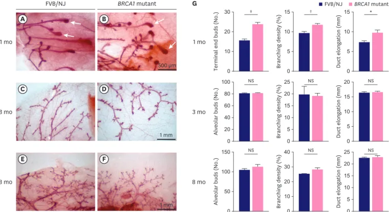
One-month-old BRCA1 mutant mice at the pubertal stage of mammary gland development showed a significantly higher number of terminal end buds (23.8 ± 1.0 vs. 15.6 ± 0.8, p = 0.0002), branching density (11.7 ± 0.4 vs. 9.6 ± 0.5%, p = 0.0082), and duct elongation (9.7 ± 0.7 vs. 7.3
± 0.4 mm, p = 0.0186), compared to FVB/NJ mice (controls) (Figure 2). All the 3-month-old mice at fertile stage from both the groups showed no difference in the number of alveolar buds (80.6 ± 1.1 vs. 80.2 ± 1.2, p = 0.8171), branching density (19.0 ± 1.2 vs. 19.7 ± 3.4%, p = 0.6411), and duct elongation (16.5 ± 0.3 vs. 16.3 ± 0.4 mm, p = 0.6386) compared to control mice. Similarly, 8-month-old mice at menopausal stage showed no significant difference in the number of alveolar buds (112.4 ± 6.7 vs. 104.6 ± 3.9, p = 0.3416), branching density (28.1
± 1.3 vs. 25.2 ± 0.4%, p = 0.06), and duct elongation (22.8 ± 0.7 vs. 22.5 ± 0.4 mm, p = 0.714) compared to control mice (Table 1).
Comparison between tumor-bearing and tumor-free BRCA1 mutant mice In the group of BRCA1 mutant mice, which was followed until 8 months, 11 out of 15 (73%) mice developed breast cancer showing histopathological features of high-grade, triple-
FVB/NJ BRCA1 mutant
G
Terminal end buds (No.) 0 10 20 30
Branching density (%)
0 5 10 15
Duct elongation (mm)
0 10 15
5
Alveolar buds (No.)
0 20 40 60 100
Branching density (%)
0 5 10 15 25
Duct elongation (mm)
0 5 10 20
80 20 15
Alveolar buds (No.)
0 50 100 150
Branching density (%)
0 10 20 40 30
Duct elongation (mm)
0 5 15 25
10 20
1 mo
3 mo
8 mo 1 mo
3 mo
8 mo
A B
C D
E F
FVB/NJ BRCA1 mutant
500 µm
1 mm
1 mm
NS NS NS
NS NS NS
‡ † *
Figure 2. Histologic images of mammary gland development between FVB/NJ (A, C, E) and BRCA1 mutant (B, D, F) mice. The quantification results at each developmental stage are shown as bar graphs (G). Arrows shown in (A) and (B) denote terminal end buds. Values are reported as the mean ± standard deviation.
NS = not significant.
*p < 0.05, †p < 0.01, ‡p < 0.001.
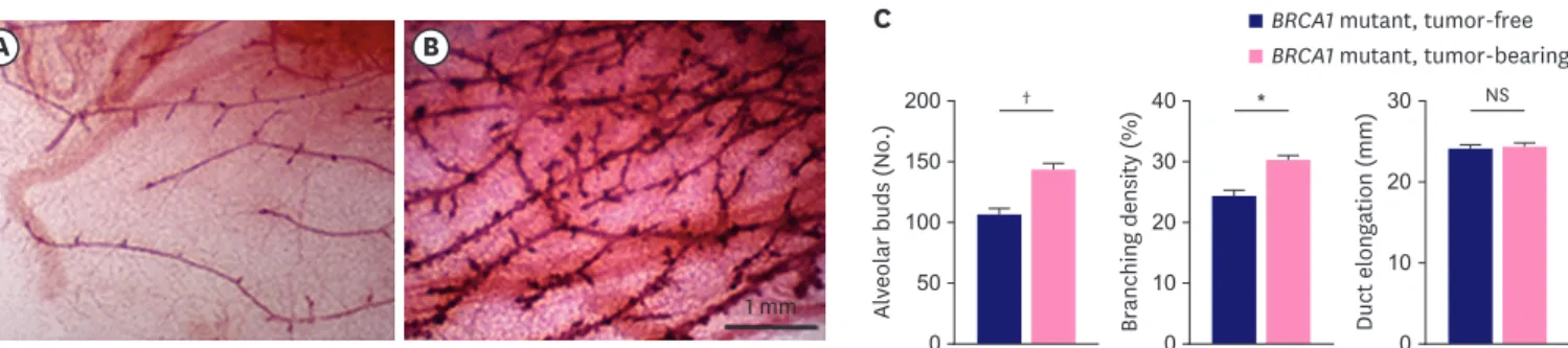
negative breast cancer. Moreover, the tumor-bearing group (11 mice) showed a significantly higher number of alveolar end buds (142.7 ± 5.5 vs. 105.5 ± 5.4, p = 0.0008) and branching density (30.0 ± 1.0 vs. 24.1 ± 1.1%, p = 0.008) compared to the tumor-free group (4 mice) (Figure 3; Table 2). However, no difference was observed in duct elongation between the two groups (23.9 ± 0.6 vs. 23.6 ± 0.6 mm, p = 0.8099).
DISCUSSION
In this study, we first quantified histological indicators of mammary gland development in both normal and BRCA1 mutant mice throughout each developmental stage to evaluate the relationship between mammary gland development and risk of breast cancer. The results showed that BRCA1 mutant mice tended to have increased terminal end buds, branching density, and ductal elongation compared with controls at one month of age, indicating precocious development of mammary glands in the BRCA1 mutant mice.
However, no difference in these characteristics was observed in the 3-month (fertile) and 8-month (menopausal)-old groups. Our results are concordant with those of previous studies regarding mammary gland development in BRCA1 mutant mice and suggest that the normal function of the BRCA1 gene is temporally and developmentally restricted [7,8].
Table 1. Comparison of mammary development parameters between BRCA1 mutant and FVB/NJ mice
Mice Number of terminal end buds
or alveolar buds Branching density (%) Duct elongation (mm)
FVB/NJ, 1-month-old (n = 5) 15.6 ± 0.8 9.6 ± 0.5 7.3 ± 0.4
BRCA1 mutant, 1-month-old (n = 5) 23.8 ± 1.0 11.7 ± 0.4 9.7 ± 0.7
p-value 0.0002 0.0082 0.0186
FVB/NJ, 3-month-old (n = 5) 80.2 ± 1.2 19.7 ± 3.4 16.3 ± 0.4
BRCA1 mutant, 3-month-old (n = 5) 80.6 ± 1.1 19.0 ± 1.2 16.5 ± 0.3
p-value 0.8171 0.6411 0.6386
FVB/NJ, 8-month-old (n = 5) 104.6 ± 3.9 25.2 ± 0.4 22.5 ± 0.4
BRCA1 mutant, 8-month-old (n = 5) 112.4 ± 6.7 28.1 ± 1.3 22.8 ± 0.7
p-value 0.3416 0.06 0.714
Values are means ± standard deviation.
BRCA1 mutant, tumor-free BRCA1 mutant, tumor-bearing
C
Alveolar buds (No.)
0 50 100 150
200 †
Branching density (%)
0 10 20 30
40 *
Duct elongation (mm)
0 10 20
30 NS
1 mm
A B
Figure 3. Histologic images of a mammary gland in 8-month-old, tumor-free (A) and tumor-bearing (B) BRCA1 mutant mice. The quantification results are shown as bar graphs (C). Values are reported as the mean ± standard deviation.
NS = not significant.
*p < 0.01, †p < 0.001.
Table 2. Comparison of mammary development parameters between tumor-free and tumor-bearing BRCA1 mutant mice
Mice Number of alveolar buds Branching density (%) Duct elongation (mm)
BRCA1 mutant, tumor-free, 8-month-old (n = 4) 105.5 ± 5.4 24.1 ± 1.1 23.6 ± 0.6
BRCA1 mutant, tumor-bearing, 8-month-old (n = 11) 142.7 ± 5.5 30.0 ± 1.0 23.9 ± 0.6
p-value 0.0008 0.008 0.8099
Values are means ± standard deviation.
The BRCA1 gene is upregulated in the breast during puberty than during menopause, and a mutation in the BRCA1 gene results in marked lobuloalveolar development, particularly in the proliferation of terminal end buds [7,10]. We followed the BRCA1 mutant mice up to the menopausal stage and compared the histological findings of normal mammary glands between tumor-bearing and tumor-free BRCA1 mutant mice. The results showed that BRCA1 mutant mice with more alveolar buds and higher branching density were more likely to develop breast cancer. This finding is concordant with the Mayo Benign Breast Disease Cohort study, which showed that reduced levels of age-related lobular involution in normal breast tissue were associated with an increased risk of breast cancer [14]. In our study, there was no difference in mammary duct elongation between tumor-bearing and tumor-free BRCA1 mutant mice. This could be explained by the fact that in both mice and humans, age- related mammary gland involution begins in the lobules and is pronounced, whereas duct elongation occurs at a later stage; therefore, it is affected to a lesser extent [22,23].
Early menarche, late menopause, nulliparity, obesity after menopause, taking hormones, family history of breast cancer, BRCA1-gene mutation, and chest radiation therapy in childhood are known breast cancer risk factors that interact with each other and determine the onset of breast cancer in a woman [2]. Our experiments support the importance of two new risk factors found in BRCA1-related breast cancer: early mammary gland development and reduced levels of age- related lobular involution. These features can be measured quantitatively using breast biopsy tissues and can be incorporated along with the estimation of mammographic breast density into risk assessment models to improve prediction of breast cancer risk [14-16,23]. Accurately identifying individuals at an increased risk for breast cancer is important, because more intense screening and specific interventions, such as risk-reducing surgery or medications, can be used more effectively [24]. In epidemiological studies, an important risk factor to be considered is an early age at thelarche, which shows a continuous trend towards early development [25].
Additional studies on the interaction of genes, hormones, and environment at puberty are needed, because our study suggests that mutations in predisposing genes related to breast cancer, such as BRCA1, could be a cause of early mammary gland development [26,27].
There are some limitations of our study. First, molecular mechanisms linking altered mammary gland development and high rates of breast cancer in BRCA1 mutant mice were not investigated. Recently, receptor activator of NF-κB (RANK) signaling by BRCA1 mutation was suggested to be a cellular mechanism required for hormone-independent abnormal proliferation of mammary luminal progenitors and an increased risk of breast cancer [28,29].
The RANK ligand is a potential target for breast cancer prevention in BRCA1 mutation carriers [30]. Second, only nulliparous mice were used in our study. Thus, the effects of pregnancy and lactation on delayed age-related mammary gland involution and breast cancer risk could not be determined. High levels of BRCA1 expression have been reported in rapidly growing alveolar buds during pregnancy [7]. Third, the effect of BRCA2 mutation that is known to be more closely related to hormone receptor-positive breast cancers was not compared in our study. In a previous study, however, no differences were observed in the expression of BRCA1 and BRCA2 genes between normal and mutant mice at pubertal and fertile stages [8].
In conclusion, pubertal mammary gland development occurred at an early stage in BRCA1 mutant mice, and delayed age-related mammary gland involution was associated with breast cancers.
If these findings are confirmed in human studies, measurement of early mammary gland
development or age-related lobular involution can be incorporated into risk assessment models,
to improve breast cancer risk prediction for individual women. Further research on the molecular
mechanisms linking mammary gland development and breast cancer risk is needed to develop a physiological approach for prevention of breast cancer in carriers of the BRCA1 mutation.
The paper is a secondary publication of the ‘Histological Findings of Mammary Gland Development and Risk of Breast Cancer in BRCA1 Mutant Mouse Models’ published by the Journal of the Korean Society for Breast Screening (16(2):45-52) in 2019 (in Korean). The Editor-in-Chiefs of the both Journals have agreed on this secondary publication for more broad readership.
ACKNOWLEDGMENTS
We thank Dr. Kyoung-Won Cho for offering her expert advice on the analysis of mouse mammary gland development.
REFERENCES
1. Kuchenbaecker KB, Hopper JL, Barnes DR, Phillips KA, Mooij TM, Roos-Blom MJ, et al. Risks of breast, ovarian, and contralateral breast cancer for BRCA1 and BRCA2 mutation carriers. JAMA 2017;317:2402-16.
PUBMED | CROSSREF
2. Narod SA. Modifiers of risk of hereditary breast cancer. Oncogene 2006;25:5832-6.
PUBMED | CROSSREF
3. Atchley DP, Albarracin CT, Lopez A, Valero V, Amos CI, Gonzalez-Angulo AM, et al. Clinical and pathologic characteristics of patients with BRCA-positive and BRCA-negative breast cancer. J Clin Oncol 2008;26:4282-8.
PUBMED | CROSSREF
4. Hall JM, Lee MK, Newman B, Morrow JE, Anderson LA, Huey B, et al. Linkage of early-onset familial breast cancer to chromosome 17q21. Science 1990;250:1684-9.
PUBMED | CROSSREF
5. Drost RM, Jonkers J. Preclinical mouse models for BRCA1-associated breast cancer. Br J Cancer 2009;101:1651-7.
PUBMED | CROSSREF
6. Liu X, Holstege H, van der Gulden H, Treur-Mulder M, Zevenhoven J, Velds A, et al. Somatic loss of BRCA1 and p53 in mice induces mammary tumors with features of human BRCA1-mutated basal-like breast cancer. Proc Natl Acad Sci U S A 2007;104:12111-6.
PUBMED | CROSSREF
7. Marquis ST, Rajan JV, Wynshaw-Boris A, Xu J, Yin GY, Abel KJ, et al. The developmental pattern of BRCA1 expression implies a role in differentiation of the breast and other tissues. Nat Genet 1995;11:17-26.
PUBMED | CROSSREF
8. Chodosh LA. Expression of BRCA1 and BRCA2 in normal and neoplastic cells. J Mammary Gland Biol Neoplasia 1998;3:389-402.
PUBMED | CROSSREF
9. Xu X, Wagner KU, Larson D, Weaver Z, Li C, Ried T, et al. Conditional mutation of BRCA1 in mammary epithelial cells results in blunted ductal morphogenesis and tumour formation. Nat Genet 1999;22:37-43.
PUBMED | CROSSREF
10. Hoshino A, Yee CJ, Campbell M, Woltjer RL, Townsend RL, van der Meer R, et al. Effects of BRCA1 transgene expression on murine mammary gland development and mutagen-induced mammary neoplasia. Int J Biol Sci 2007;3:281-91.
PUBMED | CROSSREF
11. Russo J, Tay LK, Russo IH. Differentiation of the mammary gland and susceptibility to carcinogenesis.
Breast Cancer Res Treat 1982;2:5-73.
PUBMED | CROSSREF
12. Jansen-van der Weide MC, Greuter MJ, Jansen L, Oosterwijk JC, Pijnappel RM, de Bock GH. Exposure to low-dose radiation and the risk of breast cancer among women with a familial or genetic predisposition: a meta-analysis. Eur Radiol 2010;20:2547-56.
PUBMED | CROSSREF
13. Hartmann LC, Sellers TA, Frost MH, Lingle WL, Degnim AC, Ghosh K, et al. Benign breast disease and the risk of breast cancer. N Engl J Med 2005;353:229-37.
PUBMED | CROSSREF
14. McKian KP, Reynolds CA, Visscher DW, Nassar A, Radisky DC, Vierkant RA, et al. Novel breast tissue feature strongly associated with risk of breast cancer. J Clin Oncol 2009;27:5893-8.
PUBMED | CROSSREF
15. Baer HJ, Collins LC, Connolly JL, Colditz GA, Schnitt SJ, Tamimi RM. Lobule type and subsequent breast cancer risk: results from the Nurses' Health Studies. Cancer 2009;115:1404-11.
PUBMED | CROSSREF
16. Vellal AD, Sirinukunwattan K, Kensler KH, Baker GM, Stancu AL, Pyle ME, et al. Deep learning image analysis of benign breast disease to identify subsequent risk of breast cancer. JNCI Cancer Spectr 2021;5:pkaa119.
PUBMED | CROSSREF
17. Tolg C, Cowman M, Turley EA. Mouse mammary gland whole mount preparation and analysis. Bio Protoc 2018;8:e2915.
PUBMED | CROSSREF
18. Muñoz-de-Toro M, Markey CM, Wadia PR, Luque EH, Rubin BS, Sonnenschein C, et al. Perinatal exposure to bisphenol-A alters peripubertal mammary gland development in mice. Endocrinology 2005;146:4138-47.
PUBMED | CROSSREF
19. Blacher S, Gérard C, Gallez A, Foidart JM, Noël A, Péqueux C. Quantitative assessment of mouse mammary gland morphology using automated digital image processing and TEB detection.
Endocrinology 2016;157:1709-16.
PUBMED | CROSSREF
20. Stanko JP, Easterling MR, Fenton SE. Application of Sholl analysis to quantify changes in growth and development in rat mammary gland whole mounts. Reprod Toxicol 2015;54:129-35.
PUBMED | CROSSREF
21. Paine IS, Lewis MT. The terminal end bud: the little engine that could. J Mammary Gland Biol Neoplasia 2017;22:93-108.
PUBMED | CROSSREF
22. Ghosh K, Hartmann LC, Reynolds C, Visscher DW, Brandt KR, Vierkant RA, et al. Association between mammographic density and age-related lobular involution of the breast. J Clin Oncol 2010;28:2207-12.
PUBMED | CROSSREF
23. Ghosh K, Vachon CM, Pankratz VS, Vierkant RA, Anderson SS, Brandt KR, et al. Independent association of lobular involution and mammographic breast density with breast cancer risk. J Natl Cancer Inst 2010;102:1716-23.
PUBMED | CROSSREF
24. Britt KL, Cuzick J, Phillips KA. Key steps for effective breast cancer prevention. Nat Rev Cancer 2020;20:417-36.
PUBMED | CROSSREF
25. Goldberg M, D'Aloisio AA, O'Brien KM, Zhao S, Sandler DP. Pubertal timing and breast cancer risk in the Sister Study cohort. Breast Cancer Res 2020;22:112.
PUBMED | CROSSREF
26. Terry MB, Michels KB, Brody JG, Byrne C, Chen S, Jerry DJ, et al. Environmental exposures during windows of susceptibility for breast cancer: a framework for prevention research. Breast Cancer Res 2019;21:96.
PUBMED | CROSSREF
27. Hamilton AS, Mack TM. Puberty and genetic susceptibility to breast cancer in a case-control study in twins. N Engl J Med 2003;348:2313-22.
PUBMED | CROSSREF
28. Sau A, Lau R, Cabrita MA, Nolan E, Crooks PA, Visvader JE, et al. Persistent activation of NF-κB in BRCA1- deficient mammary progenitors drives aberrant proliferation and accumulation of DNA damage. Cell Stem Cell 2016;19:52-65.
PUBMED | CROSSREF
29. Nolan E, Vaillant F, Branstetter D, Pal B, Giner G, Whitehead L, et al. RANK ligand as a potential target for breast cancer prevention in BRCA1-mutation carriers. Nat Med 2016;22:933-9.
PUBMED | CROSSREF
30. Kotsopoulos J, Singer C, Narod SA. Can we prevent BRCA1-associated breast cancer by RANKL inhibition?
Breast Cancer Res Treat 2017;161:11-6.
PUBMED | CROSSREF