1786
대한안과학회지 2016년 제 57 권 제 11 호 J Korean Ophthalmol Soc 2016;57(11):1786-1789 ISSN 0378-6471 (Print)⋅ISSN 2092-9374 (Online)
http://dx.doi.org/10.3341/jkos.2016.57.11.1786
Case Report
결막에 발생한 원발거대세포종 1예
A Case of Primary Conjunctival Giant Cell Tumor
김태훈⋅오하나
Tae Hoon Kim, MD, Ha Na Oh, MD
인제대학교 의과대학 부산백병원 안과학교실
Department of Ophthalmology, Busan Paik Hospital, Inje University College of Medicine, Busan, Korea
Purpose: To report a case of primary conjunctival giant cell tumor (GCT).
Case summary: A 67-year-old female visited our clinic with the chief complaint of a 10-year-history of conjunctival mass in the left eye. The patient had no marked changes in the mass size, and her visual acuity and intraocular pressure were within the normal range. The protruding conjunctival mass invaded the limbal area at the 8 o’clock direction in the left eye and was 5 × 4 × 2 mm in size. Moreover, the pink-colored mass had a lobulated shape with a well-defined margin. In the adjacent mass region, con- current presence of the conjunctival injection was observed. However, the patient did not exhibit pain or tenderness. We per- formed wide excision of the conjunctival mass concomitantly with amniotic membrane transplantation. Then, histopathological examinations and immunohistochemical staining of the surgical site were performed. On histopathology, the patient had findings suggestive of GCT. Additionally, immunohistochemistry was positive for CD68 and vimentin. leading to the final diagnosis of GCT.
Conclusions: To our knowledge, this is the first case of GCT of the conjunctiva, which has not been described in the literature.
Our case highlights the importance of differential diagnosis from other protuberant conjunctival tumors. A complete removal of GCT of the conjunctiva and a recovery of aesthetic outcomes can be achieved by surgical excision of the mass.
J Korean Ophthalmol Soc 2016;57(11):1786-1789
Keywords: Conjunctival mass, Primary conjunctival giant cell tumor
■Received: 2016. 6. 16. ■ Revised: 2016. 8. 7.
■Accepted: 2016. 10. 20.
■Address reprint requests to Ha Na Oh, MD
Department of Ophthalmology, Inje University Busan Paik Hospital, #75 Bokji-ro, Busanjin-gu, Busan 47392, Korea Tel: 82-51-890-6015, Fax: 82-51-890-6329
E-mail: [email protected]
ⓒ2016 The Korean Ophthalmological Society
This is an Open Access article distributed under the terms of the Creative Commons Attribution Non-Commercial License (http://creativecommons.org/licenses/by-nc/3.0/) which permits unrestricted non-commercial use, distribution, and reproduction in any medium, provided the original work is properly cited.
거대세포종은 전체 골종양의 5% 가량을 차지하는 양성 종양이다. 그러나 거대세포종은 국소적으로는 공격적으로 재발되거나 드물게 전이를 일으키기도 한다.1-3 30-40대의 젊은 성인에서 많이 발생하며, 주로 장골의 골단부에 호발 하고 드물게 골간단에 생기는 것으로 알려져 있다.3-5
그러나 거대세포종이 안구 및 안구부속기관에서 발생했 던 경우는 아직까지 국내외에 보고된 바가 없다. 이에 저자
들은 결막에 발생한 거대세포종을 광범위 절제술 및 양막 이식술을 통해 치료한 증례가 있어 문헌고찰과 함께 이를 보고하고자 한다.
증례보고
67세 여자 환자가 10년 전 발생한 좌안의 결막 종괴로 인한 불편감을 주소로 내원하였다. 종괴 크기의 갑작스런 변화는 없었다고 하며, 압통 및 통증은 없었다. 안과적으로 1년 전 증식성 당뇨망막병증으로 인한 좌안유리체 출혈과 노년성 백내장으로 본원에서 좌안 유리체절제술, 초음파수 정체유화술 및 인공수정체 삽입술을 시행 받았고, 다른 전 신질환은 없었다. 내원 당시 교정시력은 우안 0.63, 좌안
1787 - 김태훈⋅오하나 : 결막에 발생한 원발거대세포종 1예 -
A B
C D
Figure 2. Histological and Immunohistochemical examination of conjunctival giant cell tumor. (A) Multinucleated giant cells (*)
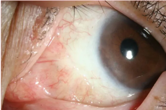
interspersed among mononuclear cells (arrows) (H&E ×200). (B) Positive staining for CD68 (arrow) (×200). (C) Positive staining for Vimentin (arrow) (×200). (D) Negative staining for smooth muscle actin (×200).Figure 1. Photograph of the conjunctival mass at medial side
of left eye. W ell circumscribed, lobulated, protruding pinkish mass.1.0이었고, 안압은 정상이었다. 세극등 검사에서 좌안 8시 방향에 각막윤부를 침범하여 5 × 4 × 2 mm 크기로 융기되 어 있는 결막 종괴가 관찰되었다. 종괴는 경계가 명확하고 분홍빛을 띤 소엽화된 형태를 보였으며 주위로 중등도의 결막 충혈이 동반되어 있었다(Fig. 1).
결막 종괴에 대한 광범위절제술 및 영구적 양막이식술을
시행하고 절제한 조직에 대해 조직병리검사를 시행하였다. Hematoxylin and eosin 염색에서 단핵세포들 사이에 산재 되어 있는 다수의 다핵 거대세포들이 관찰되었고, 추가로 시행한 면역조직화학염색 결과에서 CD68과 vimentin에는 양성, smooth muscle actin에는 음성을 나타내어(Fig. 2) 결 막에 발생한 거대세포종을 확진하였다.
결막 이외의 다른 원발병소 및 전이병소 확인을 위해 흉 부 X-ray와 정형외과에서 시행한 신체진찰에서 특이소견이 발견되지 않아 흉부 전산화단층촬영검사와 bone scan과 같 은 추가적인 검사는 시행하지 않았다. 수술 후 6개월째, 국 소재발이나 원격전이는 발생하지 않았으며 안구불편감의 증상 없이 안정적인 상태를 유지하고 있다(Fig. 3).
고 찰
거대세포종은 주로 근골격계에서 발생하는 원발성 양성 종양이나, 드물게 근골격계 이외의 위장관, 간, 췌장, 유방, 난소, 갑상선, 신장, 폐 등에서 원발병소가 발생하는 경우도 보고된 바 있다.1,6 젊은 성인에서 호발하고 대체로 여성에 서 1.5배 정도 더 많이 발생하는 것으로 알려져 있다.1,5-7 대
1788
- 대한안과학회지 2016년 제 57 권 제 11 호 -
Figure 3. Conjunctival appearance at 6months after surgical
resection. The mass was completely removed and there was no local recurrence.부분의 근골격계 거대세포종은 빠르게 자라며, 골조직을 파괴시키고 주변의 연조직을 침범하는 것이 특징이다. 또 한 거대세포종은 수술적 절제를 하게 될 경우 국소재발률 이 최대 50%에 달할 정도로 높으며, 원격전이가 1-6%에서 나타나고, 이때 주로 폐로 전이된다.3,6,7 거대세포종은 재발 후 전체 1% 미만에서 악성전환이 발생하는데, 대체로 골육 종을 비롯한 육종의 형태로 관찰되며, 이런 악성전환의 경 우 예후가 매우 불량하여 신속한 진단 및 조기치료가 중요 하다.1,6
거대세포종의 분류에는 영상의학적, 조직학적, 임상적 양 상에 기초한 Ennecking staging classification이 가장 널리 사용되고 있다. Ennecking staging classification에 따르면 골거대세포종은 골내에 국한되고 증상이 없으며 bone scan 에서 비활성을 띠는 것이 특징인 ‘잠복기’, 증상이 있으면 서 bone scan에서 활성을 띠는 ‘활성기’, 골외 전이를 일으 키고 빠른 성장을 하는 ‘공격기’로 구분되며 각각은 15%, 70%, 15%의 빈도를 갖는다.7
거대세포종의 전형적인 조직학적 소견은 다수의 다핵성 거대세포가 양성의 방추형 기질세포 사이에 흩뿌려져 있는 양상으로, 이때 단핵성 방추형 기질세포의 핵과 거대세포 의 핵은 명확히 구분된다.3 거대세포종은 CD68과 vimentin 에 양성소견을 보이며, 이는 이 종양이 중간엽 기원임을 뒷 받침하는 소견으로 알려져 있다.8,9
이 종양의 주된 치료는 수술적 제거로, 병변내소파술이 나 광범위 절제술이 표준치료법으로서 많이 시행되고 있
다.3,6,7 근골격계거대세포종의 경우, 광범위 절제술을 시행
하였을 때 재발률은 낮으나 술 후 기능적 결과가 좋지 않 고, 술 후 합병증도 심하기 때문에 Ennecking staging clas- sification 상 잠복기, 활성기에 해당할 경우 병변내소파술 및 충전물 삽입술을, 그 이상일 경우 광범위 절제술을 시행
한다.7
본 증례에서 살펴본 결막 거대세포종은 돌출 및 융기되 어 있었으며, 경계가 명확하고 분홍빛을 띤 소엽화된 형태 를 보였다. 또한 주위로 중등도의 결막 충혈이 동반되어 있 었다. 결막에서 융기된 양상을 보이는 결막종양으로는 비 정형 결막모반, 결막편평세포유두종 등이 있으며, 결막 거 대세포종은 결막에 발생하는 이러한 종양들과 육안적으로 유사하게 관찰될 수 있다. 비정형 결막모반의 경우, 경계가 명확한 돌출된 형태를 보이기는 하나, 투명한 양상을 보인 다. 또한 조직검사에서 대부분의 결막모반은 상피하의 모 반세포와 모반세포소, 낭종이 관찰되는 것이 특징이다. 결 막편평세포유두종의 경우 분홍빛의 돌출된 임상양상을 보 이나, 표면이 반짝이고 투명하여 그 아래 모세혈관이 빨간 점(red dot) 형태로 모여 있는 것이 특징이며, 조직학적 소 견에서 가시세포증이 보이고 비각화성 편평상피세포가 관 찰되는 것으로 거대세포종과 감별할 수 있다.10-13
또한 결막의 육아종 역시 결막 거대세포종과 임상적으로 유사한 융기성 종괴 형태를 띨 수 있기에 감별이 필요한 질 환이다. 그러나 육아종의 경우, 조직검사에서 림프구, 형질 세포 등의 만성염증성 세포와 조직구 등이 함께 관찰되기 때문에, 염증성 소견이 전혀 없는 거대세포와 감별이 가능 하다. 본 증례를 통해 소개한 결막에 발생한 거대세포종의 경우 조직검사 결과 악성 가능성이 낮았기에 양성 종양으 로 볼 수 있으나, 전이 가능성이 있고 국소재발률이 높기 때문에, 주 원발병소인 근골격계와 호발 전이부위인 폐 등 에 대한 주의 깊은 경과관찰이 필요할 것으로 생각된다.
REFERENCES
1) Plowman RS, Nguyen BD. Primary pulmonary giant cell tumor:
(18)F-FDG PET/CT imaging. Rev Esp Med Nucl Imagen Mol 2016;35:274-6.
2) Li C, Zheng X, Ghert M, et al. Expressions and clinical sig- nificance of factors related to giant cell tumor of bone. Int J Clin Exp Med 2015;8:22509-14. eCollection 2015.
3) Sobti A, Agrawal P, Agarwala S, Agarwal M. Giant cell tumor of bone - an overview. Arch Bone Jt Surg 2016;4:2-9.
4) Yamagishi T, Kawashima H, Ogose A, et al. Disappearance of giant cells and presence of newly formed bone in the pulmonary metastasis of a sacral giant-cell tumor following denosumab treat- ment: a case report. Oncol Lett 2016;11:243-6.
5) Yu X, Guo R, Fan C, et al. Aneurysmal bone cyst secondary to a giant cell tumor of the patella: a case report. Oncol Lett 2016;
11:1481-5.
6) Skubitz KM. Giant cell tumor of bone: current treatment options.
Curr Treat Options Oncol 2014;15:507-18.
7) López-Pousa A, Martín Broto J, Garrido T, Vázquez J. Giant cell tumour of bone: new treatments in development. Clin Transl Oncol 2015;17:419-30.
1789 - 김태훈⋅오하나 : 결막에 발생한 원발거대세포종 1예 -
= 국문초록 =
결막에 발생한 원발거대세포종 1예
목적: 결막에 발생한 원발거대세포종 1예를 보고하고자 한다.
증례요약: 67세 여자 환자가 10년 전 발생한 좌안의 결막 종괴로 내원하였다. 종괴 크기의 갑작스런 변화는 없었다고 하며, 시력 및 안압은 정상이었다. 결막 종괴는 좌안 8시 방향에 각막윤부를 침범하여 5 × 4 × 2 mm 크기로 돌출되어 있었으며 경계가 명확하고 분홍빛을 띤 소엽화된 형태를 보였다. 종괴 주위로 결막 충혈이 동반되어 있었고 압통 및 통증은 없었다. 결막 종괴에 대하여 광범위 절제술 및 양막이식술과 함께 조직검사를 시행하였다. 조직병리검사에서 다핵 거대세포가 관찰되고 추가로 실시한 면역조직화학염색 결과에서 CD68과 vimentin에서 양성을 보여 거대세포종임을 최종 확인하였다.
결론: 거대세포종이 결막에 발생한 최초의 증례로 다른 융기성 결막종양과의 감별진단이 필요할 것으로 생각되며, 수술적 치료를 통 해 거대세포종의 완전제거와 미용적 회복을 얻을 수 있다.
<대한안과학회지 2016;57(11):1786-1789>
8) Wang DD, Zheng YM, Teng LH, et al. Benign giant-cell tumor of the common bile duct: a case report. World J Gastroenterol 2014;20:15448-53.
9) Temesgen WM, Wachtel M, Dissanaike S. Osteoclastic giant cell tumor of the pancreas. Int J Surg Case Rep 2014;5:175-9.
10) Manidakis N, Polyzois I, Tsialogiannis E, et al. Metastatic malig- nant melanoma of the conjunctiva: a case report. Cases J 2009;
2:125.
11) López-Meca IC, Alcaraz-Mateos E, Córdoba-Polo C, Belmonte-
Martínez J. Atypical melanocytic nevus of the limbus. A case report. Arch Soc Esp Oftalmol 2014;89:265-8.
12) Pfister H, Fuchs PG, Völcker HE. Human papillomavirus DNA in conjunctival papilloma. Graefes Arch Clin Exp Ophthalmol 1985;223:164-7.
13) Buggage RR, Smith JA, Shen D, Chan CC. Conjunctival papil- lomas caused by human papillomavirus type 33. Arch Ophthalmol 2002;120:202-4.