• 검색 결과가 없습니다.

Titanium Ions Released from Oral Casting Alloys May Contribute to the Symptom of Burning Mouth Syndrome JOMP

N/A
N/A
Protected

Academic year: 2021

Share "Titanium Ions Released from Oral Casting Alloys May Contribute to the Symptom of Burning Mouth Syndrome JOMP"

Copied!
7
0
0

로드 중.... (전체 텍스트 보기)

전체 글

(1)

pISSN 2288-9272 eISSN 2383-8493 J Oral Med Pain 2017;42(4):102-108 https://doi.org/10.14476/jomp.2017.42.4.102

Titanium Ions Released from Oral Casting Alloys May Contribute to the Symptom of Burning Mouth Syndrome

Yang Mi Park 1 , Kyung-Hee Kim 2 , Sunhee Lee 1 , Hye-Mi Jeon 3 , Jun-Young Heo 1 , Yong-Woo Ahn 1 , Soo-Min Ok 1 , Sung-Hee Jeong 1

1 Department of Oral Medicine, School of Dentistry, Pusan National University, Dental Research Institute, Institute of Translational Dental Sciences, Yangsan, Korea

2 Department of Oral Medicine, Busan Paik Hospital, Inje University College of Medicine, Busan, Korea

3 Dental Clinic Center, Pusan National University Hospital, Busan, Korea

Received October 31, 2017 Revised November 24, 2017 Accepted November 27, 2017

Purpose: Many metal ions released from dental casting alloys have been reported to influence the intraoral symptoms of oral lichen planus (OLP) and burning mouth syndrome (BMS). The aim of this study was to investigate the relationship between salivary metal ion levels and the prosthetic duration as well as to evaluate the time-dependent morbid effects of metal ions in OLP and BMS patients.

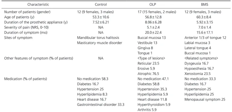
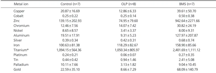
Methods: Three study groups consist of the following subjects respectively: 17 OLP patients, 12 BMS patients, and 12 patients without oral symptoms. The salivary concentrations of 13 metal ions (copper, cobalt, zinc, chromium, nickel, aluminum, silver, iron, titanium [Ti], platinum, tin, palladium, and gold) were measured by Laser Ablation Microprobe Inductively coupled Plasma Mass Spectrometry.

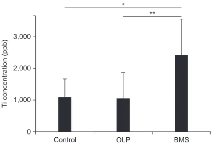
Results: The Ti ions had statistically significant differences among the groups with a pros- thetic duration of less than 5 years. There were no significant differences between all ion levels among the groups wearing dental cast alloys for over 5 years. In the BMS group, the level of Ti ions in patients with prosthetic restorations less than 5 years old were significantly high (p<0.05).

Conclusions: In the BMS group, 3-60 months during which salivary Ti levels were higher were matched with the duration of burning symptoms (15.6±17.1 months). Furthermore, Ti ions were statistically high in the oral cavity of BMS patients fitted with dental casting alloys for 5 years. These results suggest that Ti ions released from dental implants and oral prostheses could attribute to burning sensation of BMS.

Key Words: Burning mouth syndrome; Metals; Saliva; Titanium

Correspondence to:

Sung-Hee Jeong

Department of Oral Medicine, School of Dentistry, Pusan National University, 49 Busandaehak-ro, Mulgeum-eup, Yangsan 50612, Korea

Tel: +82-55-360-5242 Fax: +82-55-360-5234 E-mail: drcookie@pusan.ac.kr This study was supported by a Clinical Research Grant, Pusan National University Dental Hospital (2017).

JOMP Journal of Oral Medicine and Pain

Copyright Ⓒ 2017 Korean Academy of Orofacial Pain and Oral Medicine. All rights reserved.

CC

This is an open-access article distributed under the terms of the Creative Commons Attribution Non-Commercial License (http://creativecommons.org/licenses/by-nc/4.0/), which permits unrestricted non-commercial use, distribution, and reproduction in any medium, provided the original work is properly cited.

INTRODUCTION

Dental casting alloys placed in the oral cavity for a long time may cause adverse effects as a consequence of corro- sion. 1-3) The release of metal ions into oral cavity may in- duce cytotoxicity, allergies, and mutagenesis. Oral lichen planus (OLP) and burning mouth syndrome (BMS) are some of the common complaints reported by patients to the oral

medicine department and could be the result of metals.

OLP is a chronic immunological oral mucosal disease

with many clinical manifestations. 4,5) The exact etiology of

OLP is not known, but several factors have been proposed

for the cause including genetic background, dental mate-

rials, drugs, autoimmunity, and food allergies. 5) Previous

studies on OLP emphasized the role of contact allergy by

documenting metal sensitivities in 0% to 62% of patients. 6)

(2)

These metals included amalgam (mercury), gold, cobalt, tin, and palladium. 6-10)

BMS is characterized by burning sensations of the nor- mally visible oral mucosa. 11) Etiology of primary BMS re- mains unclear, but secondary BMS may be caused by lichen planus, candidiasis, hormonal disturbances, psychosocial stressors, vitamin or nutritional deficiencies, diabetes, dry mouth, contact allergies, galvanism, parafunctional habits, cranial nerve injuries and many other factors. 12,13) Previous studies have reported low salivary magnesium levels 14) and low serum zinc levels 15,16) in BMS patients.

The intraoral side effects from dental alloys were gingi- vitis, burning tongue, erythematous palate or tongue and lichenoid reactions. 17) These adverse effects could be influ- enced by types of casting alloys, amount of metal ions re- leased from dental alloys, prosthodontic exposure time and so on. In vitro experiments on local toxicity of casting al- loy, cell toxicity increases in time and dose-dependent pat- terns. 18,19) Alloys that release metal ions over longer periods tend to cause more toxic effects and clinically long-term released metallic ions may trigger intraoral toxic reactions at relatively low concentrations. 19,20)

The hypothesis of this study was that dental prostheses will release more metal ions over a longer period of time and that the ions will contribute to oral symptoms of OLP and BMS. Therefore, the relationship between salivary met- al ion levels and the prosthetic duration in OLP, BMS and the control group were evaluated. An analysis of the con- centration of salivary metal ions with the period of dental prostheses was also performed to explore the effects of met- al ions in OLP and BMS.

MATERIALS AND METHODS

1. Participants

This study was conducted with 41 patients who visited the Department of Oral medicine, Pusan National University Dental Hospital (Yangsan, Korea) in 2014. There were 17 patients (mean age±standard deviation [SD], 56.8±12.8 year, 15 females and 2 males) in the OLP group, 12 patients (mean age±SD, 60.3±8.4 year, 9 females and 3 males) in the BMS group and 12 patients without oral symptoms (mean age±SD, 53.3±10.6 year, 9 females and 3 males) in the

control group. Every patient had more than one prosthetic appliance on his or her teeth. There were 12 patients with amalgam restorations, 34 patients with gold crowns, 19 pa- tients with porcelain-fixed metal crowns, and 13 patients with implants. There were 40 teeth with amalgam restora- tions, 141 teeth with gold crowns, 87 porcelain-fixed metal crowns, and 45 implants (Table 1).

The patients were asked how long they had been wear- ing their fixed prostheses. For those who had more than one casting alloy, the duration was calculated based on the oldest one because the first dental alloys had been intra- orally corroded. This study focused on the relation between the concentration of oral metal ions and the exposure time.

Thus, the type, number, surface area and thickness of dental alloys were not considered.

According to the duration of oral prosthetics, patients were divided into 6 groups; OLP (≤5) group (oral prosthe- sis for less than 5 years), OLP (>5) group (oral prosthesis for more than 5 years), BMS (≤5), BMS (>5), CON (≤5), and CON (>5) group. Clinical and histopathological criteria of OLP were applied to the inclusion criteria for OLP group in this study. 4,21) The inclusion criteria for BMS group consisted of complaints of a burning sensation in oral mucosa and tongue in the normal form, normal salivary flow rate (un- stimulated whole saliva >0.1 mL/min and stimulated whole saliva >0.5-0.7 mL/min), and normal results in laboratory tests of complete blood counts, liver and renal function test (total protein, albumin, alkaline phosphatase, aspartate ami- notransferase, alanine aminotransferase, blood urea nitro- gen, and creatine), thyroid function tests (T3, T4, and thy- roid stimulating hormone), vitamin B12, folate, serum iron, ferritin, and zinc levels. 22) In the control group, patients had mandibular torus, halitosis and masticatory muscle disorder that had no oral visible lesions or oral pain. The exclusion criteria for all three groups included having serious system- ic diseases (e.g., uncontrolled medical conditions, cancer) or inability of communicating. The study protocol was ap- proved by the Institutional Review Board of Pusan National University Dental Hospital (#PNUDH-2015-015).

2. Collection of Whole Saliva Samples

Patient’s informed consents were obtained and saliva

samples were accumulated from all patients at their first

(3)

dental visit. Subjects collected their unstimulated whole sa- liva in a polypropylene tube for 3 minutes. The saliva sam- ples were immediately stored in 80 o C deep freezer until assay.

3. Analysis of Metal Ions

The 13 metal ions mainly contained in current dental prosthetic supplements were selected for this study. 19) These includes copper (Cu), cobalt (Co), zinc (Zn), chromium (Cr), nickel (Ni), aluminum (Al), silver (Ag), iron (Fe), titanium (Ti), platinum (Pt), tin (Sn), palladium (Pd), and gold (Au) ions.

To evaluate the metal levels in saliva, samples were dilut- ed with distilled water up to 3 mL. The ions in saliva were measured by Laser Ablation Microprobe Inductively coupled Plasma Mass Spectrometry (LAM-ICP-MS, Optima8300;

Perkin-Elmer, Shelton, CT, USA). As the amount of crude saliva samples were around 0.3-2.5 mL, the data was re- vised by each diluted ratio.

4. Statistical Analysis

The data did not follow the normal distribution so non- parametric tests were used in this study. The Kruskal-Wallis test was used to find meaningful differences between the

groups. The Mann-Whitney U test was used to determine sig- nificance of the OLP and BMS group compared to the control group. p-values less than 0.05 were considered statistically significant. Statistical analyses were performed using IBM SPSS Statistics ver. 20.0 (IBM Co., Armonk, NY, USA).

RESULTS

1. Prosthetic Durations of the OLP, BMS, and Control Patients

The mean±SD duration of prosthetic appliance was 8.06±6.28 years in OLP group, 5.92±3.15 years in BMS group and 7.52±6.21 years in control group (Table 1).

The mean±SD duration of oral symptom was 20.0±22.4 months in OLP group and 15.6±17 months in BMS group. Prostheses were observed in the oral cavity be- fore symptoms appeared in both the OLP and BMS groups.

Information of other clinical features and medications is given in Table 1.

2. Metal Ion Concentrations according to Prosthetic Duration

The prosthetic time of each patient was based on the

Table 1. Characteristics of the OLP, BMS, and control groups

Characteristic Control OLP BMS

Number of patients (gender) 12 (9 females, 3 males) 17 (15 females, 2 males) 12 (9 females, 3 males)

Age of patients (y) 53.3±10.6 56.8±12.8 60.3±8.4

Duration of the prosthetic appliance (y) 7.52±6.21 8.06±6.28 5.92±3.15

Severity of pain (NRS, 0-10) NA 5.1±2.4 7.0±1.4

Duration of symptom (mo) NA 20.0±22.4 15.6±17.1

Sites of symptom Mandibular torus halitosis Buccal mucosa 13 Anterior 1/3 of tongue 12

Masticatory muscle disorder Vestibule 13 Labial mucosa 3

Gingiva 8 Lateral tongue 4

Tongue 1 Buccal mucosa 1

Other features of symptom (% of patients) NA <Type of lesions> <Related symptoms>

Reticular 23.5 Dysgeusia 16.7

Erosive 5.9 Hypoesthesia 16.7

Atrophic 76.5 Xerostomia 23.5

Medication (% of patients) No medication 58.3 No medication 47.1 No medication 33.3

Diabetes 16.7 Diabetes 58.8 Diabetes 16.7

Hypertension 25 Hypertension 35.3 Hypertension 25

Hyperlipidemia 8.3 Hyperlipidemia 5.9 Hyperlipidemia 25 Heart disease 16.7 Heart disease 11.8 Menopausal symptom 25 Gastrointestinal disorder 33.3 Hyperthyroidism 5.9

Arthritis 5.9 OLP, oral lichen planus; BMS, burning mouth syndrome; NRS, numerical rating scale; NA, not applicable.

Values are presented as mean±standard deviation.

(4)

duration of the oldest casting alloy on his or her teeth.

Salivary Cu, Co, Zn, Cr, Ni, Al, Ag, Fe, Ti, Pt, Sn, Pd, and Au ions were used to check the relation between concentra- tions of metal ions and time. Ti ions showed statistical dif- ference between groups (p<0.05) (Fig. 1). There was no cor- relation between other ions and prosthetic duration.

3. Ti Ion Concentrations from Saliva Samples

In comparison of 13 ions, only Ti ions show a clear dif- ference among groups with prosthetic periods of less than 5 years (Table 2). Groups with oral dental cast alloys of over 5

years showed no difference in ion concentrations (data not shown). The Ti ions had statistically significant differences among the groups with below 5 years of prosthetic dura- tion (Fig. 2). There was no significant difference among the groups wearing dental cast alloys of more than 5 years.

4. Ti Ion Concentration in BMS (≤5) Group

To distinguish OLP and BMS groups from control group by their levels of Ti ions, three groups were analyzed by the

Table 2. The concentrations of metal ions among the groups with below 5 years of prosthetic duration

Metal ion Control (n=7) OLP (n=8) BMS (n=7)

Copper 20.87±16.69 12.86±6.33 39.61±50.70

Cobalt 0.25±0.22 0.25±0.14 0.50±0.38

Zinc 139.15±202.93 74.95±79.60 942.64±2271.66

Chromium 12.46±7.56 14.07±7.42 30.82±24.19

Nickel 8.65±8.57 3.41±3.37 8.00±9.31

Aluminum 19.51±17.91 9.31±5.23 127.97±207.87

Silver 0.39±0.34 0.42±0.31 0.68±0.74

Iron 100.63±81.38 118.29±82.67 158.90±85.66

Titanium* 1,094.15±564.36 1,050.34±805.97 2,401.69±1,111.12

Platinum 0.24±0.21 0.06±0.07 0.27±0.35

Tin 0.44±0.42 0.94±1.46 2.41±5.08

Palladium 10.11±7.66 3.13±1.82 9.04±10.45

Gold 22.59±35.10 8.66±7.29 68.09±140.79

OLP, oral lichen planus; BMS, burning mouth syndrome.

Values are presented as mean±standard deviation (ppb).

Kruskal-Wallis was performed to compare the concentrations of metal ion between groups.

*Significant difference between the groups (p<0.05).

Fig. 1. The relationship between Ti ion concentrations and the prosthetic duration. Ti ions in BMS group displayed a tendency to exhibit higher levels compared to other groups (p<0.05). Ti, titanium; OLP, oral lichen planus; BMS, burning mouth syndrome;

CONT, control.

4,000 3,000 2,000 1,000 0

Ti concentration (ppb)

0 5 10

Duration (y)

OLP BMS CONT

Fig. 2. Comparisons between Ti concentrations and period of prostheses based on 5 years. Statistically significant differences measured by Kruskal-wallis test. The Ti levels had meaningful differences among the groups with below 5 years of prosthetic duration (*p=0.016). There was no significant difference among the groups wearing dental cast alloys of more than 5 years (p=0.565).

Ti, titanium; OLP, oral lichen planus; BMS, burning mouth syndrome.

3,000 2,000 1,000 0

Ti concentration (ppb)

Control OLP BMS

<5 years >5 years

*

Control OLP BMS

(5)

Mann-Whitney U test. Ti ion concentration of the BMS (≤5) group was higher than that of the OLP (≤5) and control (≤5) groups (p<0.05) (Fig. 3).

DISCUSSION

In the oral cavity, a temperature of 37 o C and high humid- ity are maintained and electrolytes are always present in the saliva. 18) Also there are numerous chemical and physi- cal reactions as well as metabolism of about 30 species of bacteria. 23,24) Therefore, it can be said that a dental prosthe- sis is always exposed to a corrosive environment influenced by salivary pH, glucose levels and local galvanism. 18,25-27) Corrosion of dental cast alloys produces the release of metal ions, which may cause toxic effects or allergic reactions. 2,19) The intraoral symptoms were gingival swelling and ery- thema, mucosal pain and lichenoid reactions. 28) Previous studies have reported the salivary metal concentrations in patients with oral diseases, but there is no data about in vivo changes of metal ion concentrations corresponding to time. This study provided more meaningful results because the concentrations of salivary metal ions were analyzed ac- cording to period of dental prostheses in the OLP, BMS, and control group.

The data from this study reported the levels of metal ions released from dental cast alloys according to the prosthetic duration. The Ti ion concentrations in the BMS group were

significantly higher than other groups. Particularly, BMS group with prosthetic duration of lower than 5 years had significantly high Ti concentration. In BMS group, the in- creased section of 3-60 months coincided with the dura- tion of burning symptoms (15.6±17.1 months). Many other studies support this suggestion. Patients with complaints or symptoms in the oral cavity in relation to dental cast alloys mainly reported burning mouth (72%), metal taste (56%), and electric sensation (44%). 17) In other studies, subjective oral complaints like burning mouth and metal taste were related to dental alloy components, too. 29-32)

When dental cast appliances were fitted in to the oral cavity for 5 years, salivary concentrations of Ti ions in the BMS group were statistically higher than those of the OLP and control groups. As Ti has high corrosion resistance compatible with physiologic conditions, it is widely ap- proved as an excellent metal for in vivo implantation. 33-37) However, if the protective oxide layer of Ti is peeled, it can become corrosive like many other metals. The Ti ions and particles may function as an allergen that can cause a hy- peractive reaction in adjacent oral tissue and even in other remote other organs and biological systems such as lymph nodes, lungs and serum. 38,39) The inflammatory response to metallic and polymeric debris in lymph nodes is followed by the immune activation of macrophages and the associ- ated production of cytokines. 40) In sheep model, failed den- tal implants showed higher levels of Ti in lungs (2.2-3.8 times the mean of the controls) and regional lymph nodes (7-9.4 times the levels in controls). 33) In vitro studies using Memory Lymphocyte Immuno-Stimulation Assay (MELISA) demonstrate that Ti can induce clinically relevant hypersen- sitivity in some patients chronically exposed via dental or endoprosthetic implants. 41)

Systemic Ti ion levels in dental or endoprosthetic im- plants have been reported in various studies. It’s concentra- tions in peri-implant tissues have been described as 100- 300 ppm, often accompanied by discoloration or type IV hypersensitivity reactions. 23,35,42-44) Serum Ti ion levels in 52 preoperative patients were between 1.68-6.97 ppb, and 3-years after operation of mandibular dental implants, the concentration were between 1.03-4.98 ppb. 45) In other stud- ies serum, Ti ion levels ranged from 2.1-7.9 ppb. 46,47) The data from this study shows a range of 7.7-3,654.9 ppb

Fig. 3. The mean concentration of Ti ions in unstimulated whole saliva from OLP, BMS and control groups with fewer than 5 years wearing prosthetic appliances. The Mann-Whitney test was used for comparison (*p=0.017, **p=0.021). The Ti levels in BMS group were higher than the OLP and control groups. Ti, titanium; OLP, oral lichen planus; BMS, burning mouth syndrome.

3,000 2,000 1,000 0

Ti concentration (ppb)

Control OLP BMS

* **

(6)

(1,303.1±808.1 ppb) for salivary Ti ion concentrations.

These values are higher than the known serum ones. The patients in this study had no systemic Ti implants such as those for total joint arthroplasty, endovascular intervention and bone substitution. The differences may be the cause of saliva directly contacting with Ti implants and cast- ing alloys frequently containing acidic and sugared food, microbial plaque and prophylactic fluoride ions that have been reported as much more corrosive environments for Ti alloys. 48-50)

This study has several limitations. Since the prosthetic duration was determined by only patient’s report, it might not be sufficiently validated. The sample number of patients was too small and having more patients would have given a more statistically significant result. Lastly, dental cast ap- pliances were characterized by their type and duration of exposure, so their components, number, surface area, and thickness were not considered. To have information of the metal ion, particularly Ti’s contribution to BMS, further studies would be needed using a large sample size, oral prosthetic components, and salivary conditions that could affect metal corrosion rate.

As time passes, dental casting alloys in oral cavity release metal ions that could be contributors of many intraoral signs and symptoms. This present research mentioned that in the BMS group, salivary metal ions from dental prosthe- sis have high concentrations compared to patients with no oral symptoms. Particularly, Ti ions from 5-year-old pros- thetic restorations are statistically higher in the oral cavity of BMS patients. These results suggest that Ti ions released from old dental implants and Ti casting alloys could attri- bute to burning sensation of BMS patients.

CONFLICT OF INTEREST

No potential conflict of interest relevant to this article was reported.

REFERENCES

1. Baričević M, Mravak-Stipetić M, Stanimirović A, et al. Salivary concentrations of nickel and chromium in patients with burning mouth syndrome. Acta Dermatovenerol Croat 2011;19:2-5.

2. Garhammer P, Schmalz G, Hiller KA, Reitinger T. Metal con-

tent of biopsies adjacent to dental cast alloys. Clin Oral Investig 2003;7:92-97.

3. Geurtsen W. Biocompatibility of dental casting alloys. Crit Rev Oral Biol Med 2002;13:71-84.

4. Chainani-Wu N, Silverman S Jr, Lozada-Nur F, Mayer P, Watson JJ. Oral lichen planus: patient profile, disease progression and treatment responses. J Am Dent Assoc 2001;132:901-909.

5. Roopashree MR, Gondhalekar RV, Shashikanth MC, George J, Thippeswamy SH, Shukla A. Pathogenesis of oral lichen planus--a review. J Oral Pathol Med 2010;39:729-734.

6. Yiannias JA, el-Azhary RA, Hand JH, Pakzad SY, Rogers RS 3rd.

Relevant contact sensitivities in patients with the diagnosis of oral lichen planus. J Am Acad Dermatol 2000;42:177-182.

7. Athavale PN, Shum KW, Yeoman CM, Gawkrodger DJ. Oral li- chenoid lesions and contact allergy to dental mercury and gold.

Contact Dermatitis 2003;49:264-265.

8. Ditrichova D, Kapralova S, Tichy M, et al. Oral lichenoid le- sions and allergy to dental materials. Biomed Pap Med Fac Univ Palacky Olomouc Czech Repub 2007;151:333-339.

9. Gawkrodger DJ. Investigation of reactions to dental materials. Br J Dermatol 2005;153:479-485.

10. Laeijendecker R, Dekker SK, Burger PM, Mulder PG, Van Joost T, Neumann MH. Oral lichen planus and allergy to dental amalgam restorations. Arch Dermatol 2004;140:1434-1438.

11. Vučićević-Boras V, Topić B, Zadro R, Stavljenić-Rukavina A.

Quantity of salivary immunoglobulin A, lysozyme and magne- sium in patients with burning mouth syndrome and xerostomia.

Acta Stomat Croat 2001;35:211-214.

12. López-Jornet P, Juan H, Alvaro PF. Mineral and trace element analysis of saliva from patients with BMS: a cross-sectional pro- spective controlled clinical study. J Oral Pathol Med 2014;43:111- 116.

13. Scala A, Checchi L, Montevecchi M, Marini I, Giamberardino MA.

Update on burning mouth syndrome: overview and patient man- agement. Crit Rev Oral Biol Med 2003;14:275-291.

14. Pekiner FN, Gümrü B, Demirel GY, Ozbayrak S. Burning mouth syndrome and saliva: detection of salivary trace elements and cytokines. J Oral Pathol Med 2009;38:269-275.

15. Cho GS, Han MW, Lee B, et al. Zinc deficiency may be a cause of burning mouth syndrome as zinc replacement therapy has thera- peutic effects. J Oral Pathol Med 2010;39:722-727.

16. Maragou P, Ivanyi L. Serum zinc levels in patients with burning mouth syndrome. Oral Surg Oral Med Oral Pathol 1991;71:447- 450.

17. Garhammer P, Schmalz G, Hiller KA, Reitinger T, Stolz W. Pa- tients with local adverse effects from dental alloys: frequency, complaints, symptoms, allergy. Clin Oral Investig 2001;5:240- 249.

18. Lee JJ, Song KY, Ahn SG, Choi JY, Seo JM, Park JM. Evaluation of effect of galvanic corrosion between nickel-chromium metal and titanium on ion release and cell toxicity. J Adv Prosthodont 2015;7:172-177.

19. Wataha JC. Biocompatibility of dental casting alloys: a review. J Prosthet Dent 2000;83:223-234.

20. Wataha JC, Hanks CT, Craig RG. In vitro synergistic, antagonistic,

(7)

and duration of exposure effects of metal cations on eukaryotic cells. J Biomed Mater Res 1992;26:1297-1309.

21. van der Meij EH, van der Waal I. Lack of clinicopathologic cor- relation in the diagnosis of oral lichen planus based on the pres- ently available diagnostic criteria and suggestions for modifica- tions. J Oral Pathol Med 2003;32:507-512.

22. Kim HI, Kim YY, Chang JY, Ko JY, Kho HS. Salivary corti- sol, 17β-estradiol, progesterone, dehydroepiandrosterone, and α-amylase in patients with burning mouth syndrome. Oral Dis 2012;18:613-620.

23. Chaturvedi T. Allergy related to dental implant and its clinical significance. Clin Cosmet Investig Dent 2013;5:57-61.

24. Chaturvedi TP. An overview of the corrosion aspect of dental im- plants (titanium and its alloys). Indian J Dent Res 2009;20:91-98.

25. Elshahawy W, Watanabe I, Koike M. Elemental ion release from four different fixed prosthodontic materials. Dent Mater 2009;25:976-981.

26. Mutlu-Sagesen L, Ergun G, Karabulut E. Ion release from metal-ceramic alloys in three different media. Dent Mater J 2011;30:598-610.

27. Tamam E, Turkyilmaz I. Effects of pH and elevated glucose levels on the electrochemical behavior of dental implants. J Oral Im- plantol 2014;40:153-159.

28. Schmalz G, Garhammer P. Biological interactions of dental cast alloys with oral tissues. Dent Mater 2002;18:396-406.

29. Kratzenstein B, Sauer KH, Weber H. In-vivo corrosion phenome- na of cast restorations and their interactions with the oral cavity.

Dtsch Zahnarztl Z 1988;43:343-348.

30. Marcusson JA. Contact allergies to nickel sulfate, gold sodium thiosulfate and palladium chloride in patients claiming side- effects from dental alloy components. Contact Dermatitis 1996;34:320-323.

31. Sperl K. Risikominimierung dentaler Legierungen. Rundschreiben der Interessengemeinschaft der Zahnmetallgeschädigten eV vom Dezember: Zahnmetallgeschädigten e.V. vom; 1995.

32. Wirz J, Jäger K, Schmidli F. Clinical corrosion. Effect of fissures on an incompatible nonprecious-metal crown alloy. Schweiz Monatsschr Zahnmed 1987;97:1151-1158.

33. Frisken KW, Dandie GW, Lugowski S, Jordan G. A study of tita- nium release into body organs following the insertion of single threaded screw implants into the mandibles of sheep. Aust Dent J 2002;47:214-217.

34. Goutam M, Giriyapura C, Mishra SK, Gupta S. Titanium allergy:

a literature review. Indian J Dermatol 2014;59:630.

35. Sicilia A, Cuesta S, Coma G, et al. Titanium allergy in dental im- plant patients: a clinical study on 1500 consecutive patients. Clin Oral Implants Res 2008;19:823-835.

36. Smith DC, Lugowski S, McHugh A, Deporter D, Watson PA, Chip- man M. Systemic metal ion levels in dental implant patients. Int J Oral Maxillofac Implants 1997;12:828-834.

37. Takeuchi M, Abe Y, Yoshida Y, Nakayama Y, Okazaki M, Ak-

agawa Y. Acid pretreatment of titanium implants. Biomaterials 2003;24:1821-1827.

38. Schliephake H, Lehmann H, Kunz U, Schmelzeisen R. Ultrastruc- tural findings in soft tissues adjacent to titanium plates used in jaw fracture treatment. Int J Oral Maxillofac Surg 1993;22:20-25.

39. Weingart D, Steinemann S, Schilli W, et al. Titanium deposition in regional lymph nodes after insertion of titanium screw implants in maxillofacial region. Int J Oral Maxillofac Surg 1994;23:450- 452.

40. Hicks DG, Judkins AR, Sickel JZ, Rosier RN, Puzas JE, O’Keefe RJ.

Granular histiocytosis of pelvic lymph nodes following total hip arthroplasty. The presence of wear debris, cytokine production, and immunologically activated macrophages. J Bone Joint Surg Am 1996;78:482-496.

41. Valentine-Thon E, Müller K, Guzzi G, Kreisel S, Ohnsorge P, Sandkamp M. LTT-MELISA is clinically relevant for detecting and monitoring metal sensitivity. Neuro Endocrinol Lett 2006;27 Suppl 1:17-24.

42. Bilhan H, Bural C, Geckili O. Titanium hypersensitivity. A hidden threat for dental implant patients? N Y State Dent J 2013;79:38- 43.

43. Katou F, Andoh N, Motegi K, Nagura H. Immuno-inflammatory responses in the tissue adjacent to titanium miniplates used in the treatment of mandibular fractures. J Craniomaxillofac Surg 1996;24:155-162.

44. Matthew IR, Frame JW. Ultrastructural analysis of metal particles released from stainless steel and titanium miniplate components in an animal model. J Oral Maxillofac Surg 1998;56:45-50.

45. Jacobs JJ, Skipor AK, Patterson LM, et al. Metal release in pa- tients who have had a primary total hip arthroplasty. A pro- spective, controlled, longitudinal study. J Bone Joint Surg Am 1998;80:1447-1458.

46. Jacobs JJ, Skipor AK, Black J, Urban Rm, Galante JO. Release and excretion of metal in patients who have a total hip-replacement component made of titanium-base alloy. J Bone Joint Surg Am 1991;73:1475-1486.

47. Levine BR, Hsu AR, Skipor AK, et al. Ten-year outcome of se- rum metal ion levels after primary total hip arthroplasty: a concise follow-up of a previous report*. J Bone Joint Surg Am 2013;95:512-518.

48. Hirayama T, Koike M, Kurogi T, et al. Ion release from casts of commercially pure titanium in mixed solutions of organic acids contained in human saliva. Nihon Hotetsu Shika Gakkai Zasshi 2008;52:501-506.

49. Maruthamuthu S, Rajasekar A, Sathiyanarayanan S, Muthuku- mar N, Palaniswamy N. Electrochemical behaviour of microbes on orthodontic wires. Curr Sci 2005;89:988-996.

50. Rosalbino F, Delsante S, Borzone G, Scavino G. Influence of

noble metals alloying additions on the corrosion behaviour of

titanium in a fluoride-containing environment. J Mater Sci Mater

Med 2012;23:1129-1137.

수치

Table 1. Characteristics of the OLP, BMS, and control groups
Fig. 2. Comparisons between Ti concentrations and period of  prostheses based on 5 years
Fig. 3. The mean concentration of Ti ions in unstimulated whole  saliva from OLP, BMS and control groups with fewer than 5 years  wearing prosthetic appliances

참조

관련 문서

It has been observed that different kinds of images have varying texture and structure complexions Therefore, the fixed patch size may not provide equally good results.

In this study, surface characterization of Mg-doped hydroxyapatite coatings on Ti-25Ta-xHf alloys after plasma electrolytic oxidation(PEO) for dental implants

The station names have been rewritten with newly introduced Romanization Reference for Korean Place Names and Cultural Terms from January 2001.. As the observation elements

The station names have been rewritten with newly introduced Romanization Reference for Korean Place Names and Cultural Terms from January 2001.. As the observation elements

In this regard, this paper attempts to prove its legitimacy as to why the Difference Principle and Neutralizing Luck, which have not been sufficiently

Considering that most studies have been conducted in such a way so far, this study was to create a lesson plan including teaching resources to guide

Various studies have been performed to improve properties of material, electrical and mechanical properties of railway train according to the increase of the speed in the

(A) Interestingly, this has often been the case with English animal expressions.. As you may have observed, they make irregular twists and turns in their