Primary progressive aphasia (PPA) is a clinical syndrome, encompassing a group of patients who show slowly progressing dementia relatively restricted to the language area of the brain.
1The logopenic variant of PPA is the most recently identified subtype, characterized by slow word retrieval, impaired sentence repetition, and frequent word-finding pauses.
On the other hand, motor speech, grammar, and single-word comprehension are often spared.
2Although logopenic PPA is diagnosed clinically, most cases share an underlying Alzheimer's disease (AD) pathology.
3In this article, we report on a case of logopenic primary progressive aphasia with AD pathology, which developed into dementia caused by AD in the course of time.
A 59-year-old female patient first visited Seoul National University Bundang Hospital (SNUBH) in September 2015. She first experienced difficulties in finding appropriate words in 2009 and had developed stuttering and slow speech by May 2012. In 2015, she complained of further difficulties in verbal speech. In the clinic, she presented with impaired comprehension and repetition of complex sentences, difficulty in confrontation naming and phonological paraphasia. On the other hand, single-word comprehension, single-word repetition, and object knowledge were spared. Although spontaneous speech was impaired, frank agrammatism was absent. Such a language profile satisfies the diagnostic criteria of logopenic PPA (Table 1). Along with the language disturbance, she also complained of memory loss of recent events, difficulties in continuing a task after an interruption, spatial- orientation disturbance, and mild limitation in activities of daily living. Basic neurological examinations revealed no focal neurological signs. She had been previously diagnosed with hypertension, hyperlipidemia, and chronic kidney disease. She received an operation for gastric cancer in 2002, but her vitamin B12 levels (1,376 pg/mL) and folate levels (73 ng/mL) were higher than normal. She is right-handed. She had received college-level education and had previously worked as a dietitian for approximately 2 years.
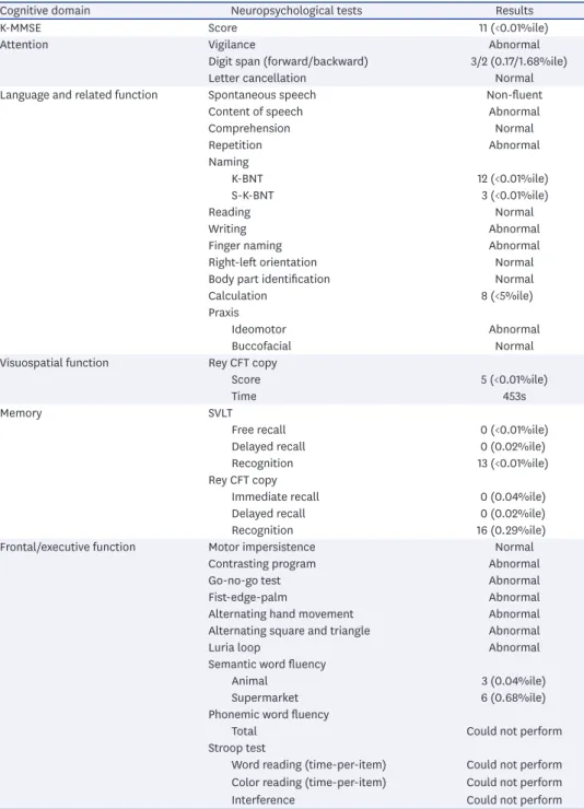
In the neuropsychological evaluation in 2015, the patient scored 11/30 in Korean version of the Mini-Mental Status Examination (K-MMSE). A detailed neuropsychological study using Seoul Neuropsychological Screening Battery-II (SNSB-II)
4revealed a marked decline in all five domains, including attention, language and related functions, visuospatial functions, memory, and frontal executive functions (Table 2). Especially in the language domain, she showed non-fluent speech, abnormal repetition, and severely impaired confrontation naming. However, comprehension of simple sentences and words was spared.
Letter to the Editor
Mi Jin Oh ,
1SangYun Kim ,
2Young Ho Park ,
2Jeewon Suh ,
2SangHak Yi
31Seoul National University College of Medicine, Seoul, Korea
2 Department of Neurology, Seoul National University College of Medicine and Clinical Neuroscience Center, Seoul National University Bundang Hospital, Seongnam, Korea
3 Department of Neurology, Wonkwang University School of Medicine and Regional Cardiocerebrovascular Center, Iksan, Korea
Early Onset Alzheimer's Disease Presenting as Logopenic Primary Progressive Aphasia
Received: Jul 1, 2018 Revised: Jul 1, 2018 Accepted: Jul 12, 2018 Correspondence to SangYun Kim, MD, PhD
Department of Neurology, Seoul National University College of Medicine and Clinical Neuroscience Center, Seoul National University Bundang Hospital, 82 Gumi-ro 173-beon-gil, Bundang-gu, Seongnam 13620, Korea.
E-mail: [email protected]
© 2018 Korean Dementia Association This is an Open Access article distributed under the terms of the Creative Commons Attribution Non-Commercial License (https://
creativecommons.org/licenses/by-nc/4.0/) which permits unrestricted non-commercial use, distribution, and reproduction in any medium, provided the original work is properly cited.
ORCID iDs Mi Jin Oh
https://orcid.org/0000-0002-5886-7868 SangYun Kim
https://orcid.org/0000-0002-9101-5704 Young Ho Park
https://orcid.org/0000-0002-2756-1786 Jeewon Suh
https://orcid.org/0000-0003-3509-6447 SangHak Yi
https://orcid.org/0000-0002-2701-9807 Conflict of Interest
The authors have no financial conflicts of interest.
Author Contributions
Conceptualization: Kim SY; Investigation: Kim SY; Supervision: Kim SY; Writing - original draft:
Oh MJ; Writing - review & editing: Kim SY, Park YH, Suh J, Yi SH.
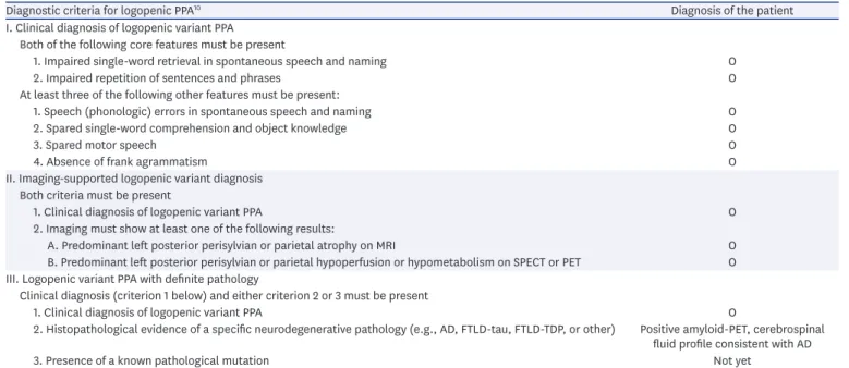
The magnetic resonance imaging (MRI) taken in 2015 showed mild focal brain atrophy, especially in the left superior temporal and left inferior parietal lobules (Fig. 1). The fluorodeoxyglucose (FDG)-positron emission tomography (PET) revealed hypometabolism in the frontal, temporal, and parietal cortices, more severe on the left temporal and parietal part (Fig. 2). These imaging results showed anatomical and functional degeneration in the temporal and parietal cortices, dominantly in the left hemisphere, which is a typical finding in logopenic PPA patients (Table 1). The amyloid-PET results showed symmetric uptake in cortical gray matter of both hemispheres, suggesting the presence of a β-amyloid pathology of AD (Fig. 3). The cerebrospinal fluid (CSF) analysis showed low amyloid β
1-42(278.1 ng/L), high total tau (534.1 ng/L), and high phosphorylated tau (86.4 ng/L), which is also consistent with the AD profile (cut-off: amyloid β
1-42<540 ng/L, total tau >350 ng/L, and phosphorylated tau >60 ng/L).
5,6Newly diagnosed as early onset Alzheimer's disease (EOAD), she was prescribed donepezil and is under regular follow-up.
Table 1. Diagnostic criteria for logopenic PPA and the diagnosis of the patient
Diagnostic criteria for logopenic PPA10 Diagnosis of the patient
I. Clinical diagnosis of logopenic variant PPA Both of the following core features must be present
1. Impaired single-word retrieval in spontaneous speech and naming O
2. Impaired repetition of sentences and phrases O
At least three of the following other features must be present:
1. Speech (phonologic) errors in spontaneous speech and naming O
2. Spared single-word comprehension and object knowledge O
3. Spared motor speech O
4. Absence of frank agrammatism O
II. Imaging-supported logopenic variant diagnosis Both criteria must be present
1. Clinical diagnosis of logopenic variant PPA O
2. Imaging must show at least one of the following results:
A. Predominant left posterior perisylvian or parietal atrophy on MRI O
B. Predominant left posterior perisylvian or parietal hypoperfusion or hypometabolism on SPECT or PET O III. Logopenic variant PPA with definite pathology
Clinical diagnosis (criterion 1 below) and either criterion 2 or 3 must be present
1. Clinical diagnosis of logopenic variant PPA O
2. Histopathological evidence of a specific neurodegenerative pathology (e.g., AD, FTLD-tau, FTLD-TDP, or other) Positive amyloid-PET, cerebrospinal fluid profile consistent with AD
3. Presence of a known pathological mutation Not yet
PPA: primary progressive aphasia, MRI: magnetic resonance imaging, SPECT: single-photon-emission computed tomography, PET: positron emission tomography, AD: Alzheimer's disease, FTLD: frontotemporal lobar degeneration, TDP: transactive response DNA-binding protein.
Table 2. Neuropsychological evaluation results in October 2015
Cognitive domain Neuropsychological tests Results
K-MMSE Score 11 (<0.01%ile)
Attention Vigilance Abnormal
Digit span (forward/backward) 3/2 (0.17/1.68%ile)
Letter cancellation Normal
Language and related function Spontaneous speech Non-fluent
Content of speech Abnormal
Comprehension Normal
Repetition Abnormal
Naming
K-BNT 12 (<0.01%ile)
S-K-BNT 3 (<0.01%ile)
Reading Normal
Writing Abnormal
Finger naming Abnormal
Right-left orientation Normal
Body part identification Normal
Calculation 8 (<5%ile)
Praxis
Ideomotor Abnormal
Buccofacial Normal
Visuospatial function Rey CFT copy
Score 5 (<0.01%ile)
Time 453s
Memory SVLT
Free recall 0 (<0.01%ile)
Delayed recall 0 (0.02%ile)
Recognition 13 (<0.01%ile)
Rey CFT copy
Immediate recall 0 (0.04%ile)
Delayed recall 0 (0.02%ile)
Recognition 16 (0.29%ile)
Frontal/executive function Motor impersistence Normal
Contrasting program Abnormal
Go-no-go test Abnormal
Fist-edge-palm Abnormal
Alternating hand movement Abnormal
Alternating square and triangle Abnormal
Luria loop Abnormal
Semantic word fluency
Animal 3 (0.04%ile)
Supermarket 6 (0.68%ile)
Phonemic word fluency
Total Could not perform
Stroop test
Word reading (time-per-item) Could not perform Color reading (time-per-item) Could not perform
Interference Could not perform
Tests not included in the SNSB, but added in the SNSB-II.
K-MMSE: Korean version of the Mini-Mental State Examination, K-BNT: Korean version of Boston Naming Test, S-K-BNT: short form of the Korean version of Boston Naming Test, Rey CFT: Rey Complex Figure Test, SVLT: Seoul Verbal Learning Test, SNSB: Seoul Neuropsychological Screening Battery.
Many recent studies have shown that AD pathology is the most common underlying pathology of logopenic PPA. There were several observational studies of logopenic PPA patients that revealed cortical amyloid binding on amyloid-PET and temporoparietal atrophy on MRI,
7,8and pathological confirmation was made in the study by Mesulam et al.
3in 2008.
This article discusses a patient who initially presented with language symptoms and was revealed to have an AD pathology shown both by amyloid-PET and CSF analysis. This is a
A B
Fig. 2. FDG-PET image of the patient, taken in December 2015. (A, B) Axial FDG-PET images each in different planes.
FDG: fluorodeoxyglucose, PET: positron emission tomography.
A B
Fig. 3. Amyloid-PET image of the patient taken in December 2015, showing extended uptake to the cortical gray matter. (A) Axial image and (B) coronal image.
PET: positron emission tomography.
REFERENCES
1. Mesulam MM. Slowly progressive aphasia without generalized dementia. Ann Neurol 1982;11:592-598.
PUBMED | CROSSREF
2. Gorno-Tempini ML, Brambati SM, Ginex V, Ogar J, Dronkers NF, Marcone A, et al. The logopenic/
phonological variant of primary progressive aphasia. Neurology 2008;71:1227-1234.
PUBMED | CROSSREF
3. Mesulam M, Wicklund A, Johnson N, Rogalski E, Léger GC, Rademaker A, et al. Alzheimer and frontotemporal pathology in subsets of primary progressive aphasia. Ann Neurol 2008;63:709-719.
PUBMED | CROSSREF
4. Kang YW, Jang SM, Na DL. Seoul Neuropsychological Screening Battery-II. Seoul: Human Brain Research &
Consulting Co., 2012.
5. Skillbäck T, Farahmand BY, Rosén C, Mattsson N, Nägga K, Kilander L, et al. Cerebrospinal fluid tau and amyloid-β1-42 in patients with dementia. Brain 2015;138:2716-2731.
PUBMED | CROSSREF
6. Hansson O, Zetterberg H, Buchhave P, Londos E, Blennow K, Minthon L. Association between CSF biomarkers and incipient Alzheimer's disease in patients with mild cognitive impairment: a follow-up study. Lancet Neurol 2006;5:228-234.
PUBMED | CROSSREF
7. Matías-Guiu JA, Cabrera-Martín MN, Moreno-Ramos T, Valles-Salgado M, Fernandez-Matarrubia M, Carreras JL, et al. Amyloid and FDG-PET study of logopenic primary progressive aphasia: evidence for the existence of two subtypes. J Neurol 2015;262:1463-1472.
PUBMED | CROSSREF
8. Teichmann M, Kas A, Boutet C, Ferrieux S, Nogues M, Samri D, et al. Deciphering logopenic primary progressive aphasia: a clinical, imaging and biomarker investigation. Brain 2013;136:3474-3488.
PUBMED | CROSSREF
9. Henry ML, Gorno-Tempini ML. The logopenic variant of primary progressive aphasia. Curr Opin Neurol 2010;23:633-637.
PUBMED | CROSSREF
10. Gorno-Tempini ML, Hillis AE, Weintraub S, Kertesz A, Mendez M, Cappa SF, et al. Classification of primary progressive aphasia and its variants. Neurology 2011;76:1006-1014.
PUBMED | CROSSREF