대흉외지 2006;39:332-334 □ 증례보고 □
- 332 - 증 례
12세 남자 어린이가 특이 병력과 증상 없이 지내던 중 학교 건강 검진 중 단순 흉부 방사선에서 좌상엽에 4 cm 크기의 응어리가 발견되어 본원 소아과로 내원하였다. 내 원 당일 촬영한 단순 흉부 방사선 촬영과 흉부 전산화 단 층촬영 소견 상 폐 좌상엽에 위치한 가장자리가 불분명한 4×3.8 cm 크기의 종괴가 발견되었고 결핵종을 동반하는 기질화된 폐렴이나 염증성 가성종양을 의심하였다(Fig. 1).
소아과에서 별 치료없이 2개월 뒤에 추적 관찰하여 단순 흉부촬영에서 결절의 크기의 변화가 없어 수술을 위해서 흉부외과로 입원하였다. 술 전 혈액검사에서 헤모글로빈 수치가 13.2 g/dL으로 약간 감소된 수치를 보였고 적혈구 침강속도는 18 mm/hr로 약간 증가하였으며 그 외 다른 검
Inflammatory Myofibroblastic Tumor of the Lung in a Child
-A case report-
Hee Jung Kim, M.D.*, Chang Ryul Park, M.D.*, Jong Pil Jung, M.D.*, Je Kyoun Shin, M.D.*
Inflammatory myofibroblastic tumor in the lung is a rare tumor. The etiology is not clear. This tumor in children is a benign tumor rarely presented with local invasiveness, recurrence, distant metastasis or malignant changes can occur. The complete surgical resection is chosen as the optimal management. A 12-years-old boy visited the outpatient clinic with a 4 cm sized pulmonary mass in left upper lung field. The patient underwent left upper lobectomy. Histopathologically, inflammatory myofibroblastic tumor was confirmed. The patient was discharged without any problems and there was no evidence of recurrence during 3 months follow-up.
(Korean J Thorac Cardiovasc Surg 2006;39:332-334)
ꠏꠏꠏꠏꠏꠏꠏꠏꠏꠏꠏꠏꠏꠏꠏꠏꠏꠏꠏꠏꠏꠏꠏꠏꠏꠏꠏꠏꠏꠏꠏꠏꠏꠏꠏꠏꠏꠏꠏꠏꠏꠏꠏꠏꠏꠏꠏꠏꠏꠏꠏꠏꠏꠏꠏꠏꠏꠏꠏꠏꠏꠏꠏꠏꠏꠏꠏꠏꠏꠏꠏꠏꠏꠏꠏꠏꠏꠏꠏꠏꠏꠏꠏꠏꠏꠏꠏꠏꠏꠏꠏꠏ
Key words:1. Lung neoplasms
2. Myofibroblasts
소아에서 발생한 폐 염증성 근섬유아세포종
-1예 보고-
김희중*․박창률*․정종필*․신제균*
*울산대학교 의과대학 울산대학교병원 흉부외과
Department of Thoracic and Cardiovascular Surgery, Ulsan University Hospital, Ulsan University College of Medicine 논문접수일:2005년 9월 16일, 심사통과일:2006년 1월 30일
책임저자:신제균 (682-060) 울산시 전하동 290-3, 울산대학교병원 흉부외과 (Tel) 052-250-7140, (Fax) 052-250-8070, E-mail: [email protected] 본 논문의 저작권 및 전자매체의 지적소유권은 대한흉부외과학회에 있다.
Fig. 1. Preoperative Chest CT with contrast enhancement shows irregular lung mass (4×3.8 cm) with spiculated border, calcifi- cation, and slight enhancement in anterior segment of left upper lobe.
김희중 외 소아의 폐 염증성 근섬유아세포종
- 333 -
사에서는 이상 소견은 없었다.수술은 전신마취 하에 우측 측와위 자세를 취하고, 좌 측 후측방 개흉술을 시행하였다. 흉강 안의 협착이나 흉 수는 관찰되지 않았다. 종양은 좌상엽 앞 분절 부위에서 장측 흉막을 침범하여 있었고 5 cm 정도의 크기였다. 종 양은 견고하였고, 노란색을 띠며 주위 폐실질조직과 분리 되지 않았다(Fig. 2). 종양의 일부분을 절제하여 동결 절편 검사를 시행하여 양성소견이 보고되었다. 종양의 크기가 크고 좌폐엽 기관지 근위부에 위치하고 있어 완전한 절제 를 위해 좌상엽 절제술을 시행하였다.
수술 후 조직병리 소견은 염증성 근섬유아세포종(inflam- matory myofibroblastic tumor)으로 진단되었다(Fig. 3). 환아 는 술 후 8일째 합병증 없이 퇴원하였으며 현재 술 후 3개 월째 재발소견 없이 외래 추적 중에 있다.
고 찰
염증성 가성종양(inflammatory pseudotumor)으로도 알려 져 있는 염증성 근섬유아세포종(inflammatory myofibrobla- stic tumor)은 폐에서는 매우 드물게 발견되는 종양으로 일 반흉부 수술의 0.04% 내지 0.7% 정도로 보고되고 있다[1].
염증성 근섬유아세포종은 양성종양으로 분류되나 드물게 국소 침윤이나 재발, 원격전이와 같은 악성종양의 형태를 가지거나 악성종양으로 전이될 수 있는 종양이다[2]. 원인 은 확실하게 밝혀지진 않았으나 감염이나 면역작용에 의 해 생겼다는 증례보고가 있고 원발성 종양으로 보는 견해 도 있다. 최근 연구결과로는 근섬유아세포종에서 염색체 이상이 보여서 이차적 원인보다는 원발 종양의 일종으로
생각되고 있다. 즉, anaplastic lymphoma kinase (ALK) 수용 체의 tyrosine-kinase 지역(염색체 2p23)에서 염색체 재배열 이 일어나거나 DNA 이수성염색체(aneuploidy)가 발견되어 양성종양 혹은 낮은 분화도의 암일 가능성이 있다고 보고 되어 있다[3].
페의 염증성 근섬유아세포종은 위치에 따라 기침이나 흉통 등이 발생할 수 있으나 주된 임상증상이 없고 혈액 검사상 저색소성 소적혈구성 빈혈, 면역 글로부린의 증가, 혈소판 증가, 적혈구침강속도의 증가 등이 나타날 수 있 다. 또한, 방사선 소견으로 국한성의 단순 종괴의 형태로 보이기도 하나 최종적인 진단은 조직병리학적으로 확진 된다[2]. 조직병리학적으로 염증성 근섬유아세포종은 형 질세포, 조직구, 림프구 및 방추형 세포 등을 포함한 다양 한 염증세포와 간엽세포가 혼합되어 있는데 특징적인 염 증세포보다는 주로 증식성 근섬유아세포와 섬유아세포로 되어 있다. 방추형 세포의 대부분은 근섬유아세포로서 유 사분열은 없고 세포 이형성 정도가 낮은 형태이고, 염증 세포에서도 유사분열이나 세포 이형성은 없는 상태이다.
폐조직에서는 염증세포의 침윤에 의해 조직학적인 구조 파괴가 나타난다[4].
치료는 외과적인 완전 절제가 가장 적합한 치료방법으 로 되어 있다. 불완전 절제시 종양의 재발의 가능성이 60% 정도까지 보고되어 있으므로 외과적 치료시 완전절 제가 매우 중요하다[1,2,4]. 종양의 위치나 크기에 따라 부 분절제나 구역절제도 가능하나 완전한 절제를 위해 폐엽 절제술과 경우에 따라서는 전폐적출술을 필요로 하기도 한다[1,5]. 외과적 치료 외에 방사선 치료, 항암치료나 스 Fig. 2. The cut surface shows relatively well demarcated whitish
solid firm to rubbery mass, which is subbronchial to pleura.
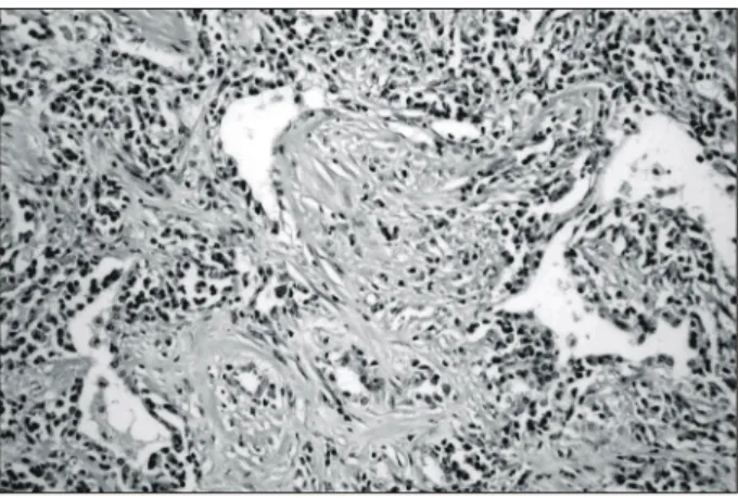
Fig. 3. The tumor contains a mixture of spindle cells showing fibroblastic and myofibroblastic differentiation arrayed in fas- cicles, or storiform architecture. Prominent plasma cells infiltrate among myofibroblastic cells in this lesion (H&E, ×100).
대흉외지 2006;39:332-334
- 334 -
테로이드 사용은 그 효과가 확실하게 밝혀지지 않았으나 최근 연구에 흉막 비후와 동반된 근섬유아세포종에서 스 테로이드를 사용하여 완치된 증례가 보고되어 있었다[6].이 증례는 12세 소아의 폐에서 발견된 염증성 근섬유아세 포종으로 폐엽절제술을 통하여 완전 절제된 경우이지만 종양의 특성상 재발의 가능성이 있으므로 정기적인 외래 추적 관찰이 필요할 것이다.
참 고 문 헌
1. Cerfolio RJ, Allen MS, Nascimento AG, et al. Inflammatory pseudotumors of the lung. Ann Thorac Surg 1999;67:933-6.
2. Kamak I, Senocak ME, Ciftci AO, et al. Inflammatory my- ofibroblastic tumor in children: diagnosis and treatment. J Pediatr Surg 2001;36:908-12.
3. Lawrence B, Perez-Atayde A, Hibbard MK, et al. TPM3- ALK and TPM4-ALK oncogenes in inflammatory myofibro- blastic tumors. Am J Pathol 2000;157:377-84.
4. Sakurai H, Hasegawa T, Watanabe S, Suzuki K, Asamura H, Tsuchiya R. Inflammatory myofibroblastic tumor of the lung.
Eur J Cardiothorac Surg 2004;25:155-9.
5. Na KJ, Yu U, Hong SB, et al. A case of pulmonary inflam- matory myofibroblastic tumor. Korean J Thorac Cardiovasc Surg 2004;37:102-4.
6. Ishioka S, Maeda A, Yamasaki M, Yamakido M. Inflamma- tory pseudotumor of the lung with pleural thickening treated with corticosteroids. Chest 2000;117:923.
=국문 초록=
폐에서 발생하는 염증성 근섬유아세포종은 원인은 정확히 밝혀져 있지 않은 매우 드문 종양이다. 소 아에서 발생한 이 종양은 양성종양으로 분류되어 있으나 드물게 국소침윤이나 재발, 원격전이 및 악 성변이가 나타날 수 있다. 외과적 완전 절제술이 가장 적절한 치료 방법으로 되어 있다. 12세 남자가 단순흉부엑스선 사진에서 우연히 발견된 좌측 폐 상부의 4 cm 크기의 응어리소견을 주소로 내원하였 다. 환아는 좌상엽 절제수술을 받은 후 진단병리학적으로 염증성 근섬유아세포종으로 확진되었다. 환 아는 술 후 3개월째 재발 소견없이 외래 추적 관찰 중이다.
중심 단어:1. 폐종양 2. 근섬유아세포