INTRODUCTION
Ovarian cancer is a very lethal type of gynecologic cancer with over 100,000 people dying every year from this disease around the world [1]. Among patients diagnosed with ovarian
cancer, 75% of the cases are initially detected at the dissemi- nated (stage III/IV) form of the disease and the 5-year survival rate is less than 25%. Primary treatment for ovarian cancer is cytoreductive surgery followed by combination chemother- apy. More than 70% of patients typically respond to primary treatment [2]. However, more than 85% of those responders will relapse despite surgical debulking, followed by platinum (e.g., cisplatin or carboplatin) and taxane regimens [2]. Despite the initial response, relapse is mainly caused by resistance to chemotherapeutic agents. At the time of recurrence, ad- ditional therapy options are very limited because secondary chemotherapeutic agents cause more serious side effects and
The relationship between cisplatin resistance and histone deacetylase isoform overexpression in epithelial ovarian cancer cell lines
Min-Gyun Kim1, Jhang Ho Pak2, Won Ho Choi2, Jeong-Yeol Park1, Joo-Hyun Nam1, Jong-Hyeok Kim1
1Department of Obstetrics and Gynecology, 2Asan Institute for Life Sciences, Asan Medical Center, University of Ulsan College of Medicine, Seoul, Korea
Received Nov 1, 2011, Revised Mar 6, 2012, Accepted Mar 24, 2012 Correspondence to Jong-Hyeok Kim
Department of Obstetrics and Gynecology, Asan Medical Center, University of Ulsan College of Medicine, 88 Olympic-ro 43-gil, Songpa-gu, Seoul 138- 736, Korea. Tel: 82-2-3010-3643, Fax: 82-2-3010-6944, E-mail: hyeokkim@
amc.seoul.kr
Objective: To investigate the relationship between cisplatin resistance and histone deacetylase (HDAC) isoform overexpression in ovarian cancer cell lines.
Methods: Expression of four HDAC isoforms (HDAC 1, 2, 3, and 4) in two ovarian cancer cell lines, SKOV3 and OVCAR3, exposed to various concentrations of cisplatin was examined by western blot analyses. Cells were transfected with plasmid DNA of each HDAC. The overexpression of protein and mRNA of each HDAC was confirmed by western blot and reverse transcriptase- polymerase chain reaction analyses, respectively. The cell viability of the SKOV3 and OVCAR3 cells transfected with HDAC plasmid DNA was measured using the cell counting kit-8 assay after treatment with cisplatin.
Results: The 50% inhibitory concentration of the SKOV3 and OVCAR3 cells can be determined 15-24 hours after treatment with 15 µg/mL cisplatin. The expression level of acetylated histone 3 protein in SKOV3 cells increased after exposure to cisplatin.
Compared with control cells at 24 hours after cisplatin exposure, the viability of SKOV3 cells overexpressing HDAC 1 and 3 increased by 15% and 13% (p<0.05), respectively. On the other hand, OVCAR3 cells that overexpressed HDAC 2 and 4 exhibited increased cell viability by 23% and 20% (p<0.05), respectively, compared with control cells 24 hours after exposure to cisplatin.
Conclusion: In SKOV3 and OVCAR3 epithelial ovarian cancer cell lines, the correlation between HDAC overexpression and cisplatin resistance was confirmed. However, the specific HDAC isoform associated with resistance to cisplatin varied depending on the ovarian cancer cell line. These results may suggest that each HDAC isoform conveys cisplatin resistance via different mechanisms.
Keywords: Cisplatin resistance, Epithelial ovarian cancer cell lines, Histone deacetylase
stronger toxicity. Reducing resistance to chemotherapeutic agents may greatly decrease the mortality of ovarian cancer and relapse of this devastating disease.
The primary role of transcription is to regulate the expression of specific target genes. Epigenetic mechanisms like histone modification and DNA methylation have been known to play a key role in the regulation of gene transcription. Variation of epigenetic regulation is one of the known causes of abnormal gene expression in a malignant tumor resistant to chemo- therapy. Specially, acetylation of DNA-bound core histones is associated with up-regulation of transcription and is regulated by two opposing classes of enzymes, histone acetyltransfer- ases (HATs) and histone deacetylases (HDACs). Acetylation of DNA sequence-specific transcription factors also regulates gene transcription. HDACs are emerging as critical regulators of cell growth, differentiation, and apoptosis. Recently, several HDAC inhibitors were studied and demonstrated consider- able anti-tumor effect in various types of cancers including colonic carcinoma, pancreatic cancer, and hepatoma [3-5]. For example, a phase I clinical trial of suberoylanilide hydroxamic acid (SAHA), an HDAC inhibitor that suppresses the activity of HDAC class I and II, demonstrated anti-tumor activity in solid and hematologic tumors [6]. In addition, several clinical trials of HDAC inhibitors that included desipeptide, MS-275, LAQ-824, LBH-589, and MGCD 0103, were reported to have a strong anti-tumor activity in various types of cancer [7-11].
For ovarian cancer, if resistance to chemotherapeutic agents is removed, mortality and morbidity would decrease and the use of more toxic secondary agents may be reduced. The overexpression of HDAC 1, 2, and 3 has previously been re- ported in ovarian cancer tissues [12]. We hypothesized that the mechanism of resistance to chemotherapeutic agents is associated with overexpressed HDACs in ovarian cancer cells.
In this study, we investigated the relationship between cis- platin resistance and HDAC overexpression in two epithelial ovarian cancer cell lines, SKOV3 and OVCAR3. HDAC isoforms associated with cisplatin resistance in each cell line were com- pared.
MATERIALS AND METHODS 1. Materials
Components for cell culture media were purchased from Life Technologies (Gaithersburg, MD, USA) unless otherwise noted. The SKOV-3 and OVCAR3 human epithelial ovarian cancer cell lines were obtained from the Korean Cell Line Bank (Seoul, Korea). The Taq DNA polymerase PCR system was purchased from Takara Bio Inc. (Shiga, Japan). Polyclonal anti-
bodies to HDAC 1-4 and acetylated histone 3 were purchased from Cell Signaling (Danvers, MA, USA) and horseradish-per- oxidase conjugated secondary antibodies were obtained from Jackson ImmunoResearch Laboratory (West Grove, PA, USA).
From Sigma-Aldrich (St. Louis, MO, USA) we obtained mouse monoclonal antibody to β-actin, complete protease inhibitor cocktail, and cisplatin. AC 1-4 expression vectors were kindly provided by Dr. Hyun Kook from the Department of Pharma- cology, Chonnam University Medical School, Gwangju, Korea.
All biotechnology-grade chemicals were purchased from Am- resco Inc. (Solon, OH, USA).
2. Cell culture
OVCAR3 and SKOV3 cells were cultured in RPMI 1640 me- dia supplemented with 100 units/mL penicillin, 100 µg/mL streptomycin, and 10% fetal bovine serum (FBS) at 37oC in a 5% CO2 incubator. Cells exposed to cisplatin were plated at 3
×105 cells per well in a 6-well culture dish for 24 hours before use. Cells were treated with various concentrations of cisplatin and were incubated for a specific length of time.
3. Cytotoxicity assay
Cell viability was examined using the colorimetric cell count- ing kit-8 (CCK-8) assay (Dojindo Lab., Kumamoto, Japan) ac- cording to manufacturer instructions. Cells were seeded at a density of 3×10³ per well in a 96-well plate and were cultured as described above. Cells were then treated with 15 µg/mL cisplatin for 0, 15, 24, 36, and 48 hours. The amount of forma- zan dye generated by cellular dehydrogenase activity was measured by absorbance at 450 nm using an ELISA reader (Molecular Devices Corporation, Sunnyvale, CA, USA). Absor- bance values were converted to percentages for comparison with untreated control. We used IC50 Calculate software (LOGIT method) for calculating the 50% inhibitory concentration (IC50).
4. Transfection
Cells were plated 24 hours prior to transfection at a density of 2×10⁵ cells per 35 mm culture dish. Cells were transiently transfected using Lipofectamine LTX (Invitrogen, Carlsbad, CA, USA) and 2.5 µg of each HDAC isoform expression vector or mock vector as a control. Twenty-four hours after transfection, the cells were treated with cisplatin for the indicated length of time.
5. Nuclear protein extraction for the detection of histone 3 (H3) acetylation
Cells were harvested and nuclear proteins were isolated us- ing Cellytic Nuclear TM Extraction Kit (Sigma-Aldrich) accord-
ing to manufacturer instructions. Nuclear extract protein lev- els were measured and the extracts were stored at -20oC until use.
6. Western blot analysis
Cells were washed twice with phosphate-buffered saline (PBS) on ice and were disrupted with RIPA lysis buffer (50 mM Tris-HCl [pH 7.4], 150 mM NaCl, 1% NP-40, 0.25% Na- deoxychalate, 1 mM EDTA, 1 mM NaVO4, 2 mM NaF, and com- plete protease inhibitor cocktail). The extracts were homog- enized by sonication and centrifuged at 13,000 rpm for 20 minutes at 4oC. Protein concentration was measured using the BCA Protein Assay (Pierce, Rockford, IL, USA). Protein samples (30 µg per lane) were resolved on SDS-polyacrylamide gel and electrophoretically transferred to nitrocellulose membranes (GE Healthcare Biosciences, Uppsala, Sweden). After blocking with 5% skim milk in TBS-T buffer (50 mM Tris-HCl [pH 8.0], 150 mM NaCl, 0.5% Tween-20), the membranes were probed with primary antibodies that correspond to target proteins.
Membranes were then washed three times with TBS-T buffer followed by incubation with horse radish-conjugated second- ary antibody (Jackson ImmunoResearch Lab). The reaction was detected with enhanced chemiluminescence (ECL; GE Healthcare Biosciences) and exposed to X-ray film. To normal- ize for protein loading, the membranes were washed with stripping solution (62.5 mM Tris-HCl [pH 6.8], 100 mM β-mer- captoethanol, 2% SDS) at 55oC and reprobed with a monoclo- nal antibody to β-actin.
7. Total RNA extraction and RT-PCR analysis
Cells were washed twice with cold PBS and total RNA was extracted using TRIzol (Invitrogen) according to manufacturer instructions. The RNA samples were quantified, aliquoted, and stored at -80oC until use. Approximately 1 µg of total RNA was used to synthesize first-stranded cDNA using the SuperScript
II First-Strand System (Invitrogen). The PCR was performed for 30 cycles of denaturation at 94oC for 1 minute, anneal- ing at 55oC for 45 seconds, extension at 72oC for 45 seconds.
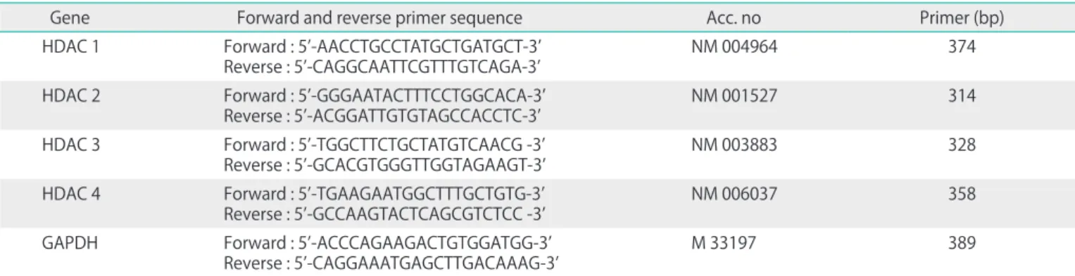
After completing 30 cycles, a final extension cycle of 72oC for 2 minutes was carried out. Table 1 illustrates a pair of DNA oli- gonucleotides designed with PrimerDesigner program, based on the cDNA sequences of each human HDAC isoform and glyceraldehyde-3-phosphate dehydrogenase (GAPDH) in the GenBank database. The PCR products were separated on a 1%
agarose gel and images were captured using an ultraviolet plate.
8. Statistical analysis
Cell survival rates for the control and test groups after treat- ment with cisplatin were presented as average±standard deviation. The statistical analysis was performed using the Mann-Whitney U-test employing the SPSS ver. 12.0 (SPSS Inc., Chicago, IL, USA). A value of p<0.05 was considered statisti- cally significant.
RESULTS
1. Cytotoxicity test
After treatment with 15 µg/mL cisplatin, the survival rate of SKOV3 and OVCAR3 cells decreased with time and both groups reached IC50 between 15 and 24 hours (Fig. 1). Al- though there were no significant differences in the survival rates of SKOV3 and OVCAR3 cells until 15 hours, SKOV3 cells did show more survival compared with OVCAR3. It shows that SKOV3 has more resistance than OVCAR3 to cisplatin.
2. Histone acetylation and deacetylation by cisplatin After treatment of SKOV3 cells with 15 µg/mL cisplatin, nuclear protein was separated and the level of expression of
Table 1. Primer sequences of HDAC isoforms for RT-PCR
Gene Forward and reverse primer sequence Acc. no Primer (bp)
HDAC 1 Forward : 5’-AACCTGCCTATGCTGATGCT-3’
Reverse : 5’-CAGGCAATTCGTTTGTCAGA-3’ NM 004964 374
HDAC 2 Forward : 5’-GGGAATACTTTCCTGGCACA-3’
Reverse : 5’-ACGGATTGTGTAGCCACCTC-3’ NM 001527 314
HDAC 3 Forward : 5’-TGGCTTCTGCTATGTCAACG -3’
Reverse : 5’-GCACGTGGGTTGGTAGAAGT-3’ NM 003883 328
HDAC 4 Forward : 5’-TGAAGAATGGCTTTGCTGTG-3’
Reverse : 5’-GCCAAGTACTCAGCGTCTCC -3’ NM 006037 358
GAPDH Forward : 5’-ACCCAGAAGACTGTGGATGG-3’
Reverse : 5’-CAGGAAATGAGCTTGACAAAG-3’ M 33197 389
GAPDH, glyceraldehydes-3-phosphate dehydrogenase; HDAC, histone deacetylase; RT-PCR, reverse transcriptase-polymerase chain reaction.
acetylated H3 was measured. The level of H3 expression was increased, especially around 24 hours (Fig. 2). After treatment with cisplatin for 24 hours, we compared the protein expres- sion levels of HDAC 1, 2, 3, and 4. No meaningful changes in the expression level were observed for HDAC 2 and 3 despite an increase in cisplatin exposure. At the concentration of 15 µg/mL cisplatin, HDAC 1 expression significantly decreased.
HDAC 4 expression markedly decreased from the concentra- tion of 4 µg/mL cisplatin and was nearly undetected above 4 µg/mL cisplatin (Fig. 3). From these results, it may be esti- mated that HDAC 1-3 are associated primarily with histone deacetylation in SKOV3 cells.
3. Overexpression of HDACs
SKOV3 cells transfected with HDAC 1, 2, 3, and 4 plasmid DNA showed significantly higher expression of HDACs genes Fig. 2. The expression of acetylated histone 3 in SKOV3 cells treated
with cisplatin. Cells were treated with 15 µg/mL cisplatin and harvested between 9 and 49 hours, followed by western blot analysis.
Western blot for β-actin is shown as a control for protein loading.
Fig. 3. Expression levels of histone deacetylase (HDAC) isoforms in SKOV3 cells treated with various concentrations of cisplatin. 30 µg of total soluble protein from each lysate was immunoblotted with anti- HDAC 1, 2, 3, and 4 polyclonal antibodies. Western blot for β-actin is shown as a control for protein loading.
Fig. 4. Expression levels of histone deacetylase (HDAC) mRNA in SKOV3 cells transfected with each HDAC isoform.
Fig. 5. Protein expression levels of histone deacetylase (HDAC) isoforms in SKOV3 and OVCAR3 cells transfected with HDAC isoform expression vectors or not transfected as a control. Expression of β- actin was used as a loading control. endo, endogenous HDAC 1; exo, exogenous HDAC 1 which was transfected.
Fig. 1. A comparison of cisplatin resistance in SKOV3 and OVCAR3 cells treated with 15 µg/mL cisplatin. The number of viable cells was measured by CCK-8 assay. *p<0.05.
(Fig. 4). We also identified similar results with the overexpres- sion of HDAC proteins in transfected SKOV3 and OVCAR3 cells (Fig. 5).
4. Relationship between overexpression of HDAC proteins and resistance to cisplatin
After overexpressing HDACs in SKOV3 and OVCAR3 cells treated with 15 µg/mL cisplatin, we compared differences in cell viability among the HDAC isoforms. In the case of SKOV3, the IC50 of the control group was observed at 24 hours. The viability of cells overexpressing HDAC 1 and 3 increased up to 15% and 13%, respectively, compared with control cells at 24 hours (p<0.05) (Fig. 6). In the case of OVCAR3, the viability of cells overexpressing HDAC 2 and 4 increased up to 23% and 20%, respectively, compared with control at cells at 24 hours (p<0.05) (Fig. 7). These results suggest that increased expres- sion of various types of HDAC increased cisplatin resistance for both cell lines. In addition, the resistance conferred by differ- ent HDAC isoforms depended on the type of ovarian cancer cell line that was transfected.
DISCUSSION
Gene expression is not determined only by the DNA code but also by epigenetic modifications. Epigenetic modifications can be mitotically inherited for more than one generation [13].
However, epigenetic mechanisms can be modified by chemi- cal agents and not by DNA sequence alterations. Despite extensive research into the biological functions of HDACs in various types of cancer, there are few reports on the relation- ship of HDACs and anti-cancer drug resistance. Previous stud- ies from our institution demonstrated that HDACs 1-3 were elevated in ovarian cancer tissue at both transcriptional and translational levels. It is suspected that overexpressed HDACs may play a significant role in ovarian carcinogenesis. Recently, Muscolini et al. [14] showed that an HDAC inhibitor induces apoptosis in cisplatin-resistant ovarian cancer cells.
In this study, we showed that overexpression of HDACs is associated with resistance to cisplatin, a chemotherapeu- tic agent used for treating ovarian cancer. Furthermore, we demonstrated that the specific HDAC isoform associated with resistance to cisplatin was different according to the specific type of ovarian cancer cell line. Our study may provide the scientific basis for the clinical application of HDAC inhibitors in cisplatin-resistant ovarian cancer.
The epigenetic regulation of genes can be divided into two different mechanisms: structural modification of chromatin, and covalent modification of histone protein or DNA methyla- tion. These mechanisms are proven in fields of research such as; cancer biology [15], viral latency [16], somatic gene therapy [17], genomic imprinting [18], etc. The human genome is packaged as chromatin which consists of DNA, histone and non-histone protein. Chromatin remodeling is important in Fig. 6. Effect of overexpressed histone deacetylase (HDAC) isoform
on cisplatin resistance in SKOV3 cells. SKOV3 cells transfected with a unique HDAC isoform and nontransfected control cells were treated with 15 µg/mL cisplatin for 24 hours. Cell survival rate was measured by cell counting kit-8 (CCK-8) assay. The values were normalized to time 0 as a starting point and the survival rate was calculated by dividing the average number of treated cells by the average number of untreated cells. Each data set represents a relative percentage of nontransfected control values. Values are expressed as mean±SE for three independent experiments. *p<0.05 compared with control.
Fig. 7. Effect of overexpressed histone deacetylase (HDAC) isoforms on cisplatin resistance in OVCAR3 cells. OVCAR3 cells transfected with a unique HDAC isoform and nontransfected control cells were treated with 15 µg/mL cisplatin for 24 hours. Cell survival rate was measured by cell counting kit-8 (CCK-8) assay. The values were normalized to time 0 as a starting point and the survival rate was calculated by dividing the average number of treated cells by the average number of untreated cells. Each data set represents a relative percentage of nontransfected control values. Values are presented as mean±SE for three independent experiments. *p<0.05 compared with control.
epigenetic regulation of gene expression. The tail regions of both H3 and H4 are covalently modified where the ε-amino group of lysine in the N-terminal region of histone acts as a substrate for HDAC and HAT. The histone tails protruded from the chromatin polymer and are very sensitive to protease.
This modification is commonly called the histone code or epi- genetic code and may be controlled by HAT, HDAC, histone methyltransferase (HMT), and histone kinase (HK).
Hypoacetylated histone containing positive charges within the nucleic acid may be combined with a phosphate back- bone to cause transcriptional silencing of the chromatin.
At that time, acetylation of histone may neutralize positive charges, changing the structure of the chromatin and mak- ing it easier to access transcription factors, the transcription regulatory complex and RNA polymerase, among others. In contrast, histone deacetylation may restore positive charges of histone lysine, and turn the chromatin to a very supercoiled state and cause genetic silencing [19]. HDAC and HAT can af- fect transcription by selectively acetylating or deacetylating the ε-amino group at the amino terminus in core histone.
HDACs modify core histones and participate in large regula- tory complexes that both suppress and enhance transcription.
There are 18 HDACs identified in humans which can be classi- fied into 4 categories on the basis of homology to yeast HDACs and phylogenic analysis [20,21]. Class I HDACs are homologous to yeast RpD3 and are located primarily in the nucleus. Class I HDACs include HDAC 1-3 and 8. As an exception, HDAC 3 moves between the cytoplasm and the nucleus. Class I HDACs appear to play a primary role in cell survival and proliferation and are mainly associated with transcriptional repressors and cofactors. HDAC 1 and 3 are known to associate with transcrip- tion factor and hypoxia inducible factor (HIF)-1α, as well as af- fect angiogenesis [22]. HDAC 2 can modulate transcriptional activity through interaction with p53. HDAC 4 serves as a nuclear co-repressor involved in regulating bone and muscle development, and regulates neuronal survival in retinas [23].
Class II HDACs are homologous to yeast Hda1 and include HDAC 4-7, 9, and 10. Class II HDACs exist both in the nucleus and the cytoplasm. Class II HDACs have tissue-specific roles.
HDAC 6 and 10 have two catalytic domains, C-terminal and N- terminal. Class III HDACs are homologous to yeast Sir 2. Class III HDACs are sirtuins 1-7 which have an absolute requirement for NAD+, while class I, II, and IV HDACs are zinc dependent.
Class IV HDAC (HDAC 11) has both features of class I and class II. Class I and II HDACs are sensitive to the already known HDAC inhibitors [24]. However, the functions of most class III HDACs and class IV HDAC (HDAC 11) remains to be elucidated.
It has already been proven that epigenetic alteration plays an important role in carcinogenesis, tumor growth, and
progression. Aberration of DNA methylation and histone deacetylation may be a significant cause of resistance to che- motherapeutic agents. This aberration occurs in the course of apoptosis and differentiation. The resensitization of tumor cells to chemotherapeutic agents could be achieved by re- forming this abnormal modification. Thus, as epigenetic che- motherapy and conventional chemotherapy are combined, tumor cells that were resistant to chemotherapeutic agents are resensitized and would once again respond to first-line chemotherapeutic agents such as taxane and cisplatin.
We demonstrated that cancer cells that overexpress HDACs show resistance to cisplatin. HDAC 1 and 3 were involved in SKOV3 resistance to cisplatin, while HDAC 2 and 4 were involved in OVCAR3 resistance to cisplatin. The difference of HDAC isoforms may be due to the features of cell lines themselves or to the different mechanism of action for each HDAC isoform. In SKOV3, the p53 gene, which is important in the process of carcinogenesis, is deleted while in OVCAR3 it is mutated. The p53 gene activates proapoptotic proteins like Bax, Bam, Puma, and Noxa through a variety of pathways and suppresses antiapoptotic proteins like Bcl2. Thus, p53 plays an important role in inducing apoptosis and cell cycle arrest [25]. HDAC 1 suppresses p53-mediated apoptosis by sup- pressing the activity of p53 on the Bax promoter while HDAC 2 suppresses the DNA-binding activity of p53 [26]. Moreover, HDAC 3 and 4 are implicated in transcription suppression, with HDAC 3 suppressing the p53-p21 pathway and HDAC 4 causing posttranslational modification (acetylation or meth- ylation of the C-terminal lysine of p53 protein) [27,28]. In ad- dition, HDACs can also take part in apoptosis through other pathways, regardless of p53. However, the function of HDACs on p53 still remains unclear. Based on this study, HDAC 1 and 3 may provide resistance to apoptosis independent of p53 via another pathway, and HDAC 2 and 4 may increase resistance via p53 mutation or another p53-mediated pathway in addi- tion to another p53 non-mediated pathway.
In this study, it was identified that when HDAC 1, 2, 3, and 4 were overexpressed in SKOV3 and OVCAR3 ovarian cancer cell lines, resistance to cisplatin was conveyed when compared with a control group not containing overexpressed HDACs.
HDAC 1 and 3 in SKOV3, and HDAC 2 and 4 in OVCAR3 were strongly associated with cisplatin resistance. These results sug- gest that the HDAC isoforms resistant to cisplatin in the two cell lines are different from each other. If we could find the specific HDAC isoforms associated with cisplatin resistance in ovarian carcinoma, combination therapy with HDAC inhibitors may magnify the effects of primary chemotherapeutic agents in the treatment of ovarian carcinoma while reducing the tox- icity of secondary anti-cancer drugs.
CONFLICT OF INTEREST
No potential conflict of interest relevant to this article was reported.
ACKNOWLEDGMENTS
This study was supported by a grant (2007-203) from the Asan Institute for Life Sciences, Seoul, Korea.
REFERENCES
1. Parkin DM, Bray F, Ferlay J, Pisani P. Estimating the world cancer burden: Globocan 2000. Int J Cancer 2001;94:153- 6.
2. Ozols RF. Treatment goals in ovarian cancer. Int J Gynecol Cancer 2005;15 Suppl 1:3-11.
3. Chen Z, Clark S, Birkeland M, Sung CM, Lago A, Liu R, et al.
Induction and superinduction of growth arrest and DNA damage gene 45 (GADD45) alpha and beta messenger RNAs by histone deacetylase inhibitors trichostatin A (TSA) and butyrate in SW620 human colon carcinoma cells. Cancer Lett 2002;188:127-40.
4. Sato N, Ohta T, Kitagawa H, Kayahara M, Ninomiya I, Fushida S, et al. FR901228, a novel histone deacetylase inhibitor, induces cell cycle arrest and subsequent apoptosis in refractory human pancreatic cancer cells. Int J Oncol 2004;24:679-85.
5. Yamashita Y, Shimada M, Harimoto N, Rikimaru T, Shirabe K, Tanaka S, et al. Histone deacetylase inhibitor tricho statin A induces cell-cycle arrest/apoptosis and hepatocyte differentiation in human hepatoma cells. Int J Cancer 2003;103:572-6.
6. Kelly WK, Richon VM, O'Connor O, Curley T, MacGregor- Curtelli B, Tong W, et al. Phase I clinical trial of histone deacetylase inhibitor: suberoylanilide hydroxamic acid administered intravenously. Clin Cancer Res 2003;9:3578- 88.
7. Edwards A, Li J, Atadja P, Bhalla K, Haura EB. Effect of the histone deacetylase inhibitor LBH589 against epidermal growth factor receptor-dependent human lung cancer cells. Mol Cancer Ther 2007;6:2515-24.
8. Fuino L, Bali P, Wittmann S, Donapaty S, Guo F, Yamaguchi H, et al. Histone deacetylase inhibitor LAQ824 down- regulates Her-2 and sensitizes human breast cancer cells to trastuzumab, taxotere, gemcitabine, and epothilone B.
Mol Cancer Ther 2003;2:971-84.
9. Kell J. Drug evaluation: MGCD-0103, a histone deacetylase inhibitor for the treatment of cancer. Curr Opin Investig Drugs 2007;8:485-92.
10. Saito A, Yamashita T, Mariko Y, Nosaka Y, Tsuchiya K, Ando T, et al. A synthetic inhibitor of histone deacetylase, MS- 27-275, with marked in vivo antitumor activity against human tumors. Proc Natl Acad Sci U S A 1999;96:4592-7.
11. Sandor V, Bakke S, Robey RW, Kang MH, Blagosklonny MV, Bender J, et al. Phase I trial of the histone deacetylase inhibitor, depsipeptide (FR901228, NSC 630176), in patients with refractory neoplasms. Clin Cancer Res 2002;8:718-28.
12. Jin KL, Pak JH, Park JY, Choi WH, Lee JY, Kim JH, et al.
Expression profile of histone deacetylases 1, 2 and 3 in ovarian cancer tissues. J Gynecol Oncol 2008;19:185-90.
13. Lengauer C, Issa JP. The role of epigenetics in cancer.
DNA Methylation, Imprinting and the Epigenetics of Cancer: An American Association for Cancer Research Special Conference. Las Croabas, Puerto Rico, 12-16 1997 December. Mol Med Today 1998;4:102-3.
14. Muscolini M, Cianfrocca R, Sajeva A, Mozzetti S, Ferrandina G, Costanzo A, et al. Trichostatin A up-regulates p73 and induces Bax-dependent apoptosis in cisplatin-resistant ovarian cancer cells. Mol Cancer Ther 2008;7:1410-9.
15. Jones PA, Baylin SB. The fundamental role of epigenetic events in cancer. Nat Rev Genet 2002;3:415-28.
16. Takacs M, Salamon D, Myohanen S, Li H, Segesdi J, Ujvari D, et al. Epigenetics of latent Epstein-Barr virus genomes:
high resolution methylation analysis of the bidirectional promoter region of latent membrane protein 1 and 2B genes. Biol Chem 2001;382:699-705.
17. Zuccotti M, Garagna S, Redi CA. Nuclear transfer, genome reprogramming and novel opportunities in cell therapy. J Endocrinol Invest 2000;23:623-9.
18. Howell CY, Bestor TH, Ding F, Latham KE, Mertineit C, Trasler JM, et al. Genomic imprinting disrupted by a maternal effect mutation in the Dnmt1 gene. Cell 2001;
104:829-38.
19. Davie JR. Covalent modifications of histones: expression from chromatin templates. Curr Opin Genet Dev 1998;
8:173-8.
20. de Ruijter AJ, van Gennip AH, Caron HN, Kemp S, van Kuilenburg AB. Histone deacetylases (HDACs): charac- terization of the classical HDAC family. Biochem J 2003;
370:737-49.
21. Gregoretti IV, Lee YM, Goodson HV. Molecular evolution of the histone deacetylase family: functional implications of phylogenetic analysis. J Mol Biol 2004;338:17-31.
22. Yang QC, Zeng BF, Shi ZM, Dong Y, Jiang ZM, Huang J,
et al. Inhibition of hypoxia-induced angiogenesis by tri- chostatin A via suppression of HIF-1a activity in human osteosarcoma. J Exp Clin Cancer Res 2006;25:593-9.
23. Chen B, Cepko CL. HDAC4 regulates neuronal survival in normal and diseased retinas. Science 2009;323:256-9.
24. Thiagalingam S, Cheng KH, Lee HJ, Mineva N, Thiagalingam A, Ponte JF. Histone deacetylases: unique players in shaping the epigenetic histone code. Ann N Y Acad Sci 2003;983:84-100.
25. Bargonetti J, Manfredi JJ. Multiple roles of the tumor suppressor p53. Curr Opin Oncol 2002;14:86-91.
26. Bandyopadhyay D, Mishra A, Medrano EE. Overexpression
of histone deacetylase 1 confers resistance to sodium butyrate-mediated apoptosis in melanoma cells through a p53-mediated pathway. Cancer Res 2004;64:7706-10.
27. Basile V, Mantovani R, Imbriano C. DNA damage promotes histone deacetylase 4 nuclear localization and repression of G2/M promoters, via p53 C-terminal lysines. J Biol Chem 2006;281:2347-57.
28. Zeng L, Xiao Q, Margariti A, Zhang Z, Zampetaki A, Patel S, et al. HDAC3 is crucial in shear- and VEGF-induced stem cell differentiation toward endothelial cells. J Cell Biol 2006;174:1059-69.