Introduction
The biceps brachii muscle (BB) is located in the anterior compartment of the arm. It has 2 heads; the short head orig- inates from the coracoid process of the scapula, and the long head originates from the supraglenoid tubercle of the sca- pula. The 2 heads join together distally and insert into the radial tuberosity of the radius bone. When the elbow is ex- tended, the BB is a simple flexor of the forearm; however, when the elbow is flexed and more power is required against resistance, the BB is the primary supinator of the forearm.
Several research papers have discussed the presence of
an additional head of the BB and have used different terms to describe it, such as the third head, accessory head, or humeral head [1-13]. In this article, this additional head will be referred to as the third head of the BB (THBB).
According to previous studies, the THBB can be corre- lated to clinical practice by 2 different aspects. The first is that if the humerus is fractured, fragmented bone may ex- hibit a pattern of unusual bone displacement, posing a threat to surrounding structures such as vessels or nerves. The second is that THBB may be a predisposing factor in par- tial nerve entrapment, which can be confused with patho- logical conditions that occur during shoulder surgery.
Various authors have reported the incidence of THBB to range from 1.3% to 20.5% in different populations [1-13].
In the Asian population, an approximate 10% difference in THBB incidence was reported between the Chinese and Japanese populations [4]. No study has investigated the frequency of occurrence of THBB in the Korean popula- tion. The goal of this study was to investigate the incidence of THBB for understanding unexpected cases in clinical practice.
The Third Head of Biceps Brachii Muscle in Korean:
Anatomical Study
Je-Hun Lee
Department of Anatomy, College of Medicine, Konyang University of Korea
(Received 8 August 2013, revised 19 August 2013, accepted 12 September 2013, Published Online 30 September 2013)
Abstract : The goal of this study was to investigate the incidence of third head of biceps brachii (THBB) for understanding unexpected cases in clinical practice.
The sample consisted of 214 upper extremities from 107 adult cadavers donated to science (70 males and 37 females; age, 51-87 years). Cases with pathological changes or trauma to the upper limbs were excluded.
THBB was found in 14 of the 214 upper extremities. The incidence of variation was approximately 6.5%. THBB was found in 10 male limbs and 4 female limbs, and none of the cases was bilateral. THBB was a flat muscular belly-like structure that originated from the anteromedial surface of the humerus between the insertion of the coracobrachialis and the origin of the brachialis.
This finding may be clinically important because the musculocutaneous nerve is subjected to compression by the bulky third head.
Keywords:Third head, Biceps brachii, Musculocutaneous nerve, Anatomy
*We thank Seung-Ho Han, Department of Anatomy, College of Medicine, Chungang University of Korea, for help in processing the research.
*This research was supported by basic science research program through the national research foundation of Korea (NRF) funded by the Ministry of Education, Science and Technology (NO.2012R1A1A2007099).
The author (s) agree to abide by the good publication practice guideline for medical journals.
The author (s) declare that there are no conflicts of interest.
Correspondence to : Je-Hun Lee (The Konyang University of Korea, Daejeon 302-718, Korea)
E-mail : [email protected] 대한체질인류학회지 제26권 제3호
Korean J Phys Anthropol Vol. 26, No. 3 (2013) pp. 97~100
http://dx.doi.org/10.11637/kjpa.2013.26.3.97 Original Article
Materials and Methods
The sample consisted of 214 upper extremities from 107 adult cadavers (fresh and fixed) donated to science (70 males and 37 females; age, 51-87 years). Cases with pathological changes or trauma to the upper limbs were excluded. Dis- sections were performed in the supine position; the incision was made at the shoulder to just below the elbow joint. Dis- section was carried out to expose the origin and insertion of muscles; nerve innervations and blood supply were deter- mined if THBB was found. In this study, nerve innerva- tions were examined in 4 cases of all specimens.
Results
THBB was found in 14 of the 214 upper extremities. The incidence of variation was approximately 6.5%. THBB was
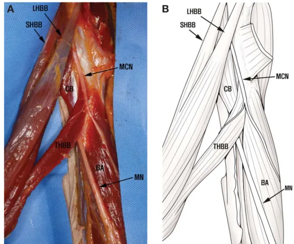
found in 10 male limbs (7.1%) and 4 female limbs (5.4%), and none of the cases was bilateral. The 4 cases of THBB in the found limbs were innervated by the musculocuta- neous nerve. THBB was a flat muscular belly-like structure that originated from the anteromedial surface of the hume- rus between the insertion of the coracobrachialis and the origin of the brachialis (Fig. 1). The biceps brachii with third head was supplied by the small branches of brachial artery, thus suggesting that the third head was a muscle of the anterior compartment of the arm.
Discussion
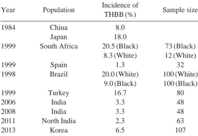
THBB has been reported in various populations with various incidence rates according to population, from 1.3%
for Spain to 20.5% for South Africa. In other countries, variation in the incidence of THBB according to the pop- 98 Je-Hun Lee
A B
Fig. 1. The third head of the biceps brachii. The distal part of the biceps brachii is detached from the bone to show the third head. LHBB:
long head of biceps brachii, SHBB: short head of biceps brachii, THBB: third head of biceps brachii, CB: coracobrachialis muscle, BA:
brachialis muscle, MCN: musculocutaneous nerve, MN: median nerve.
ulation can be a serious problem because of the clinical aspects of this condition. Therefore, the author of this arti- cle investigated the variation in the incidence of THBB in the Korean population and found an incidence of 6.5%.
This finding will be helpful in understanding the approach to the brachii region (Table 1).
Before the year 2000, the incidence of THBB was ¤8.0%, except in the Spanish population; however, after the year 2000, the incidence was ⁄3.3%. The current study showed a lower incidence of 6.5%, comparable with the findings before the year 2000. These trends are noticeable that THBB was found less than the findings before the year 2000.
The present study found that the incidence of THBB is higher in males than in females and that this difference is statistically significant (p⁄0.05). These results are in agre- ement with previously reported results that the incidence of THBB is relatively rare in females [3,9], except for the findings by Rodriguez-vazquez et al [11]. The various re- sults according to the population were obtained that a cer- tain study [7] was occurred more frequently in neonates than in adults. According to Asvat et al [3], THBB was more often found on the left side in males and only 1 case was found in a female, also on the left side. Kopuz et al [7] reported that all cases of THBB were bilateral in adult males. We found 10 unilateral limbs with THBB in this study. These results are attributed the appearance of this variant to racial trends.
A few researchers [3,7,9] found that THBB allows flex- ion of the elbow joint irrespective of the position of the shoulder joint. In the 14 cases of THBB in the present
study, THBB originated from the medial shaft of the hu- merus in common with and distal to the insertion of the coracobrachialis muscle (Fig. 1). This muscle belly pattern may facilitate elbow flexion.
In the present study, nerve innervations of THBB were investigated in 4 limbs of all specimens; THBB was inner- vated by the musculocutaneous nerve in 4 cases. Other studies [8,11] have reported similar findings. This finding may be clinically important because the nerve is subjected to compression by the bulky third head.
References
1. Abu-hijleh MF. Three headed biceps brachii muscle asso- ciated with duplicated musculocutaneous nerve. Clin Anat.
2005; 18:376-9.
2. Ashwini SJ, Shashikala RL. Case report: Third head of biceps brachii muscle a case study. Biomed Res. 2011; 22:387-9.
3. Asvat R, Candler P, Sarmiento EE. High incidence of the third head of biceps brachii in South African populations. J Anat. 1993; 182:101-4.
4. Bergman RA, Thompson SA, Afifi AK. Catalogue of human variation. Urban and Schwarzenberg: Munich; 1983. p. 27-30.
5. Cheema P, Singla R. Low incidence of the third head of the biceps brachii in the North Indian population. J Clin Diag Res. 2011; 5:1323-6.
6. Fating AS, Salve VM. A third head of the biceps brachii and coexisting fused higher origin of brachioradialis. Int J Anat Vari. 2011; 4:31-3.
7. Kopuz C, Sancak B, Ozbenli S. On the incidence of third head of biceps brachii in Turkish neonates and adults. Acta Anat Nippon. 1999; 74:301-5.
8. Kumar H, Das S, Rath G. An anatomical insight into the third head of biceps brachii muscle. Bratisl Lek Listy. 2008;
109:76-8.
9. Nayak SR, Prabhu LV, Sivanandan R. Third head of biceps brachii: a rare occurrence in the Indian population. Ann Anat. 2006; 188:159-61.
10. Neto HS, Camilli JA, Andrade JCT, Filho JM, Marques MJ.
On the incidence of the biceps brachii third head in Brazilian whites and blacks. Ann Anat. 1998; 180:69-71.
11. Rodriguez-vazquez JF, Velasco-merida JR, Collado JJ. Unu- sual variation of a third head of the biceps brachii muscle.
Ann Anat. 1999; 181:573-5.
12. Swieter MG, Carmichael SW. Bilateral three-headed biceps brachii muscle. Anat Anz. 1980; 148:346-9.
13. Warner JJP, Paletta GA, Warren RF. Accessory head of the biceps brachii. Clin Orthop Relat Res. 1992; 280:179-81.
Third Head of Biceps Brachii 99
Table 1. Comparison of incidence of third head of biceps brachii Year Population Incidence of
Sample size THBB (%)
1984 China 8.0
Japan 18.0
1999 South Africa 20.5 (Black) 73 (Black) 8.3 (White) 12 (White)
1999 Spain 1.3 32
1998 Brazil 20.0 (White) 100 (White)
9.0 (Black) 100 (Black)
1999 Turkey 16.7 80
2006 India 3.3 48
2008 India 3.3 48
2011 North India 2.3 63
2013 Korea 6.5 107
THBB, third head of the biceps brachii.
한국인 위팔두갈래근의 셋째갈래: 해부학적 연구
이제훈
건양대학교 의과대학 해부학교실
간추림 : 임상에서 위팔 손상시 나타날 수 있는 다양한 경우에 대한 이해를 돕고자 시신을 이용하여 위팔두갈 래근 셋째갈래의 빈도와 형태 및 발견된 4쪽에 대한 신경분포를 조사하였다.
신선 및 포르말린 고정시신 107구, 214쪽(남자: 70, 여자: 37, 나이: 51~87세)을 해부하여 위팔두갈래근의 셋 째갈래의 존재 유무를 조사하였다. 또한 위팔에 병변 및 비정상적인 소견이 의심되는 시신은 연구에서 제외시 켰다.
위팔두갈래근의 셋째갈래는 전체 경우 중 14쪽이 발견되었고 그 발생빈도는 6.5% 이었다. 발견된 경우 중, 남 자가 10쪽이고(7.1%) 여자가 4쪽이었으며(5.4%), 양쪽에서 동시에 발견된 경우는 없었다. 이 셋째갈래는 위팔근 의 이는곳과 부리위팔근의 닿는곳 사이 뼈에 부착하였고 지배신경은 근육피부신경이었다.
이 연구의 결과는 위팔뼈가 부러진 경우 또는 위팔 주변 신경의 죄임증후군과 어깨관절움직임을 이해하는 데 도움이 될 것이다.
찾아보기 낱말 : 셋째갈래, 위팔두갈래근, 근육피부신경, 해부 100 Je-Hun Lee
교신저자 : 이제훈(건양대학교 의과대학 해부학교실) 전자우편 : [email protected]