pISSN: 2234-8646 eISSN: 2234-8840 https://doi.org/10.5223/pghn.2018.21.1.68
Pediatr Gastroenterol Hepatol Nutr 2018 January 21(1):68-71
PGHN
Case Report
PEDIATRIC GASTROENTEROLOGY, HEPATOLOGY & NUTRITION
Colonic Angioectasia in an Adolescent Boy with
Hoyeraal-Hreidarsson on Long-Term Anabolic Steroid Therapy
Racha Khalaf and Carmen Cuffari*
Department of Pediatrics, Johns Hopkins All Children’s Hospital, The Johns Hopkins University School of Medicine, St.
Petersburg, FL, *Division of Pediatric Gastroenterology, The Johns Hopkins Hospital, Baltimore, MD, USA
Androgen therapy has proven efficacy in treating patients with bone marrow failure who are not candidates for bone marrow transplantation. Herein, we report on a case of colonic angioectasia secondary to oxymetholone use in an adolescent patient with Hoyeraal-Hreidarsson syndrome (HHS). A 13-year-old Caucasian male with HHS charac- terized by cerebellar hypoplasia, developmental delay, microcephaly, esophageal strictures and myelodysplasia presented with severe hematochezia from colonic angioectasia secondary to long-term oxymetholone therapy.
These vascular lesions resolved spontaneously once this anabolic steroid was discontinued. While androgen therapy is often recommended for certain anemias and myelodysplastic syndromes, clinicians should be aware of the poten- tial complication in developing these perceived uncommon colonic angioectasias. Moreover, pediatric gastro- enterologists should familiarize themselves in identifying these vascular lesions by colonoscopy, especially among the high risk groups on long-term anabolic steroid therapy.
Key Words: Hoyeraal-Hreidarsson syndrome, Colon, Angioectasia, Testosterone congeners
Received:June 16, 2017, Revised:July 11, 2017, Accepted:July 15, 2017
Corresponding author: Racha Khalaf, Department of Pediatrics, Johns Hopkins All Children’s Hospital, 601 Fifth Street South, Suite 608, St.
Petersburg, FL 33701, USA. Tel: +1-727-767-4106, Fax: +1-727-767-8804, E-mail: [email protected] Copyright ⓒ 2018 by The Korean Society of Pediatric Gastroenterology, Hepatology and Nutrition
This is an openaccess article distributed under the terms of the Creative Commons Attribution NonCommercial License (http://creativecommons.org/licenses/by-nc/4.0/) which permits unrestricted noncommercial use, distribution, and reproduction in any medium, provided the original work is properly cited.
INTRODUCTION
Hoyeraal-Hreidarsson syndrome (HHS), is a rare severe variant of dyskeratosis congenita (DKC) char- acterized by cerebellar hypoplasia, microcephaly, myelodysplasia and immunodeficiency [1,2]. The multi-systemic nature of this genetic disorder often precludes the application of hematopoietic stem cell transplantation among those patients with bone marrow failure. In these patients, androgen therapy
has provided an evidenced base therapeutic option [3-5].
Oxymetholone is a synthetic anabolic steroid that stimulates erythropoiesis through the increased pro- duction and secretion of erythropoietin. It has pro- ven efficacy in treating patients with myelodys- plastic syndromes, including Fanconi’s anemia [5].
Other studies have described a 70% success rate of oxymetholone therapy in patients with DKC [3].
Adverse effects associated with use of oxymetholone
www.pghn.org 69
Racha Khalaf and Carmen Cuffari:Colonic Angioectaisa in an Adolescent Body with Hoyeraal-Hreidarsson Syndrome
include: hepatotoxicity, cholestatic jaundice which is dose and duration dependent, pelosis hepatitis, and benign and malignant hepatic tumors [6].
Oxymetholone has also been linked to decreased an- ticoagulant tolerance and hyperlipidemia. Other common side effects associated with the long-term use of anabolic steroids include: virilization in fe- males, gynecomastia, amenorrhea, changes in libido and impaired thyroid function [4,6]. To our knowl- edge, this is the first case report in the literature at- tributing colonic angioectasia secondary to oxy- metholone in a pediatric patient with HHS. Written informed consent was obtained from the patient for publication of this case report and accompanying images.
CASE REPORT
Our patient is a 13-year-old Caucasian male with cerebellar hypoplasia, skin dyskeratosis with hyper- pigmentation, nail atrophy, esophageal strictures, urethral meatal stricture, developmental delay, and bone marrow failure who presented with hema- tochezia. He was initially diagnosed with devel- opmental delay and failure to thrive as an infant.
Features of DKC, and ultimate diagnosis with HHS was not made until he was about 8 years old when he presented with notable anemia, mild thrombocyto- penia, and mild leukopenia. He was diagnosed with HHS based on his classical clinical features. Genetic testing was negative for mutations associated with DKC but revealed a telomere length less than the first percentile. When he was diagnosed with bone marrow failure syndrome, he was placed on a trial of androgens that notably improved his counts so that he was transfusion independent.
At age 11 years, a national shortage of androgens led to an interruption in oxymetholone therapy last- ing two years. When the treatment course was re- sumed with 50 mg of daily oxymethalone, the pa- tient failed to respond to therapy, at which point he was evaluated for bone marrow transplantation. He presented to an outlying facility with low grade fe- ver, neutropenia and severe hematochezia. A flexible
sigmoidoscopy showed friable colonic tissue, and two small rectal ulcers with an overlying eschar. No biopsies were obtained secondary to the elevated risk of bleeding. In addition, no esophagogastroduo- denoscopy was performed due to an esophageal stricture. Stool cultures were negative for all in- fectious causes of hemorrhagic colitis, including cytomegalovirus. The suspicion for inflammatory bowel disease was low.
One week thereafter, the patient developed anoth- er episode of bright red blood per rectum associated with neutropenia and fever. Upon admission to Johns Hopkins All Children's Hospital, the patient had a platelet count of 22/μL, hemoglobin was 8.1 g/dL, white blood cell count was 0.72×10³/μL, and the international normalized ratio was 1.2. Physical examination of the abdomen was benign. Once again, an infectious workup, including stool ad- enovirus, cytomegalovirus, norovirus, enterovirus, cryptosporidium, giardia, clostridium difficile, Escherichia coli and other bacterial stool pathogens were negative. The patient continued to have profuse hematochezia that required multiple transfusions of packed red blood cells. Bleeding was bright red would suggesting a colonic source for the bleeding.
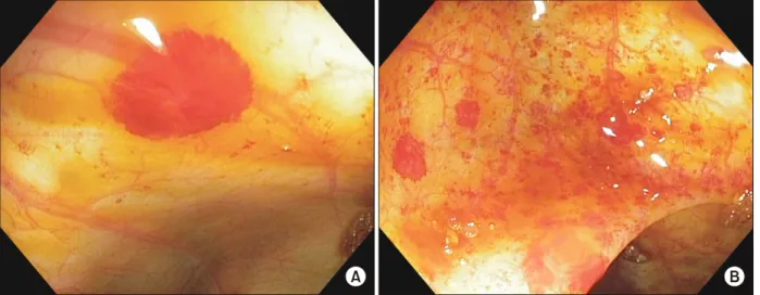
An abdominal ultrasonography and doppler was normal. A sedated abdominal and pelvic magnetic resonance enterography was unremarkable. On co- lonoscopy, several large angioectasias were identi- fied at the hepatic flexure and cecum (Fig. 1). No le- sions were actively bleeding and biopsies were not obtained due to the high risk of re-bleeding. In addi- tion, the patient had no evidence of liver cirrhosis.
Since the patient had been evaluated for bone marrow transplantation, the oxymetholone was dis- continued. Interestingly, the hematochezia sponta- neously resolved and the neutropenia improved. The patient was then discharged home. A follow-up colo- noscopy, performed 40 days post colonoscopy showed complete resolution of the colonic angioectasias. The planned laser ablation of these angioectasias was thus aborted. Alternative therapeutic options to laser ablation include placement of endoscopic hemoclips and thermal therapy. The patient did not have any
70 Vol. 21, No. 1, January 2018 Pediatr Gastroenterol Hepatol Nutr
Fig. 1. (A) Angioectasia at the hepatic flexure, (B) diffuse angioectasias of the ascending colon.
further episode of hematochezia and no further in- tervention was indicated.
DISCUSSION
To the best of our knowledge, this is the first report of a case of colonic angioectasia secondary to oxy- metholone in a pediatric patient with HHS. This ge- netic syndrome is unlikely to be the source of these abnormalities since the lesions disappeared sponta- neously once the anabolic steroid was discontinued.
A review of the literature revealed one case of colitis associated with DKC. In this case, the patient was re- ported to have idiopathic colitis, and portal hyper- tension [7]. No similar cases have been identified in patients with HHS. In our patient, the abdominal ul- trasonography with Doppler did not show portal hypertension.
One study described an association between the androgen danazol and hemorrhagic cystitis. In this case series of 69 patients with angioneurotic edema, 13 had developed hematuria while on danazol therapy.
In these patients, cystoscopic evaluation showed a nonspecific pattern of erythema, submucosal te- langiectasia and neovascularity. Interestingly, as in our patient, these vascular lesions also sponta- neously resolved once the anabolic steroid was dis- continued [8]. Using the Naranjo adverse drug re-
action probability scale, the yield shows a score of 7 which is indicative of a probable association between oxymetholone and the development of angioectasia.
With the increased use of anabolic steroids to treat patients with bone marrow failure, the potential risk in developing angioectasias must be underscored.
Moreover, pediatric gastroenterologists should also familiarize themselves with these vascular lesions since they are perceived as uncommon causes of in- testinal bleeding in children. Further studies are needed to elucidate the pathogenesis of anabolic ste- roid induced angioectasia.
REFERENCES
1. Glousker G, Touzot F, Revy P, Tzfati Y, Savage SA.
Unraveling the pathogenesis of Hoyeraal-Hreidarsson syndrome, a complex telomere biology disorder. Br J Haematol 2015;170:457-71.
2. Ohga S, Kai T, Honda K, Nakayama H, Inamitsu T, Ueda K. What are the essential symptoms in the Hoyeraal-Hreidarsson syndrome? Eur J Pediatr 1997;
156:80-1.
3. Islam A, Rafiq S, Kirwan M, Walne A, Cavenagh J, Vulliamy T, et al. Haematological recovery in dysker- atosis congenita patients treated with danazol. Br J Haematol 2013;162:854-6.
4. Khincha PP, Wentzensen IM, Giri N, Alter BP, Savage SA. Response to androgen therapy in patients with dys- keratosis congenita. Br J Haematol 2014;165:349-57.
www.pghn.org 71
Racha Khalaf and Carmen Cuffari:Colonic Angioectaisa in an Adolescent Body with Hoyeraal-Hreidarsson Syndrome
5. Shimamura A, Alter BP. Pathophysiology and manage- ment of inherited bone marrow failure syndromes.
Blood Rev 2010;24:101-22.
6. Pavlatos AM, Fultz O, Monberg MJ, Vootkur A, Pharmd. Review of oxymetholone: a 17alpha-alkylated anabolic-androgenic steroid. Clin Ther 2001;23:789- 801; discussion 771.
7. Brown KE, Kelly TE, Myers BM. Gastrointestinal in-
volvement in a woman with dyskeratosis congenita. Dig Dis Sci 1993;38:181-4.
8. Andriole GL, Brickman C, Lack EE, Sesterhenn IA, Javadpour N, Linehan WM, et al. Danazol-induced cys- titis: an undescribed source of hematuria in patients with hereditary angioneurotic edema. J Urol 1986;
135:44-6.