INTRODUCTION
Electrical injury can cause cardiac arrhythmias, myocardial/
valvular rupture, structural changes in coronary arteries, peri- cardial effusion, and various electrocardiographic changes. In this report, we present a patient who developed sick sinus syn- drome (SSS) as a result of electrical injury.
CASE REPORT
A 20-yr-old male patient suffering from dizziness and syn- copal attacks was referred to our hospital. He had a history of high-voltage electrical injury at the age of 12, and other- wise showed no prior history of cardiac illness. After a care- ful interview of the patient, we learned that the symptoms, such as dizziness and transient loss of consciousness of less than 1-min duration, developed after the accident associated with electrical energy and were precipitated by exercise.
On physical examination, the arterial blood pressure and heart rate were 120/80 mmHg and 44 beats per minute, res- pectively. Cardiovascular assessment was normal. On chest ex- amination, there was a large cutaneous burn scar extending from the right subclavicular to the left subscapular area on his chest wall. Other systemic examinations were normal. Sur- face electrocardiogram demonstrated a junctional rhythm (Fig.
1). Echocardiographic assessment revealed a normal left ven- tricular size and function, and no valvular pathology. Holter monitoring showed sinus bradycardia with a sinus pause of 4.6 sec and concomitant dizziness. Routine laboratory tests,
including serologic tests for collagen tissue diseases, were in normal values.
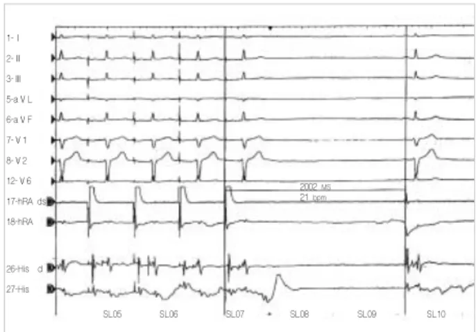
After 8-hr-fasting, a written informed consent was obtained, electrophysiological study (EP) was performed for the evalua- tion of the functions of sinoatrial (SA) and atrioventricular (AV) nodes. The findings were as follows: baseline cycle length (BC L), 1,200 msec; atrial-His interval (AH), 90 msec; His-ven- tricle interval (HV), 48 msec; Wenckebach point, 480 msec;
sinoatrial conduction time, 300 msec; sinus-node recovery time (SNRT), 2,800 msec; corrected sinus node recovery time (cSN- RT), 1,600 msec (Fig. 2). An injection of 2 mg of atropine failed to increase the sinus rate over 90 beats per minute, sug- gesting chronotropic incompetence. SNRT and cSNRT did not shorten after atropine injection.
We decided that the sinus node was not functioning nor- mally. This abnormal function of sinus node, associated with clinical symptoms, led us to a diagnosis of SSS. Coronary angio- graphy was performed to exclude possible coronary artery dis- ease, even if there was no family history related to coronary heart disease or other risk. The angiography showed a normal coronary anatomy. Since the patient had chronotropic incom- petence, we implanted a DDD-R pacemaker (Kappa, Medtro- nic, Inc., Minneapolis, MN, U.S.A.) to the left subclavian region. However, during implantation procedure, the pacing thresholds of both ventricle and atrium were higher than ex- pected. The values measured during the procedure were as follows: p-wave amplitude, 5.6 mV; atrial pacing threshold, 1.5 V; atrial lead impedance, 476 Ohm; R-wave amplitude, 15.2 mV; ventricle pacing threshold, 1.2 V; and ventricle lead impedance, 538 Ohm.
114 J Korean Med Sci 2003; 18: 114-5
ISSN 1011-8934
Copyright � The Korean Academy of Medical Sciences
Electrical Injury As A Possible Cause of Sick Sinus Syndrome
Electrical injury is a serious public health problem. Heart is one of the most fre- quently affected organs. Electrical injury can cause life-threatening cardiac com- plications such as asystole, ventricular fibrillation, and myocardial rupture. In this case report, we present a 20-yr-old male patient with sick sinus syndrome that devel- oped years after electrical injury.
Key Word : Electric Injuries; Side Sinus Syndrome; Turkey
Sedat Kose, Atila Iyisoy,
Hurkan Kursaklioglu, Ertan Demirtas
Department of Cardiology, Gulhane Military Medical Academy, Ankara, Turkey
Address for correspondence Sedat KOSE, M.D.
Gulhane Askeri Tip Akademisi Kardiyoloji AD Etlik, Ankara, TURKEY 06018
Tel : +90-312-304 2352, Fax : +90-312-304 4250 E-mail: [email protected]
Received : 27 December 2001 Accepted : 2 May 2002
Sick-Sinus-Syndrome after Electrical Injury 115
DISCUSSION
SSS is a disease frequently encountered in the elderly popu- lation, especially in the 6th and 7th decades. It may frequent- ly result from decreased pace cells in SA node with aging, and fibrotic and calcific process of SA and AV node regions. Other etiologies may include hypothyroidism, ischemic heart dis- ease, a number of pharmacologic agents such as digoxin, cal- cium-channel blockers, beta-blockers, class-I anti-arrhythmic agents, and amiodarone, prior cardiac operation, and heart transplantation.
Heart is one of the most vulnerable organs against electric- ity. Various myocardial manifestations develop at the time of injury. These include asystole, ventricular fibrillation, which may cause immediate death, QT-prolongation, right bundle branch block, complete AV block, valvular or myocardial rup- ture, CK-MB elevations caused by myocardial injury, struc- tural changes in the small coronary vessels, and pericardial effusion (1).
In young patients, SSS is usually related to congenital heart disease (CHD) or seen after operation for correcting congenital heart disease (2, 3). In our case, there was no history of CHD or operation for CHD.
James et al. (4) reported autopsy findings of 4 patients died from electrical injury. They noticed an increased myocardial thickness in two of them, fibromuscular changes in the coro- nary arteries in one of them, and fatty infiltration in the vicini- ty of both SA and AV regions in the last one. We speculated that a probable infiltration in the SA node could be responsi- ble for the clinical manifestations of our case. The probable myocardial injury due to electricity may explain why the levels of thresholds of both atrial and ventricular pacing were higher than expected during pacemaker implantation. Unfortunate- ly, the patient refused endomyocardial biopsy.
Bognolo et al. (5) reported a 50-yr old patient with SSS, sug-
gesting non-penetrating chest trauma as a causative factor. As described previously, electricity can cause mechanical trau- ma to the body at the same time. This kind of trauma could also cause SSS. Carleton (6) recommended that all patients in- jured by electrical energy should be followed because of the risk of developing cardiac manifestations for at least one year, and avoid any cardiac operation during the first six months. In our case, the symptoms developed several months after the accident and unfortunately, the diagnosis was confirmed 8 yr after the accident. This case merits attention in that there has been no case report on SSS caused by electrical injury.
In conclusion, electrical injury can infrequently cause SSS.
In such cases, high pacing threshold levels can be encountered during pacemaker implantation.
REFERENCES
1. Solem LD, Fischer RP, Strate RG. The natural history of electrical injury. J Trauma 1977; 17: 487-92.
2. Marcus B, Gillette PC, Garson A Jr. Electrophysiologic evaluation of sinus node dysfunction in postoperative children and young adults utilizing combined autonomic blockade. Clin Cardiol 1991; 14: 33-40.
3. Albin G, Hayes DL, Holmes DR Jr. Sinus node dysfunction in pedi- atric and young adult patients: treatment by implantation of a per- manent pacemaker in 39 cases. Mayo Clin Proc 1985; 60: 667-72.
4. James TN, Riddick L, Embry JH. Cardiac abnormalities demonstrat- ed postmortem in four cases of accidental electrocution and their potential significance relative to nonfatal electrical injuries of the heart. Am Heart J 1990; 120: 143-57.
5. Bognolo DA, Rabow FI, Vijayanagar RR, Eckstein PF. Traumatic sinus node dysfunction. Ann Emerg Med 1982; 11: 319-21.
6. Carleton SC. Cardiac problems associated with electrical injury. Car- diol Clin 1995; 13: 263-6.
Fig. 1.Surface 12-lead ECG recorded at rest in the patient. Fig. 2.Sinus node recovery time during electrophysiologic study (paper speed: 50 mm/sec). hRA: high right atrium; His: His bun- dle.
1-Ⅰ 2-Ⅱ 3-Ⅲ 5-aⅤL 6-aⅤF 7-Ⅴ1 8-Ⅴ2 12-Ⅴ6 17-hRA ds 18-hRA
26-His d 27-His
SL05 SL06 SL07 SL08 SL09 SL10
2002 MS 21 bpm