내측두엽 경화증(mesial temporal sclerosis) 또는 해마 경 화증(hippocampal sclerosis)은 성인에서 난치성 측두엽 간질 환자의 가장 흔한 병리학 및 영상 진단 소견으로, 측두엽 간질 로 수술 받은 환자 중 60-75%에 달한다 (1). 내측두엽 경화 증의 가장 중요한 자기공명영상 소견은 해마의 경화를 반영하 는 T2 신호 증가와 용적의 감소이다. 이 외에도 Papez회로 (Papez circuit)와 연관된 해마외 변연계(extrahippocampal limbic system) 및 다른 뇌구조물의 신호 변화나 형태 변화가 보고되고 있으며, 자기공명영상에서 확인 가능한 해마외 구조 의 이상 소견으로는 전방 측두엽 피질(anterior temporal lobe cortex), 편도핵(amygdala), 부해마회(parahippocampal gyrus)의 백질(collateral white matter) 등의 신호 변화와 측 두엽, 편도핵의 형태변화 등이 있다 (2-9). 그러나 이런 해마 외 구조의 이상은 해마 경화증의 이차적인 소견으로 여겨지고 있으며, 그 진단적 가치나 의의는 아직까지 주목을 받지 못했 다.
자기공명영상은 뇌에서 뛰어난 공간해상능력과 조직 대조도 를 보이므로 간질 발작을 초래하는 병소를 진단 및 국소화하 는데 매우 유용하여, 현재 간질환자 특히, 난치성 간질 환자의 평가에 있어 표준화된 진단 방법이 되고 있다 (10). 그러나 아 직까지 국내에서는 측두엽 간질 환자들을 대상으로 한 내측두 엽 경화증의 자기공명영상 소견에 대한 문헌은 매우 적다 (9, 11-13). 이에 저자는 우리나라 환자들을 대상으로 내측두엽 경화증으로 진단된 환자들의 측두엽 자기공명영상을 분석하여 보고자 하였다.
대상과 방법
1994년 7월 15일에서 1999년 7월 14일까지 5년간 본원에 서 복합부분발작을 주소로 측두엽 자기공명영상을 시행하였던 733명의 환자들 중에서 외상성, 혈관성, 또는 다른 기질적 원 인이 있는 예를 제외하고, 최종진단이 내측두엽 경화증인 환자 116명(남자 64명, 여자 52명, 연령 분포 16세-72세, 평균 31 세)을 대상으로 하여 이들의 자기공명영상 소견을 후향적으로
내측두엽 경화증 환자의 자기공명영상 소견
1김 민 정・김 선 용・서 정 호
목적: 자기공명영상으로 내측두엽 경화증으로 진단된 환자들의 자기공명영상 이상소견을 알아 보고자 하였다.
대상과 방법: 복합부분발작을 주소로 측두엽 자기공명영상을 시행하였던 환자들 중에서 내측두
엽 경화증으로 최종진단된 환자 116명(수술로 내측두엽 경화증이 확진된 18명 포함)의 자기 공명영상 소견을 후향적으로 분석하였다. 자기공명영상에서 해마의 이상여부 및 해마외 뇌구 조의 비정상적 소견을 다양한 영상기법을 통하여 분석하였다.
결과: 측두엽 자기공명영상에서 내측두엽 경화증으로 진단된 116명 중에서 해마의 이상소견을 보인 예가 113명(97.4%), 해마외 구조의 이상소견을 보인 예가 20명(17.2%)이었다. 해마의 이상 소견으로는 95명(82.8%)에서 해마의 용적이 감소하였고 해마의 신호 변화는 53명(45.7%) 에서 보였는데, T1 신호 감소가 13명(11.2%)에서, T2 신호 증가가 50명(43.1%)에서 나타났 으며 이 중 T1과 T2신호가 모두 변한 경우는 10명(8.6%)이었다. 해마외 구조의 이상이 있는 20명 중에서 변연계의 이상으로는 원개 위축(n=10), 편도 위축(n=10), 유두체 위축(n=8), 전 방 시상핵의 위축과 T2 신호 증가(n=2), 그리고 대상 회전의 위축(n=2) 등이 관찰되었다. 그 외 구조의 변화로는 전방 측두엽 피질의 위축 또는 신호 증가(n=8)와 대뇌반구의 편측 위축 (n=4)이 나타났다.
결론: 내측두엽 경화증의 가장 중요한 자기공명영상 소견은 해마의 위축과 T2 신호강도의 증가 이지만 해마외 변연계 및 측두엽, 대뇌 피질, 편도 등 연관된 뇌구조에도 이상 소견을 동반할 수 있으므로, 자기공명영상에서 해마의 경화와 동반된 여러 이상 소견을 주의 깊게 살펴야 한다.
1아주대학교 의과대학 진단방사선과학교실
이 논문은 2000년 1월 17일 접수하여 2000년 5월 4일에 채택되었음.
분석하였다. 이들 중에서 측두엽 절제술 또는 해마 절제술 후 병리적으로 내측두엽 경화증이 확진된 환자는 18명이었다.
사용한 기기는 1.5T 자기공명영상기기(Signa, GE medical system, Milwakee, Wisconsin, U.S.A.)였다. 시상면 T1 강조 영상(TR/TE, 500msec/12-20msec), 축상면 및 관상면 T2 강조영상(TR/TE, 4000msec/120msec), 축상면 양자 밀도 (proton density)강조영상(TR/TE, 4000msec/15-20msec), 축상면 및 관상면 FLAIR(fluid attenuated inversion recovery) 영상(TR/TE, 10,000msec/120-140msec) 및 3D- FGR(three-dimensional fast gradient recalled echo) 또는 3D-IR(inversion recovery)영상 등의 기법을 이용하여 자기 공명영상을 얻었다.
자기공명영상에서 해마의 이상여부는 신호의 변화(T2 신호 증가, T1 신호 감소)와 형태의 변화(용적 감소, 내부구조의 파 괴)로 판단하였으며 원개(fornix), 유두체(mammillary body), 전방 시상핵(anterior thalamic nuclei) 및 대상 회전(cingulate gyrus) 등의 변연계(limbic system)와 전방 측두엽피질, 편도 (amygdala), 및 반대측 소뇌반구(contralateral cerebellar hemisphere) 등 해마외 구조물(extrahippocampal structure)의 비정상적 소견에 대하여 분석하였다. 해마의 용적감소를 포함 한 모든 이상 소견은 육안적 진단에 의존하였다.
병리적으로 내측두엽 경화증이 확진된 18명 환자들의 수술 후 치료 결과는 임상 의무 기록지를 토대로 Engel 등급에 따 라 분류하였다 (14).
결 과
측두엽 자기공명영상으로 내측두엽 경화증이 진단된 116환 자들의 자기공명영상을 분석한 결과는 Table 1에 요약하였다.
이들 중에서 일측성 내측두엽 경화증으로 진단된 환자는 112 명으로, 오른쪽에 이상 소견을 보인 경우는 64명, 왼쪽에 이상 소견을 보인 경우는 48명이었고, 양측성 내측두엽 경화증으로 진단된 환자는 4명(3.4%)이었다. 해마의 이상 소견으로는 전 체 116예 중 113명(97.4%)에서 나타났다. 95예(82.8%)에서 해마의 용적이 감소하였고 (Figs. 1A, 2, 4A), 해마의 신호 변 화는 53명(45.7%)에서 보였는데, T1 신호 감소가 13명 (11.2%)에서, T2 신호 증가(Figs. 1B, 2, 4A)가 50명(43.1%)
에서 나타났으며, 이 중 T1과 T2신호가 모두 변한 경우는 10 명(8.6%)이었다. 해마외 구조의 변화는 모두 20명(17.2%)에 서 보였다. 해마외 변연계의 이상 소견으로는 원개 위축이 10 명(8.6%), 유두체 위축(Fig. 2)이 8명(6.9%), 편도 위축(Fig.
3)이 10명(8.6%), 전방 시상핵의 위축(Fig. 4B)과 T2신호 증 가가 2명(1.7%), 그리고 대상 회전의 위축이 2명(1.7%)에서 나타났다. 그 외 구조의 변화로는 측두엽 피질의 위축 또는 T2 신호 증가(Fig. 4A)가 8예(6.9%)에서, 대뇌반구의 편측 위축 (cerebral hemiatrophy)이 4명(3.4%)에서 보였다.
수술 후 1년에서 5년 사이 기간동안의 임상 의무 기록지를 검토하여 판정한 수술 치료 결과는 Engel Class I(seizure free, with or without auras: 전구증상의 유무와는 상관없이 경련이 없어진 경우)이 10명(55.6%), Engel Class II(rare seizures (≤2/year): 경련 회수가 1년에 2회 이하로 감소)가 5명 (27.8%), Engel Class III(worthwhile improvement (>90%
decrease in seizure frequency): 경련 회수가 90%이상 감소) 가 2명(11.1%), Engel Class IV(no worthwhile improvement
Fig. 1. Coronal T1-weighted fast gradient recalled echo MR image through anterior hippocampus (A) shows volume loss and distortion of internal architecture on the right hippocampus (arrow).
Coronal T2-weighted fast spin- echo MR image (B) shows in- creased signal intensity of the right hippocampus (arrow).
A B
Table 1. MR Imaging Findings of Mesial Temporal Sclerosis(n=116)
Finding No. of case(%)
Hippocampus abnormalities 113(97.4%)
Changes in signal intensity 053(45.7%) Decreased T1-weighted signal intensity 013(11.2%) Increased T2-weighted signal intensity 050(43.1%)
Morphological changes 096(82.8%)
Decreased volume 095(81.9%)
Disruption of the internal hippocampal
structure 013(11.2%)
Extrahippocapal abnormalities 020(17.2%)
Atrophy of fornix 1000(8.6%)
Atrophy of mammillary body 0080(6.9%) Increased signal intensity or atrophy of Anterior
thalamic nucleus 0020(1.7%)
Atrophy of cingulate gyrus 0020(1.7%)
Cerebral hemiatrophy 0040(3.4%)
Increased signal intensity of anterior temporal
lobe cortex 0080(6.9%)
Increased signal intensity or atrophy of amygdala0100(8.6%) Atrophy of contralateral cerebellar hemisphere 0000(0%)
(<90% decrease in seizure frequency): 경련 회수의 감소가 90%미만)가 1명(5.6%)으로 나타났다 (14).
고 찰
내측두엽 경화증으로 인해 유발되는 난치성 측두엽 간질은 수술 대상을 신중하게 선택한 경우에 수술 성공률이 70-90%
에 이를 정도로 수술 결과가 매우 좋다 (15). 수술적 치료를 고려할 때 반드시 확인해야 할 점은 그 정확한 간질병소의 확 인으로, 이는 수술 결과와 서로 유의한 상관관계가 있기 때문 이다. 수술 전 검사에서 자기공명영상과 뇌파 검사가 일치하는 간질 발생 병소의 편측화(concordant lateralization)가 이루어 진 경우에는 수술 성공률이 97%인 반면, 자기공명영상으로 편 측화를 할 수 없었던 경우(nonlateralization)는 수술 성공률이 42%, 자기공명영상과 뇌파 검사에서 편측화가 서로 반대로 이 루어진 경우(disconcordant lateralization)에는 성공률이 33%
로 보고 되었다 (16).
내측두엽 경화증의 조직병리 소견으로는 주로 해마 아몬각 1(Cornu Ammonis 1: 이하 CA1)과 문(Hilum)(혹은 CA3과 CA4)에서 신경원 소실(neuronal loss)과 신경교증(gliosis)이 보이고 CA2와 치상회(Dentate gyrus)에서는 경미한 변화만이 보인다고 알려져 있으나, 심한 경우 해마의 모든 분획, 치상회, 그리고 편도와 부해마회 등의 해마외 구조에도 변화가 나타날 수 있다 (17-19).
난치성 간질환자의 자기공명영상에서 관찰되는 가장 흔하고 중요한 소견은 해마의 T2 신호 증가나 용적 감소로 밝혀져 있 다. 해마의 위축은 신경원 소실에 해당하는 소견이며 T2 신호 증가는 신경교증에 해당하는 변화이다 (8, 20). 초기에는 내측 두엽 경화증의 8%에서만 자기공명영상의 이상 소견을 발견할 수 있었으나(21), 고속스핀에코 자기공명영상 기법의 도입으 로 70-80% 정도의 신호 증가가 보고되었고 해마의 위축이나 T2 강조영상에서의 신호 증가는 이후 간질 병소의 위치 결정 에 있어서 민감한 방법으로 인정 받고 있다 (6, 22, 23). 정량 적인 분석 방법으로는 자기공명영상을 통한 해마의 용적 측정 술(MR volumetry)과 T2 Relaxometry가 있다. 최근에는 자기 공명분광법(MRS ; magnetic resonance spectroscopy)을 이 용하여 간질 병소쪽의 뇌조직에서 N-acetylaspartate라는 물 질이 감소한다는 것이 확인되었다 (24).
내측두엽 경화증에서의 해마외 변연계 변화에 대한 연구는 Margerison과 Corsellis(1966년)가 만성 측두엽 간질 환자들 의 뇌를 부검하여 해마뿐만 아니라 변연계의 다른 구조들과 내 측두엽에서도 이상 소견을 발견했다고 보고함으로써 시작되었 다 (17). 이들은 난치성 측두엽 간질 환자들에서 간질에 의한 해마의 신경원 소실과 신경교증을 보고하였으며, 또한 전형적 인 내측두엽 경화증 22예 중 20예에서 시상(50%), 편도(60%), 및 소뇌(65%) 등 해마외 구조에서 조직학적 이상소견을 확인 했다고 보고했다 (17). 내측두엽 경화증에서 보이는 이러한 해 마외 변연계 구조의 변화는 1937년에 제안된 Papez 회로로 설
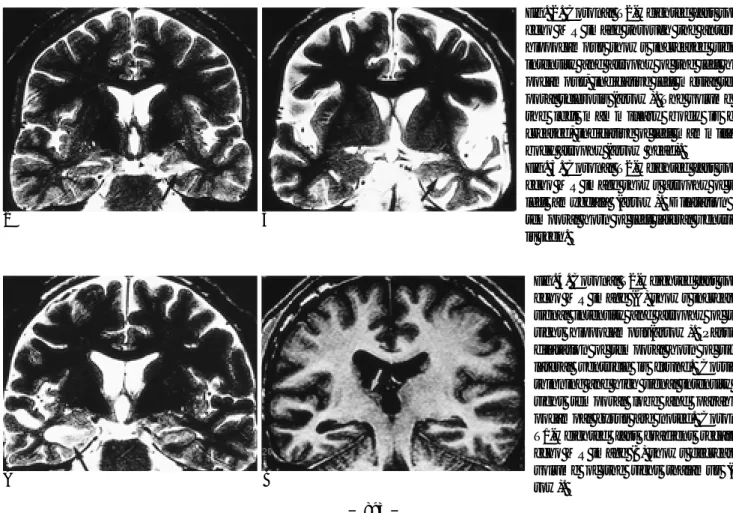
Fig. 2. Coronal T2-weighted fast spin- echo MR image through the anterior hippocampus shows increased signal intensity and atrophy of the left hip- pocampus, indicative left mesial tem- poral sclerosis (arrow). The volume of the left mammillary body is de- creased, indicative of left mammillary body atrophy (arrow head).
Fig. 3. Coronal T2-weighted fast spin- echo MR image shows atrophy of the left amygdala (arrow). Dilatation of temporal horn of left lateral ventricle is seen.
2 3
Fig. 4. Coronal T2-weighted fast spin- echo MR image (A) shows increased signal intensity and atrophy of the right hippocampus(arrow). Passive dilatation of temporal horn of right lateral ventricle is found. Cortical thinning and high signal intensity in right temporal lobe and parahip- pocampal gyrus are noted. Coronal T1-weighted fast gradient recalled echo MR image (B) shows decreased volume of the right thalamus (ar- row).
A B
명할 수 있다. Papez는 대상 회전과 해마체(hippocampal formation), 원개 및 동측 시상하부의 유두체로의 연결을 통해 대뇌 피질이 시상하부에 영향을 미친다는 회로 개념을 도입하 였다 (25). 유두체에서 유두체시상경로(mammillothalamic tract)를 통해 전방 시상핵이 연결되고, 시상대상섬유 (thalamocingulate fibers)를 통해 전방 시상핵에서 대상 회전 이 연결됨으로써 이 회로는 완성된다 (26). 이후로 많은 해부 학적 연구가 Papez의 변연계 개념을 뒷받침하였으며 최근 Maclean 등은 변연계 회로 내에 편도, 후두 전두엽, 및 시상하 부의 일부를 포함시켰다 (27). 최근 연구에서 내측두엽 경화 증 환자에서 이런 해마외 변연계의 이상 소견에 대한 자기공 명영상의 보고가 활발히 이루어 지고 있으며, Papez 회로를 통 해 해마와 직접 연결되는 원개나 유두체에서는 해마와 직접적 인 연결은 없다고 알려진 전방 시상핵과 대상 회전 등의 변연 계 구조보다 자기공명영상의 변화가 더 흔한 것으로 보고되었 다(28). Chan 등의 연구에서는 42명의 내측두엽 경화증 환자 중에서 Papez 회로 내의 해마외 구조의 이상으로 원개 위축이 23명(55%), 유두체 위축이 11명(26%)에서 관찰된 반면, 전 방 시상핵이나 대상 회전의 이상 소견을 보인 예는 없었고, Papez 회로 밖의 해마외 구조의 이상으로 대뇌반구의 편측 위 축이 1명, 전방 측두엽 피질의 신호증가가 16명에서 보였고, 편도의 이상이나 반대측 소뇌반구의 위축을 보인 경우는 없었 다 (28). 본 연구에서도 116명의 내측두엽 경화증 환자 중에 서 원개 위축은 10명, 유두체 위축은 8명에서 보였고 전방 시 상핵과 대상회전의 이상 소견은 각각 2명에서 보여 다른 연구 들과 마찬가지로 해마와 직접 연결되는 구조에서 동반된 변화 를 더 흔히 관찰할 수 있었다.
내측두엽 경화증에서 해마외 구조의 손상의 진단적 의의가 아직 많은 주목을 받지 못하였지만 동반된 변연계 구조의 손 상도 해마경화증과 유사한 흥분성 독성 기전에 의해 생길 수 있을 것으로 생각되며, 간질에 의한 이상 전기 활동이 Papez 회로 등과 같은 정상 생리적 경로를 따라 전파된다면 마찬가 지로 흥분성 독성 손상이 같은 경로를 따라 퍼질 수 있을 것 으로 생각된다 (28).
결론적으로, 내측두엽 경화증의 가장 중요한 자기공명영상 소견은 해마의 이상소견이지만 해마외 변연계 및 그 외 뇌 구 조에도 이상소견을 동반할 수 있으므로 고해상도 자기공명영 상에서 해마경화 유무 외에도 해마외 구조의 여러 이상 소견 등이 동반되어 있는지 주의 깊게 살펴 보아야 한다.
참 고 문 헌
1. Wartson C, Cendes F, Fuerst D, et al. Specificity of volumetric magnetic resonance imaging in detecting hippocampal sclerosis.
Arch Neurol 1997;54:67-73
2. Meiners LC, van Gils A, Jansen GH, et al. Temporal lobe epilepsy : the various MR appearances of histological proven mesial tempo- ral sclerosis. AJNR Am J Neuroradiol 1994;15:1547-1555
3. Baldwin GN, Tsuruda JS, Maravilla KR, Hamills GS, Hayes CE.
The fornix in patients with seizures caused by unilateral hip- pocampal sclerosis : detection of unilateral volume loss on MR im-
ages. AJNR Am J Neuroradiol 1994;162:1185-1189
4. Mamourian AC, Brown DB. Asymmetric mamillary bodies : MR identification. AJNR Am J Neuroradiol 1993;14:1332-1335 5. Grant R, Hadley DM, Condon B, et al. Magnetic resonance imag-
ing in the management of resistant focal epilepsy : Pathological case report and experience of 12 cases. J Neurol Neurosurg Psych 1987;50:1529-32
6. Heinz R, Crain BJ, Radtke RA, Burger PC. MR imaging in patients with temporal lobe seizures : Correlation of results with pathologic findings. AJNR Am J Neuroradiol 1990;11:827-32
7. Heinz R, Ferris N, Lee EK, et al. MR and Positron emission tomog- raphy in the diagnosis of surgically correctable temporal lobe epilepsy. AJNR Am J Neuroradiol 1994;15:1341-48
8. Jackson GD, Berkovic SF, Duncan JS, Duncan JS. Optimizing the diagnosis of hippocampal sclerosis using MR imagings. AJNR Am J Neuroradiol 1993;14:753-62
9. Cheon JE, Chang KH, Won HJ, et al. Magnetic resonance imaging findings in temporal lobe epilepsy. International Journal of Neuroradiology 1997;3:199-205
10. Kuzniecky RI, Jackson GD. Magnetic resonnance in epilepsy. New York : Raven Press, 1995:107-183
11. 김종덕, 박동우, 은충기, 정덕환, 황태규. 소아 복잡성(초점성) 부분 간질의 뇌자기공명영상 소견. 대한방사선의학회지 1992;28:631- 638
12. Cheon JE, Chang KH, Kim HD, et al. MR of hippocampal sclorosis:
comparision of qualitative and quantitative assessments. AJNR Am J Neuroradiol 1998;19:465-468
13. 손석현, 장승국, 은충기. 내측두 경화증의 진단에 있어 액체감쇠역 전회복 영상의 유용성: 고속스핀에코 T2강조영상과의 비교. 대한방 사선의학회지 1999;41:1071-1076
14. Engel J Jr. Outcome with respect to epileptic seizures. In: Enjel J Jr, eds. Surgical Treatment of the epilepsies. New York : Raven Press, 1987:553-571
15. Engel J Jr, Van Ness P, Rassmussen TB, Ojemann LM. Outcome with respect to epileptic seizures. In: Enjel J Jr, 2nd eds. Surgical Treatment of the epilepsies. New York : Raven Press, 1993:609-621 16. Jack CR, Sharbrough FW, Cascino GD, Hirschorn KA, O’Brien
PC, Marsh WR. Magnetic resonance imaging-based hippocampal volumetry: Correlation with outcome after temporal lobectomy.
Ann Neurol 1992;31:138-46
17. Magerison JH, Corsellis JAN. Epilepsy and the temporal lobes : a clinical, electroencephalographic and neuropathological study of the brain in epilepsy, with particular reference to the temporal lobes. Brain Res1966;89:499-530
18. Sagar HJ, Oxbury JM. Hippocampal neuron loss in temporal lobe epilepsy : correlation with early childhood convulsions. Ann Neurol 1987;22:334-340
19. Babb TL, BrownWJ. Pathological findings in epilpesy. In Engel J Jr.
Surgical Treatment of the Epilepsies. New York : Raven Press, 1987:
511-540
20. Bronen RA, Cheung G, Charles JT, et al. Imaging findings in hip- pocampal sclerosis : Correlation with pathology. AJNR Am J Neuroradiol 1991;12: 933-40
21. Brooks BS, King DW, EI Gammal T, et al. MR imaging in patients with intractable complex partial epileptic seizure. AJNR Am J Neuroradiol 1990;11: 93-99
22. Tien RD, Felsberg GJ, Castro CC, et al. Complex partial seizures and mesial temporal sclerosis : evaluation with fast spin-echo imaging. Radiology 1990;189:835-842
23. Berkovic SF, Anderman F, Ethier R, et al. Hippocampal sclerosis in temporal lobe epilepsy demonstration by magnetic resonance imaging. Ann Neurol 1991;29:175-182
24. Cross JH, Gordon I, Connelly A, et al. Interictal 99m-Tc-HMPAO SPECT and 1H MRS in children with temporal lobe epilepsy.
Epilepsia 1997;38:338-45
25. Papez JW. A proposed mechanism of emotion. Arch Neurol Psychiatr 1937;38:725-743
26. Martin JH. Neuroanatomy: text and atlas. New York : Elsevier, 1989:375-395
27. Kupferman I. Hypothalamus and limbic system. In : Kandel ER, Schwarz JH, Jessell TM, eds. Principles of neural science. 3rd ed.
Stanford, Conn : Appleton & Lange, 1991:735-749. Cited to Stephen C, Joel KE
28. Chan S, Erickson JK, Yoon SS. Limbic system abnormalities associ- ated mesial temporal sclerosis : a model of chronic cerebral changes due to seizures. Radiographics 1997;17:1095-111
J Korean Radiol Soc 2000;42:891-895
Address reprint requests to : Sun Yong Kim, M.D., Department of Diagnostic Radiology, Ajou University, Medical Center San 5, Wonchon-Dong, Paldal-gu, Suwon, Kyunggi-do, 442-749, South Korea.
Tel. 82-31-219-5854 Fax. 82-31-219-5862
MR Imaging Findings of Patients with Mesial Temporal Sclerosis
1Min Jung Kim, M.D., Sun Yong Kim, M.D., Jung Ho Suh, M.D.
1Department of Diagnostic Radiology, Ajou University, School of Medicine P
Puurrppoossee:: To evaluate the MR imaging findings of patients with mesial temporal sclerosis.
M
Maatteerriiaallss aanndd MMeetthhooddss:: We retrospectively reviewed the MR imaging findings of 116 patients diagnosed by MRI as suffering from mesial temporal sclerosis. In 18 of these, the condition was also histologically proven.
R
Reessuullttss:: Among the 116 patients, volume loss of the hippocampus was found in 95 (81.9%) and signal changes of the hippocampus in 53 (45.7%). Decreased signal intensity in the hippocampus on T1-weighted images was found in 13 (11.2%) and increased signal on T2-weighted images in 50 (43.1%). Signal abnormality in the hip- pocampus on both T1- and T2-weighted images was found in ten, and associated extrahippocampal abnormali- ties, as follows, in 20 (17.2%): atrophy of the fornix (n=10), atrophy of the mammillary body (n=8), atrophy of the amygdala (n=10), atrophy or increased T2 signal intensity of the anterior thalamic nuclei (n=2), atrophy of the cingulate gyrus (n=2), atrophy or increased signal intensity of the anterior temporal lobe (n=8), and cere- bral hemiatrophy (n=4).
C
Coonncclluussiioonn:: A high T2 signal and atrophy of the hippocampus are the most common and important MRI find- ings of mesial temporal sclorosis. Other abnormal findings, if any, which may be found in extrahippocampal structures such as the fornix, mammillary body and temporal lobe, should, however, also be carefully ob- served.
IInnddeexx wwoorrddss :: Brain, diseases Brain, MR Epilepsy
일 정 대 회 명
내 용 일 정 장 소