Vol. 25, No. 1, 2013 113
Letter to the Editor
Received May 2, 2012, Revised May 10, 2012, Accepted for publication May 12, 2012
Corresponding author: Sung Yul Lee, Department of Dermatology, Soon Chun Hyang University Cheonan Hospital, Soon Chun Hyang University College Medicine, 31 Soonchunhyang 6-gil, Dongnam-gu, Cheonan 330-721, Korea. Tel: 82-41-570-2272, Fax: 82-41-570-2783, E-mail: [email protected]
This is an Open Access article distributed under the terms of the Creative Commons Attribution Non-Commercial License (http://
creativecommons.org/licenses/by-nc/3.0) which permits unrestricted non-commercial use, distribution, and reproduction in any medium,
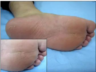
provided the original work is properly cited. Fig. 1. Before biopsy; large hyperkeratotic plaque on his left plantar surface (inset: close-up view).
http://dx.doi.org/10.5021/ad.2013.25.1.113
A Rapidly Regressed Giant Plantar Wart Following Biopsy
Sung Yul Lee
Department of Dermatology, Soon Chun Hyang University Cheonan Hospital, Soon Chun Hyang University College of Medicine, Cheonan, Korea
Dear Editor:
I read the case reported by Jin et al.1, with great interest, because I recently experienced a rapidly regressed giant plantar wart, which followed a biopsy. A 23-year-old male came to our department for a large hyperkeratotic plaque on his left plantar surface. It appeared more than 18 months prior to the presentation as multiple small pimple- like lesions, which had been slowly growing and coalescing (Fig. 1). His medical and family histories were noncontributory. A punch biopsy was taken, and the histopathological findings were acanthotic epidermis with hyperkeratosis, papillomatosis, parakeratosis, and koilo- cytes. The diagnosis of wart was made. The lesion rapidly regressed within 2 weeks of the biopsy, and it had completely disappeared when the patient presented for the follow-up after one month (Fig. 2). Warts, or verrucae, are benign proliferations of the skin and mucosa that are caused by an infection with human papillomavirus (HPV).
These viruses do not produce acute signs or symptoms, but induce a slow, focal expansion of the epithelial cells.
Lesions may remain subclinical for long periods, or may grow into large fulminating masses that persist for months or even years2. The management of warts depends on the degree of physical and emotional discomfort, the extent and duration of the lesions, the patient’s immunologic status, and the patient’s desire for therapy2. Irrespective of
the treatment modality, a host immune response is the key to achieving a complete clearance of the wart. Immuno- compromised individuals may never achieve wart clearance. The role of immunity in HPV infection is not completely understood. The decrease in the frequency of warts with age implies that the resistance to infection develops over time, and much of this resistance may be immunologic3. There are instances in which the treatment of one or more warts leads to a complete clearance in immuno-competent individuals. Specific cell-mediated immunity against viral infected keratinocytes took place in plane warts, under spontaneous regression4. CD4-positive lymphocyte predominance in the regression of genital warts had been demonstrated5. Tissue biopsy might have exposed viral antigens to the host immune system, which subsequently triggered HPV-specific immunity. It is po- ssible that the biopsy may have then exposed viral anti- gens and induced a delayed hypersensitivity reaction, which led to the rapid regression of the lesion. We suggest that a similar mechanism has been in the works in our case. There have been many cases of warts with spontaneous regression, but there has been no report of
114 Ann Dermatol Letter to the Editor
Received May 11, 2012, Revised May 17, 2012, Accepted for publication May 21, 2012
Corresponding author: Kyu Han Kim, Department of Dermatology, Seoul National University College of Medicine, 101 Daehak-ro, Jongno-gu, Seoul, 110-744, Korea. Tel: 82-2-2072-2410, Fax: 82-2-747-0611, E-mail: [email protected]
This is an Open Access article distributed under the terms of the Creative Commons Attribution Non-Commercial License (http://
creativecommons.org/licenses/by-nc/3.0) which permits unrestricted non-commercial use, distribution, and reproduction in any medium, provided the original work is properly cited.
Fig. 2. Six weeks later following biopsy, the wart had completely disappeared (inset: close-up view).
large plantar wart regression, following a small biopsy. In conclusion, wart regression can be achieved if the HPV-specific immunity can be stimulated. Therefore, inducing and boosting such a response is critical for the treatment of warts. I report here on a case of a rapidly regressed large plantar wart, following a biopsy without any additional treatment.
ACKNOWLEDGMENT
This work was supported in part by the Soon Chun Hyang University Research Fund.
REFERENCES
1. Jin SP, Jeon YK, Cho KH, Chung JH. A rapidly regressing wart following biopsy. Ann Dermatol 2011;23:123-124.
2. Elliot J, Androphy DRL. Wart. In: Wolff K, Goldsmith LA, Katz SI, Gilchrest BA, Paller AS, Leffell DJ, editors.
Fitzpatrick’s dermatology in general medicine. 7th ed. New York: McGraw-Hill, 2008:1914-1922.
3. Sterling JC, Handfield-Jones S, Hudson PM; British Associ- ation of Dermatologists. Guidelines for the management of cutaneous warts. Br J Dermatol 2001;144:4-11.
4. Iwatsuki K, Tagami H, Takigawa M, Yamada M. Plane warts under spontaneous regression. Immunopathologic study on cellular constituents leading to the inflammatory reaction.
Arch Dermatol 1986;122:655-659.
5. Coleman N, Birley HD, Renton AM, Hanna NF, Ryait BK, Byrne M, et al. Immunological events in regressing genital warts. Am J Clin Pathol 1994;102:768-774.
http://dx.doi.org/10.5021/ad.2013.25.1.114
Refractory Atopic Dermatitis in Childhood:
Improvement with Methotrexate?
Young Woon Park1, Kkot Bora Yeom1, Kyu Han Kim1,2,3
1Department of Dermatology, Seoul National University College of Medicine, 2Laboratory of Cutaneous Aging and Hair Research, Biomedical Research Institute, Seoul National University Hospital, 3Institute of Human-Environment Interface Biology, Medical Research Center, Seoul National University, Seoul, Korea
Dear Editor:
A 5-year-old boy had suffered from generalized severe atopic dermatitis (AD) for 3 years, and his symptoms were
not controlled by first-line therapeutics, that is, topical corticosteroids, topical calcineurin inhibitors, and oral antihistamines. Initially, we tried cyclosporine (5 mg/kg/d)