pISSN 2233-7903 •eISSN 2093-0488
Malignant thyroid bed mass after total thyroidectomy
Do Sung Park, Jin Seong Cho, Min Ho Park, Young Jae Ryu, Min Jung Hwang, Sun Hyung Shin, Hee Kyung Kim
1, Hyo Soon Lim
2, Ji Shin Lee
3, Jung Han Yoon
Departments of Surgery,
1Internal Medicine,
2Radiology, and
3Pathology, Chonnam National University Medical School, Gwangju, Korea
ORIGINAL ARTICLE
Purpose: Ultrasonographic (US) criteria on malignant thyroid bed mass have been suggested, including taller than wide shape, loss of echogenic hilum, abnormal vascularity, and microcalcification. The relationship between fine-needle aspiration (FNA) cytology findings and US findings on thyroid bed mass is unknown. We have retrospectively assessed the malignant thyroid bed mass after total thyroidectomy due to papillary thyroid carcinoma (PTC).
Methods: We retrospectively evaluated 2,048 patients who underwent total thyro- idec tomy due to PTC. FNA was performed in 97 patients on the thyroid bed under US surveillance. The 97 suspicious thyroid bed masses were divided into two groups:
metastatic thyroid bed group (n = 34) and nonmetastatic group (n = 63). The groups were evaluated according to various clinical, serologic, and US findings.
Results: Within a median 47.0 months of follow-up, the proportion of malignant thyroid bed mass was high in large tumor size (1.37 cm vs. 1.03 cm), isthmic position (10.3% vs. 3.9%), and previous N1a (55.9% vs. 34.9%). US findings revealed that the presence of microcalcification or macrocalcification (47.1% vs. 19.0%) and thyroid bed mass height (5.4 mm vs. 3.9 mm) were the only discriminable criteria for central compartment recurrence. But, degree of echogenicity, loss of hilum, and irregularity of margin failed to discriminate malignant thyroid bed mass.
Conclusion: US findings on malignant thyroid bed mass were different from previously reported general criteria on lateral metastatic nodes. Additional FNA cytology should be performed on patients, even low-risk patients, who present the above findings.
Journal of the Korean Surgical Society
JKSS
INTRODUCTION
According to the revised American Thyroid Association thyroid cancer guidelines [1], neck ultrasonography to evaluate the thyroid bed, and the central and lateral cervical nodal compartments should be performed 6-12 months after initial sur gery and then periodically, depending on the patient’s risk for recurrence and thyro- globulin (TG) status. Locoregional recurrence of papillary thyroid cancer (PTC) with reported rates of 15-25% occur in either cervical lymph nodes or in the thyroid bed [2,3], and careful structural evaluation of the neck is a key component of the follow- up surveillance.
In addition to identifying normal and abnormal cervical lymph nodes, ultrasono-
Corresponding Author Jin Seong Cho
Department of Surgery, Chonnam National University Medical School, 160 Baekseo-ro, Dong- gu, Gwangju 501-746, Korea
Tel: +82-62-220-6356 Fax: +82-62-227-1635 E-mail: [email protected]
Key Words
Ultrasonography, Fine-needle biopsy, Thyroidec- tomy, Papillary thyroid cancer
Received May 6, 2013 Reviewed July 2, 2013 Accepted July 3, 2013 J Korean Surg Soc 2013;85:97-103 http://dx.doi.org/10.4174/jkss.2013.85.3.97
Copyright © 2013, the Korean Surgical Society
cc Journal of the Korean Surgical Society is an Open Access Journal. All articles are distributed under the terms of the Creative Commons Attribution Non-Commercial License (http://
creativecommons.org/licenses/by-nc/3.0/) which permits unrestricted non-commercial use, distribution, and reproduction in any medium, provided the original work is properly cited.
graphy often detect small discrete nodules in the postoperative thyroid bed. Most are benign lesions such as postoperative scar or suture granulomas. However, some represent persistent thyroid cancer on the thyroid bed. The ultrasonographic (US) appearance of lateral cervical lymphadenopathy is useful in distinguishing benign from malignant cervical lymph nodes (cystic changes, taller than wide shape, abnormal vascularity, loss of echogenic hilum, and microcalcification or macrocalcification) [4-10]. However, these sonographic findings may not accurately predict small malignant thyroid bed mass [11-14].
Many studies have attempted to identify the characteristics of malignant thyroid bed mass and cervical lymphadenopathy.
US criteria have been suggested, including taller than wide shape, loss of echogenic hilum, abnormal vascularity, and microcalcification. The relationship between fine-needle aspiration (FNA) cytology findings and US findings on thyroid bed mass is unknown. So, we addressed this shortcoming by evaluating various US findings on malignant thyroid bed mass detected after total thyroidectomy.
METHODS
Between May 2004 and April 2008, 2,048 patients under- went total thyroidectomy due to thyroid malignancy Gwangju and Hwasoon Chonnam National University Hospital. Of these, 98 patients (4.56%) developed central or lateral neck recurrence. After obtaining approval from our Institutional Review Board, we retrospectively reviewed these patients’ records and US data, and investigated the US report. FNA was attempted in 97 patients on the thyroid bed and in 119 patients on the lateral neck under US surveillance. Suspicious thyroid bed masses were divided into two groups (metastatic thyroid bed group, n = 34; nonmetastatic group, n = 63) and the various clinical, serologic, and US findings were evaluated.
To be eligible, patients had to have at least one aspiration or surgically confirmed thyroid bed mass after surgery. Patients were excluded from the study for the following reasons: not enough data on follow-up or US, pediatric patients, medullary or follicular thyroid cancer patients, patients with other abnor- mal findings on lateral cervical lymph nodes such as previous lymphoma or other cancer history, and previous history of pulmonary or visceral tuberculosis.
US features of thyroid bed masses that underwent US- guided FNA included height and width, echogenicity, mar- ginal status, abnormal vascularity, and presence of micro- calcifications or macrocalcifications of thyroid bed nodules and cervical lymph nodes. Images of biopsied nodules were obtained with one image in the transverse plane and one image in the longitudinal plane. Thyroid bed masses were
classified as suspicious and biopsy was done when one or more of the aforementioned malignant suspicious US findings were evident. Echogenicity was characterized relative to strap muscles. Microcalcifications or macrocalcifications were defined as multiple punctate bright echoes with or with- out acoustic shadowing. Thyroid bed nodules considered to have increased vascularity included those with either peripheral or intranodular vascular flow. Hypoechogenicity, internal vascular flow, and microcalcifications in thyroid bed nodules were classified as suspicious sonographic features, as previously reported [12]. All of the suspicious thyroid bed masses were needle aspirated under US-guided targeting. US- FNA was performed with a 27-gauze needle attached to a disposable 10 mL syringe using a free-hand technique. Each lesion was aspirated at least three times.
We used the t-test to compare continuous variables between each group and the chi-square test for categorical variables.
Receiver operating characteristic (ROC) curve analysis was also used to compare diagnostic performance. The diag- nostic performance of each of the criteria were reported as sensitivity, specificity, positive predictive value, negative pre- dictive value, accuracy, and area under the ROC curve with 95% confidence intervals. Binary logistic regression test was used for multivariate analysis of statistically significant variables from the univariate analysis. Statistical significance was indicated by a P-value < 0.05. Results were analyzed using PASW ver. 18.0 (IBM Co., Armonk, NY, USA).
RESULTS
Incidence and FNA results of thyroid bed mass
Within a median 47.0 months of follow-up, 216 patients (10.6%) had FNA cytology on suspicious lesions in 97 central and 119 lateral necks. Of these patients, 34 (35.1%) and 63 (53.7%) were confirmed with central or lateral neck recurrence.
Thus, the positive predictive value on thyroid bed mass was significantly lower than that on lateral cervical nodes. Among the enrolled 97 thyroid bed masses, 34 had malignant cytology results, but 49 (50.5%) had benign results. Of the patients proven to have a nonmalignant thyroid bed mass, there were 30 (61.2%) reactive hyperplasia of lymph nodes, seven (14.3%) remaining thyroid or thyroiditis, four (8.2%) postoperative scar tissue containing histiocytes and neutrophils, two (4.1%) parathyroid glands, and two (4.1%) suture granulomas. Four patients were nondiagnostic due to bloody smear. Fourteen (14.4
%) were observed without FNA cytology and disappeared on follow-up US or diagnostic scans, or have been serologically negative to date.
Do Sung Park, et al: Malignant thyroid bed mass
Patient demographics and pathologic results
Clinicopathologic features of the 97 patients with thyroid bed masses detected on routine follow-up neck US after total thyroidectomy are presented in Table 1. Median age was 46.0 years. Central and lateral neck node dissection was done in 70 (72.2%) and 15 patients (15.5%), respectively, and 58.6% had central neck metastases. Hemostasis was done with absorbable tie material in 22 patients (22.7%) and with nonabsorbable silk tie material in 70 patients (77.3%). Postoperative radioactive iodine ablation was done in 46 patients (47.9%) and the mean dose was 122.6 mCi. Median time for FNA was 22 months (range, 2 to 81 months) after thyroid surgery.
Tumor and patient’s characteristics of metastatic thyroid bed mass
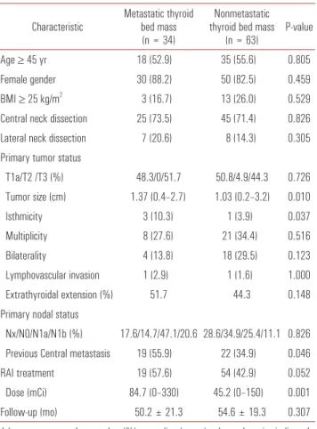
Patients were divided into two groups: metastatic thyroid bed group (n = 34) and nonmetastatic (n = 63) group. Age, sex, and body mass index were not significantly different between the two groups. The proportion of metastatic thyroid bed mass was high in patients with large primary tumor size (1.37 cm vs. 1.03 cm, respectively; P = 0.001), isthmic position (10.3% vs.
3.9%, respectively; P = 0.037), and previous central metastasis (55.9% vs. 34.9%, respectively; P = 0.046). As for other pri- mary tumor characteristics, bilaterality, marginal positivity, lymphovascular invasion, and extrathyroidal extension were not significantly different in multiplicity. Use of nonabsorbable silk tie material was not different in both groups (73.5% vs.
79.4%, respectively; P = 0.513), and larger dose (84.7 mCi vs.
45.2 mCi, respectively; P = 0.001) of radioactive iodine ablation after total thyroidectomy were related to metastatic groups.
Follow-up period and the times of FNA were not significantly different (Table 2).
Table 1. Characteristics of 97 patients who have thyroid bed mass
Characteristic Value
Age (yr)
Mean ± SD 44.9 ± 13.5
Median (range) 46.0 (15–79)
≥45, n (%) 53 (54.6)
Male gender, n (%) 15 (15.5)
Histology, n (%)
Papillary type 92 (94.8)
Follicular variant papillary type 5 (5.2) Operation, n (%)
Central neck dissection 70 (72.2)
Lateral node dissection 15 (15.5)
Central metastasis 41 (58.6)
Nonabsorbable tie 75 (77.3)
Absorbable tie material 22 (22.7)
T stage, n (%)
T1 45 (46.4)
T2 3 (3.1)
T3 42 (43.3)
N stage, n (%)
N0 27 (27.8)
N1a 31 (33.0)
N1b 14 (14.4)
Nx 24 (24.7)
Postoperative RAI 46 (47.9)
Dose, mean ± SD 122.6 ± 61.1
Dose, median (range), mCi 150 (100–330)
Follow-up (mo) 47 (10–94)
FNA interval since operation (mo) 22 (2–81)
SD, standard deviation; RAI, radioactive iodine ablation; FNA, fine-needle aspiration.
Table 2. Characteristics of metastatic vs. nonmetastatic thyroid bed mass after total thyroidectomy
Characteristic Metastatic thyroid bed mass
(n = 34)
Nonmetastatic thyroid bed mass
(n = 63) P-value
Age ≥ 45 yr 18 (52.9) 35 (55.6) 0.805
Female gender 30 (88.2) 50 (82.5) 0.459
BMI ≥ 25 kg/m2 3 (16.7) 13 (26.0) 0.529
Central neck dissection 25 (73.5) 45 (71.4) 0.826 Lateral neck dissection 7 (20.6) 8 (14.3) 0.305 Primary tumor status
T1a/T2 /T3 (%) 48.3/0/51.7 50.8/4.9/44.3 0.726 Tumor size (cm) 1.37 (0.4–2.7) 1.03 (0.2–3.2) 0.010
Isthmicity 3 (10.3) 1 (3.9) 0.037
Multiplicity 8 (27.6) 21 (34.4) 0.516
Bilaterality 4 (13.8) 18 (29.5) 0.123
Lymphovascular invasion 1 (2.9) 1 (1.6) 1.000 Extrathyroidal extension (%) 51.7 44.3 0.148 Primary nodal status
Nx/N0/N1a/N1b (%) 17.6/14.7/47.1/20.6 28.6/34.9/25.4/11.1 0.826 Previous Central metastasis 19 (55.9) 22 (34.9) 0.046
RAI treatment 19 (57.6) 54 (42.9) 0.052
Dose (mCi) 84.7 (0–330) 45.2 (0–150) 0.001
Follow-up (mo) 50.2 ± 21.3 54.6 ± 19.3 0.307
Values are presented as number (%) or median (range) unless otherwise indicated.
BMI, body mass index; RAI, radioactive iodine ablation.
US and laboratory characteristics of metastatic thyroid bed mass
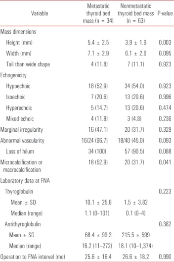
Height of the thyroid bed mass was significantly larger in
the metastatic group (5.4 mm) vs. the nonmetastatic group (3.9 mm) (P = 0.003). Other dimensions such as width or taller-than-wide shape were not different. We assumed echo- genicity, marginal irregularity, abnormal vascularity, and loss of hilum to be factors for metastatic thyroid bed mass, as is in other studies [4-10]. But, only microcalcification or macrocalcification findings were observed statistically more frequently (52.9% vs. 31.7%, respectively; P = 0.041). TG levels at suspicious findings on US were usually helpful for making decision for FNA. The proportion of elevated TG was greater but was not statistically significant (1.1 IU/mL vs. 0.1 IU/mL, respectively; P = 0.223) (Table 3).
Cutoff values of continuous variables
The cut-off values of various continuous variables in relation to malignant thyroid bed mass were analyzed through ROC curve and the area under curve (AUC). Primary tumor size over 1.0 cm (AUC, 0.715), thyroid bed mass height over 0.4 cm (AUC, 0.713), width over 0.7 cm (AUC, 0.600), and TG levels at the time of suspicious findings was 0.3 IU/mL (AUC, 0.677). Sensitivity and specificity of cutoff values of these variables proved to be good predictors (Table 4).
Prediction of metastatic thyroid bed mass after total thyroidectomy
The risk of thyroid bed metastasis in relation to primary Table 3. Ultrasonographic and laboratory characteristics between two
groups
Variable Metastatic
thyroid bed mass (n = 34)
Nonmetastatic thyroid bed mass
(n = 63) P-value Mass dimensions
Height (mm) 5.4 ± 2.5 3.9 ± 1.9 0.003
Width (mm) 7.1 ± 2.9 6.1 ± 2.6 0.095
Tall than wide shape 4 (11.8) 7 (11.1) 0.923 Echogenicity
Hypoechoic 18 (52.9) 34 (54.0) 0.923
Isoechoic 7 (20.6) 13 (20.6) 0.996
Hyperechoic 5 (14.7) 13 (20.6) 0.474
Mixed echoic 4 (11.8) 3 (4.8) 0.236
Marginal irregularity 16 (47.1) 20 (31.7) 0.329 Abnormal vascularity 16/24 (66.7) 18/40 (45.0) 0.093
Loss of hilum 34 (100) 57 (90.5) 0.088
Microcalcification or
macrocalcification 18 (52.9) 20 (31.7) 0.041 Laboratory data at FNA
Thyroglobulin 0.223
Mean ± SD 10.1 ± 25.8 1.5 ± 3.82 Median (range) 1.1 (0–101) 0.1 (0–4)
Antithyroglobulin 0.382
Mean ± SD 68.4 ± 99.3 215.5 ± 599 Median (range) 16.2 (11–272) 18.1 (10–1,374) Operation to FNA interval (mo) 25.6 ± 16.4 26.6 ± 18.2 0.990 Values are presented as mean ± SD or number (%) unless otherwise indicated.
FNA, fine-needle aspiration; SD, standard deviation.
Table 4. Cutoff value of continuous variables (n=97)
Variable AUC Cutoff value Sensitivity (%) Specificity (%)
Primary tumor size 0.715 9.5 mm 75.9 67.2
Thyroid bed mass height 0.713 3.7 mm 73.5 63.5 Thyroid bed mass width 0.600 6.8 mm 55.9 65.1 Thyroglobulin levels at FNA 0.677 0.25 IU/mL 73.3 55.2 AUC, area under curve; FNA, fine-needle aspiration cytology.
Table 5. Metastasis in the thyroid bed according to the fine-needle aspiration cytology by preoperative tumor status and ultrasonographic findings and thyroglobulin levels at that time
Variable Metastatic thyroid bed
(n = 34) Nonmetastatic thyroid bed (n = 63)
P-value
Univariate Multivariate
Tumor size over 1.0 cm 22/29 (75.9) 20/61 (32.8) 0.000 0.000
Previous central metastasis 19 (55.9) 22 (34.9) 0.046
Bed mass height over 0.4 cm 20 (58.8) 20 (31.7) 0.010 0.013
Bed mass width over 0.7 cm 18 (52.9) 22 (34.9) 0.085
Microcalcification or macrocalcification 16 (47.1) 12 (19.0) 0.004 0.008
Intranodal vascularity 16/24 (66.7) 18/40 (45.0) 0.093
TG over 0.3 IU/mL at FNA 11/15 (73.3) 12/29 (41.4) 0.044
TG, thyroglobulin; FNA, fine-needle aspiration.
Do Sung Park, et al: Malignant thyroid bed mass
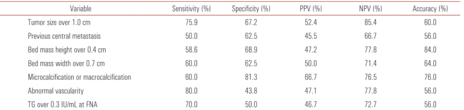
tu mor and nodal status, US findings, and TG levels at the time of suspicious findings was analyzed with categorical values. The proportion of thyroid bed metastasis in the bio- psied nodules was higher in primary tumor sizes over 1.0 cm (75.9% vs. 32.8%, respectively), previous central metastasis (55.9% vs. 34.9%, respectively), thyroid bed mass height over 0.4 cm (58.8% vs. 31.7%, respectively), microcalcification or macrocalcification (47.1% vs. 19.0%, respectively), and TG >0.3 IU/mL were significant (Table 5). In multivariate analysis, primary tumor size over 1.0 cm (P = 0.000), thyroid bed mass height over 0.4 cm (P = 0.013), and microcalcifications or macrocalcifications (P = 0.008) were significant. Also, in Cox analysis, the risk of thyroid bed metastasis was significantly higher in those with primary tumor size over 1.0 cm (odds ratio [OR], 3.372), thyroid bed mass height over 0.4 cm (OR, 2.844), and microcalcification or macrocalcification (OR, 4.935) (Table 6). These were independent predictive factors of thyroid bed metastasis. Diagnostic accuracy of various variables on pre diction of malignant thyroid bed mass was calculated.
Of these factors, thyroid bed mass height over 0.4 cm and micro calcification or macrocalcification findings exhibited an accuracy of 84.0% and 76.0% for the prediction of malignant thyroid bed mass, and negative predictive value of primary tumor size over 1.0 cm was 85.4% (Table 7). The risk of thyroid bed metastasis was better indicated by US rather than by primary tumor status or laboratory findings. Surprisingly, echogenicity, loss of hilum, and marginal irregularity poorly discriminated the malignant thyroid bed mass.
Time course of thyroid bed metastasis after total thyroi- dectomy
We recommended routine US surveillance at 6 months and 1 year after surgery, then once or twice a year thereafter. Most new onset thyroid bed metastasis was observed by 2 years (53%
of patients) and 91.2% of the metastases has occurred by 4 years. In the subsequent year, metastases developed in 8.8% of patients (Fig. 1).
DISCUSSION
US findings of metastatic lymph nodes have been well- Table 6. Cox proportional hazard ratio for metastatic thyroidectomy bed masses after total thyroidectomy
Factor b (SE) P-value Exp (b) 95% CI Exp (b)
Lower Upper
Primary tumor size over 1.0 cm 0.454 0.007 3.372 1.385 8.209
Bed mass height over 0.4 cm 0.423 0.013 2.844 1.241 6.516
Microcalcification or macrocalcification 0.416 0.037 2.384 1.055 5.387
SE, standard error; Exp (b), odds ratio; CI, confidence interval.
Table 7. Predictors of thyroidectomy bed metastasis
Variable Sensitivity (%) Specificity (%) PPV (%) NPV (%) Accuracy (%)
Tumor size over 1.0 cm 75.9 67.2 52.4 85.4 60.0
Previous central metastasis 50.0 62.5 45.5 66.7 56.0
Bed mass height over 0.4 cm 58.6 68.9 47.2 77.8 84.0
Bed mass width over 0.7 cm 60.0 62.5 50.0 71.4 64.0
Microcalcification or macrocalcification 60.0 81.3 66.7 76.5 76.0
Abnormal vascularity 80.0 43.8 47.1 77.8 56.0
TG over 0.3 IU/mL at FNA 70.0 50.0 46.7 72.7 56.0
PPV, positive predictive value; NPV, negative predictive value; TG, thyroglobulin; FNA, fine-needle aspiration.
Fig. 1. Time course of thyroid bed metastasis after total thyroidectomy.
reported [15-18]. However, thyroid bed recurrence is less clear [19,20]. Until now, diagnostic criteria for selecting the thyro idectomy bed lesions to biopsy have not been clearly established [20]. Also, prediction of malignancy cannot be made on the basis of the sonographic findings without FNA cytology [12]. In our study, positive predictive values of US- guided FNA on central or thyroid bed mass were significantly lower than that on lateral cervical nodes (35.1% vs. 53.7%).
So, we evaluated the various clinical and US findings on malignant thyroid bed mass on thyroid bed mass, rather than lateral cervical nodes.
Large tumor size, previous lymph node metastasis, and the isthmic position of tumor were associated with thy roid bed recurrences. Height and microcalcification or macro- calcification findings of thyroid bed mass were signi ficant predictors. Width, echogenicity, marginal status, loss of nodal hilum, and, especially, taller-than-wide shape of thyroid bed mass were not significant.
In a recent study, taller-than-wide shape in malignant thy- roid nodules and the reverse situation in benign nodules were related to the ability of the probe to compress the thyroid nodule during the US examination. Since benign nodules and cystic nodules are softer and infiltrate less into the surrounding tissue, benign nodules are more easily compressed than malignant nodules [21]. We assumed that the compressibility on malignant thyroid bed mass would disappear due to lack of the thyroid gland after thyroidectomy.
Thyroid bed masses are commonly seen in the routine follow-up after total thyroidectomy. Although the revised American Thyroid Association guidelines allow for obser- vation without FNA cytology of small abnormal cervical lymph nodes [1], there are no specific recommendations re- gar ding the management of small thyroid bed nodules. This is significantly higher than those who have thyroid bed mass height over 0.4 cm (accuracy, 84.0%), those who have microcalcification or macrocalcifications on sonographic sur veil lance (accuracy, 76.0%), and those who have primary tumor size over 1.0 cm (negative predictive value, 85.4%).
These were independent predictive factors of malignant thyroid bed metastasis. Unfortunately, echogenicity, loss of hilum, and marginal irregularity poorly discriminated the malignant thyroid bed mass, although these were good pre- dictors for lateral metastatic nodes.
Ultrasound is more sensitive than serum TG levels and radioactive iodine scans for detection of recurrence, and ultra sound should be indicated, even in low-risk thyroid cancer patients [19]. In our study, TG levels at suspicious findings did not achieve as high accuracy (only 56%) as could have been expected. Regular US evaluations seem to be a very reasonable approach to management. The absence of
suspicious sonographic findings combined with the absence of other abnormal cervical lymph nodes and rising serum TG can provide clinicians with a strong predictor for clinical quiescence over several years of follow-up.
This study is limited by its retrospective nature and thyroid bed masses without definite cytological results were included.
Neck ultrasonography on nondiagnostic or nonaspirated thyroid bed mass was followed until the end of this study, but all were followed without any evidence of clinical, sero- logic, and other diagnostic scan or positron emission tomo- graphy. Secondly, the diagnosis of thyroid bed mass and recommendation for US-guided FNA is usually based on the combination of suspicious features of radiologic findings and serologic criteria such as TG over 1.0 mIU/mL, and included hot uptake on previous diagnostic scans. But, we had a large number of negative lesions on FNA cytology. The relatively large number of negative results suggests that the diagnostic criteria used to determine whether a lesion should be biopsied were based on traditional US findings on lymph nodes.
In our study, US findings on malignant thyroid bed mass were different from previously reported general criteria on lateral metastatic nodes. Additional FNA cytology should be performed on patients presenting with these findings, even low-risk patients. Postoperative follow-up could be guided more easily by using the US criteria as well as the clinical and serologic monitoring.
CONFLICTS OF INTEREST
No potential conflict of interest relevant to this article was reported.
REFERENCES
1. American Thyroid Association (ATA) Guidelines Taskforce on Thyroid Nodules and Differentiated Thyroid Cancer, Cooper DS, Doherty GM, Haugen BR, Kloos RT, Lee SL, et al. Revised American Thyroid Association management guidelines for patients with thyroid nodules and differentiated thyroid cancer.
Thyroid 2009;19:1167-214.
2. Mazzaferri EL, Kloos RT. Clinical review 128: current appro- aches to primary therapy for papillary and follicular thyroid cancer. J Clin Endocrinol Metab 2001;86:1447-63.
3. Hay ID, Thompson GB, Grant CS, Bergstralh EJ, Dvorak CE, Gorman CA, et al. Papillary thyroid carcinoma managed at the Mayo Clinic during six decades (1940-1999): temporal trends in initial therapy and long-term outcome in 2444 consecutively treated patients. World J Surg 2002;26:879-85.
4. Fish SA, Langer JE, Mandel SJ. Sonographic imaging of thyroid nodules and cervical lymph nodes. Endocrinol Metab Clin North
Do Sung Park, et al: Malignant thyroid bed mass
Am 2008;37:401-17.
5. Rosario PW, de Faria S, Bicalho L, Alves MF, Borges MA, Purisch S, et al. Ultrasonographic differentiation between meta- static and benign lymph nodes in patients with papillary thyroid carcinoma. J Ultrasound Med 2005;24:1385-9.
6. Takashima S, Sone S, Nomura N, Tomiyama N, Kobayashi T, Nakamura H. Nonpalpable lymph nodes of the neck: assessment with US and US-guided fine-needle aspiration biopsy. J Clin Ultra sound 1997;25:283-92.
7. Ying M, Ahuja A, Metreweli C. Diagnostic accuracy of sono- graphic criteria for evaluation of cervical lymphadenopathy. J Ultrasound Med 1998;17:437-45.
8. Lyshchik A, Higashi T, Asato R, Tanaka S, Ito J, Hiraoka M, et al. Cervical lymph node metastases: diagnosis at sonoelastography: initial experience. Radiology 2007;243:258-67.
9. Kuna SK, Bracic I, Tesic V, Kuna K, Herceg GH, Dodig D.
Ultrasonographic differentiation of benign from malignant neck lymphadenopathy in thyroid cancer. J Ultrasound Med 2006;25:
1531-7.
10. Leboulleux S, Girard E, Rose M, Travagli JP, Sabbah N, Caillou B, et al. Ultrasound criteria of malignancy for cervical lymph nodes in patients followed up for differentiated thyroid cancer. J Clin Endocrinol Metab 2007;92:3590-4.
11. Kim JH, Lee JH, Shong YK, Hong SJ, Ko MS, Lee DH, et al.
Ultrasound features of suture granulomas in the thyroid bed after thyroidectomy for papillary thyroid carcinoma with an emphasis on their differentiation from locally recurrent thyroid carcinomas. Ultrasound Med Biol 2009;35:1452-7.
12. Shin JH, Han BK, Ko EY, Kang SS. Sonographic findings in the surgical bed after thyroidectomy: comparison of recur- rent tumors and nonrecurrent lesions. J Ultrasound Med 2007;
26:1359-66.
13. Lee JH, Lee HK, Lee DH, Choi CG, Gong G, Shong YK, et al.
Ultrasonographic findings of a newly detected nodule on the thyroid bed in postoperative patients for thyroid carcinoma: cor- relation with the results of ultrasonography-guided fine-needle aspiration biopsy. Clin Imaging 2007;31:109-13.
14. Frates MC. Ultrasound in recurrent thyroid disease. Otolaryngol Clin North Am 2008;41:1107-16.
15. Sohn YM, Kwak JY, Kim EK, Moon HJ, Kim SJ, Kim MJ.
Diagnostic approach for evaluation of lymph node metastasis from thyroid cancer using ultrasound and fine-needle aspiration biopsy. AJR Am J Roentgenol 2010;194:38-43.
16. Ahuja AT, Chow L, Chick W, King W, Metreweli C. Metastatic cervical nodes in papillary carcinoma of the thyroid: ultrasound and histological correlation. Clin Radiol 1995;50:229-31.
17. Ahuja AT, Ying M, Yuen HY, Metreweli C. Power Doppler sonography of metastatic nodes from papillary carcinoma of the thyroid. Clin Radiol 2001;56:284-8.
18. Antonelli A, Miccoli P, Ferdeghini M, Di Coscio G, Alberti B, Iacconi P, et al. Role of neck ultrasonography in the follow-up of patients operated on for thyroid cancer. Thyroid 1995;5:25-8.
19. Frasoldati A, Pesenti M, Gallo M, Caroggio A, Salvo D, Valcavi R. Diagnosis of neck recurrences in patients with differentiated thyroid carcinoma. Cancer 2003;97:90-6.
20. Kamaya A, Gross M, Akatsu H, Jeffrey RB. Recurrence in the thyroidectomy bed: sonographic findings. AJR Am J Roentgenol 2011;196:66-70.
21. Yoon SJ, Yoon DY, Chang SK, Seo YL, Yun EJ, Choi CS, et al. "Taller-than-wide sign" of thyroid malignancy: compa- rison between ultrasound and CT. AJR Am J Roentgenol 2010;
194:W420-4.