Copyright © 2012, the Korean Surgical Society J Korean Surg Soc 2012;83:321-324
http://dx.doi.org/10.4174/jkss.2012.83.5.321
CASE REPORT
Journal of the Korean Surgical Society
JKSS
pISSN 2233-7903ㆍeISSN 2093-0488
Received March 5, 2012, Revised June 29, 2012, Accepted July 30, 2012 Correspondence to: Shin Young Park
Department of Pathology, Konyang University Hospital, 158 Gwanjeodong-ro, Seo-gu, Daejeon 302-718, Korea Tel: +82-42-600-9302, Fax: +82-42-600-9280, E-mail: [email protected]
cc Journal of the Korean Surgical Society is an Open Access Journal. All articles are distributed under the terms of the Creative Commons Attribution Non-Commercial License (http://creativecommons.org/licenses/by-nc/3.0/) which permits unrestricted non-commercial use, distribution, and reproduction in any medium, provided the original work is properly cited.
Sarcomatoid carcinoma of the small intestine: a rare and highly aggressive tumor
Sang Eok Lee, Shin Young Park
1Departments of Surgery and 1Pathology, Konyang University Hospital, Daejeon, Korea
Sarcomatoid carcinoma of the small intestine is an extremely rare malignant neoplasm that usually has a poor prognosis. We report a case of sarcomatoid carcinoma arising in the small intestine in a 62-year-old man who was hospitalized for abdomi- nal pain. Computed tomography revealed wall thickening of the small intestine. The resected specimen showed a gray-whit- ish solid mass with hemorrhage and necrosis. Microscopically, the tumor was composed of pleomorphic spindle and dis- cohesive polygonal cells with frequent mitosis. No carcinomatous component was recognized. Immunohistochemistry re- vealed coexpression of cytokeratin and vimentin by the tumor cells, whereas expressions of C-kit, CD34, HMB-45, smooth muscle actin, and desmin were negative. The diagnosis was sarcomatoid carcinoma of the small intestine.
Key Words: Small intestine; Sarcomatoid carcinoma
INTRODUCTION
Sarcomatoid carcinoma is an uncommon tumor in the gastrointestinal tract [1]. These tumors have been reported in diverse sites including gallbladder [2], stomach [3], esophagus [4] and colon [5], but are only rarely reported in the small intestine [1,6,7]. Outcome is poor in patients with sarcomatoid carcinoma, because patients usually present with large tumor at extended stages [1]. We report a case of a 62-year-old man with sarcomatoid carcinoma involving the small intestine and review the relevant literature.
CASE REPORT
A 62-year-old man was admitted to Konyang University Hospital due to abdominal pain experienced for one month. His personal and family histories were unremark- able. Physical examination revealed generalized abdomi- nal tenderness. Laboratory tests revealed anemia (hemo- globin, 5.9 g/dL; hematocrit, 18.7%) and neutrophilia (13.0
× 103/μL). Liver and renal function tests were unre- markable.
Examination by esophagogastroduodenoscopy and co- lonoscopy showed no abnormal findings. Abdominal computed tomography (CT) scan showed wall thickening of the small intestine. A small bowel series showed com- plete segmental fold effacement of the ileum (Fig. 1). No
Sang Eok Lee and Shin Young Park
322 thesurgery.or.kr
Fig. 1. Abdominal computed tomography image showing 10 cm sized mass in small intestine.
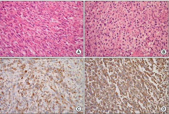
Fig. 2. Microscopically, tumor is composed of atypical spindle (A: H&E, ×200) and discohesive giant polygonal cells (B: H&E, ×200) in inflammatory background. Immunohistochemical staining, tumor cells are positive for cytokeratin (C: cytokeratin, ×200) and vimentin (D:
vimentin, ×200).
distant metastasis was detected after the patient under- went a positron emission tomography (PET)-CT scan.
Segmental resection revealed a mass located at the ileum, 40 cm from the ileocecal junction. The resected specimen showed an ulcerofungating mass that measured 15 × 9 × 2 cm in dimension. The cut surface revealed a gray- ish white, solid, and firm mass, with areas of hemorrhage and necrosis. The mass extended through the entire thick- ness of the wall of the small intestine, with invasion into the mesenteric fat. The gross and microscopic margins
were widely free of involvement. Microscopically, the tu- mor consisted of spindle and polygonal giant cells.
Spindle cells were arranged haphazardly in a fascicular growth pattern, with nuclear hyperchromasia and distinct nucleoli. Discohesive polygonal giant cells have dense eo- sinophilic cytoplasm and pleomorphic nuclei. The num- bers of mitotic figures averaged 15 per 10 high power fields. Lymphatic invasion was present, but the surround- ing lymph nodes were free of tumors. No carcinomatous component was recognized. Immunohistochemistry re- vealed diffuse, strong positive staining for cytokeratin and vimentin. Epithelial membrane antigen and CD68 were focal positive. Smooth muscle actin, S-100, CD34, HMB-45, Desmin and C-kit were negative (Fig. 2). The final diag- nosis was sarcomatoid carcinoma of the small intestine.
The patient received 2 cycles of chemotherapy includ- ing doxorubicin and dacarbazine. Three months later, PET-CT showed a recurrent mass in the pelvic cavity. After resection of recurrent small bowel mass, the patient re- started chemotherapy with doxorubicin, dacarbazine, ifosfamide and mensa.
Sarcomatoid carcinoma in small bowel
thesurgery.or.kr 323
DISCUSSION
Sarcomatoid carcinoma is a controversial and rare tu- mor that displays both carcinomatous and sarcomatous features. It has variety of names including carcinosarco- ma, metaplastic carcinoma, spindle cell carcinoma, and pleomorphic carcinoma [1]. For the small intestine, sarco- matoid carcinoma was first described by Dikman and Toker [8] in 1973. Less than 30 cases of small intestinal sar- comatoid carcinoma have been reported in the English language literature to date [1,6,7]. In Korea, only one case of small intestinal sarcomatoid carcinoma has been re- ported in the duodenum [9].
Clinically, sarcomatoid carcinoma in the small intestine seems to be a disease that affects elderly patients, present- ing at a mean age of 57 years [1]. Presenting symptoms in- clude: abdominal pain, intestinal obstruction, palpable ab- dominal mass, gastrointestinal bleeding, and anemia [6,7,10]. In the present case, the patient had abdominal pain and anemia.
Histologically, sarcomatoid carcinoma may appear with a biphasic or monophasic pattern [1]. A mixture of ep- ithelial-looking and mesenchymal-like cells characterized the typical biphasic pattern. Monophasic tumors show a predominance of the mesenchymal-like component, with minimal to absent epithelioid areas [1]. The presenting case was predominantly composed of spindle cells, with some polygonal cells. No conventional carcinomatous component was present. Monophagic sarcomatoid carci- noma may be confused with other sarcomas due to the ab- sence of carcinomatous features. A wide panel of im- munohistochemical markers was performed to sort out a diagnosis from a wide range of differentials, including leiomyosarcoma, epithelioid angiosarcoma, epithelioid malignant peripheral nerve sheath tumor (MPNST), gas- trointestinal stromal tumor (GIST), and melanoma. The absence of S-100 and HMB-45 ruled out the possibilities of an epithelioid MPNST and melanoma. A leiomyosarcoma was ruled out in view of smooth muscle actin and desmin negativity. Lack of C-kit and CD34 ruled out the possi- bility of a GIST and an epithelioid angiosarcoma. Most of the pleomorphic spindle cells expressed both cytokeratin and vimentin. This tumor was suggested to be epithelial in
origin and had transformed to a sarcomatous tumor.
The first choice of therapy for a solid tumor is always surgical resection. Surgery is an effective treatment for sar- comatoid carcinoma of the small intestine [1,7]. Neither ra- diotherapy nor chemotherapy contributes to the survival rate [1]. However, due to the high malignancy of sarcoma- toid carcinoma, many patients are diagnosed with the tu- mor at a late stage and die due to metastasis [1,7,11]. The duration of survival is generally only a few months [1,7].
In this case, although the patient was treated with ad- juvant chemotherapy, the tumor recurred within three months after surgery. Informing the patient of the ag- gressive biological behavior of the tumor and careful fol- low-up would be necessary for patients.
In summary, we present a case of sarcomatoid carcino- ma in the small intestine. As small intestinal sarcomatoid carcinoma demonstrates highly aggressive behavior, radi- cal surgery and short-term follow up are recommended.
CONFLICTS OF INTEREST
No potential conflict of interest relevant to this article was reported.
REFERENCES
1. Reid-Nicholson M, Idrees M, Perino G, Hytiroglou P.
Sarcomatoid carcinoma of the small intestine: a case report and review of the literature. Arch Pathol Lab Med 2004;
128:918-21.
2. Kim MJ, Yu E, Ro JY. Sarcomatoid carcinoma of the gall- bladder with a rhabdoid tumor component. Arch Pathol Lab Med 2003;127:e406-8.
3. Khan AR. Sarcomatoid carcinoma of the stomach with het- erologous elements. Ann Saudi Med 1999;19:135-6.
4. Raza MA, Mazzara PF. Sarcomatoid carcinoma of esopha- gus. Arch Pathol Lab Med 2011;135:945-8.
5. Choi YY, Jeen YM, Kim YJ. Sarcomatoid carcinoma of co- lon: extremely poor prognosis. J Korean Surg Soc 2011;
80(Suppl 1):S26-30.
6. Yucel AF, Kocakusak A, Arikan S, Demirbag N, Tarlaci A, Batur S. A rare cause of acute abdomen: perforated pri- mary sarcomatoid carcinoma of the small intestine: report of a case, with a brief review of the literature. J Cancer Res Ther 2011;7:348-50.
7. Moriwaki Y, Sugiyama M. Severe anemia inducing pre-
Sang Eok Lee and Shin Young Park
324 thesurgery.or.kr
shock caused by sarcomatoid carcinoma of the small intestine. Int Surg 2009;94:164-70.
8. Dikman SH, Toker C. Enteroblastoma complicating re- gional enteritis. Gastroenterology 1973;65:462-6.
9. Paik HJ, Choi YM. A case of carcinosarcoma in duodenum.
J Korean Surg Soc 1991;41:549-53.
10. Lam KY, Leung CY, Ho JW. Sarcomatoid carcinoma of the small intestine. Aust N Z J Surg 1996;66:636-9.