The Effects of the Position of Ipsilateral Neck Rotation on the Inhibition of the Upper Trapezius Muscle During
Lower Trapezius Exercises
Se-in Park, BHSc, PT, Ji-yeong Chae, BHSc, PT, Hyeong-hwi Kim, BHSc, PT, Yu-geoung Cho, BHSc, PT, Kyue-nam Park, PhD, PT
Dept. of Physical Therapy, College of Medical Science, Jeonju University
Abstract
Background: The unilateral prone arm lift (UPAL) is commonly used to exercise the lower trapezius muscle. However, overactivation of the upper trapezius can induce pain during UPAL exercises in subjects with upper trapezius tenderness.
Objects: The purpose of this study was to investigate the effects of position of ipsilateral neck rotation (INR) on the inhibition of upper trapezius muscle activity and the facilitation of the lower trapezius muscle when performing UPAL exercises.
Methods: In total, 19 subjects with upper trapezius tenderness were recruited for the study.
Electromyographic (EMG) activity was measured in the upper, middle, and lower trapezius muscles during UPAL with and without INR position. Wilcoxon signed-rank test was used to compare EMG activity in the trapezius muscles and the muscle ratios.
Results: EMG activity in the upper trapezius muscles was decreased significantly in the INR condition compared to without the position with INR during UPAL exercises (p<.05). EMG activity in the middle and lower trapezius was not significantly different between the with and without INR conditions (p>.05).
However, the ratio of lower to upper trapezius activation showed a significant increase in the INR condition compared to the without INR condition (p<.05), indicating greater lower trapezius activation relative to the upper trapezius in the INR position than in the without INR position.
Conclusions: The EMG results obtained in this study suggest that the position with INR reduced overactivation in the upper trapezius and improved muscle imbalance during lower trapezius exercises in individuals with upper trapezius tenderness.
Key Words: Electromyography; Ipsilateral neck rotation; Lower trapezius; Upper trapezius.
Introduction
Prolonged overactivity or tightness of the upper trapezius muscle can induce trigger point formation in the upper trapezius muscle (Gerwin et al, 2004).
Additionally, overuse of the upper trapezius muscle can lead to weakness in the middle and lower tra- pezius muscles, resulting in neck-shoulder pain and postural adaption (Kelley, 1995; Pink and Tibone, 2000). Janda stated that muscle imbalance, also called upper crossed syndrome, refers to tightness of the upper trapezius and pectoralis muscles and weakness
of the lower trapezius and cervical flexors (Page et al, 2010). Patients with upper crossed syndrome show specific postural changes, such as thoracic ky- phosis as well as abduction and winging of the scapula. These postural changes in the thorax and scapula decrease glenohumeral stability. Loss of sta- bility can be compensated for by increased activation of the upper trapezius muscle (Page et al, 2010).
Repeated compensatory activation can induce pain and hyperalgesia in the upper trapezius and de- creased participation of the lower trapezius (Page et al, 2010). Thus, exercise that improves the imbalance Corresponding author: Kyue-nam Park [email protected]
in trapezius muscles has been suggested to increase the activation ratio of the lower trapezius relative to the upper trapezius (Reinold et al, 2009).
The unilateral prone arm lift (UPAL) exercise has been used to improve activation of the lower tra- pezius muscle, which is performed with the shoulder abducted at 120° or 135° in a prone position (Ekstrom et al, 2003; Reinold et al, 2009). The UPAL exercise focuses on the action of the lower trapezius, which is involved in scapular depression and upward rotation (Reinold et al, 2009). However, in people with muscle imbalance in the neck and shoulder, there is a ten- dency for overactivation of the upper trapezius rather than the lower trapezius during full shoulder abduction, leading to scapular elevation instead of upward rota- tion and depression (Cools et al, 2003). Thus, during the UPAL exercise, unwanted scapular elevation with less upward rotation should be prevented.
Elongation of the muscle can lead to a decrease in its activation (Andriacchi et al, 1984; Heckathorne and Childress, 1981; Lunnen et al, 1981). In positions where the upper trapezius is stretched, depression, downward rotation of the scapula, ipsilateral rotation, and contralateral lateral bending of the neck are in opposition to upper trapezius action (Kendall et al, 2005). This study used ipsilateral neck rotation (INR) as a stretch position for the upper trapezius muscle because other stretch positions of the scapula (depression, downward rotation) cannot be performed simultaneously during the UPAL exercise, which should involve scapula upward rotation.
Although the UPAL is a beneficial exercise to ac- tivate the lower trapezius (Reinold et al, 2009), no study has investigated how to train the lower tra- pezius muscle effectively by preventing the over- activation of upper trapezius in subjects with upper trapezius tenderness during UPAL. Thus, the primary purpose of this study was to investigate the effects of INR position on inhibition of the upper trapezius during the UPAL exercise in subjects with upper trapezius tenderness, versus performance of the UPAL exercise without INR. We also compared the
ratios of middle to upper trapezius activation, and lower to upper trapezius activation, between the with and without the position with INR during UPAL ex- ercise conditions, to ascertain the extent of improve- ment in muscle imbalance in each condition.
Methods
Subjects
In total, 19 subjects (mean age, 22.1±1.4 years;
mean height, 170.5±9.3 ㎝; mean weight, 61.3±10.6
㎏; body mass index, 21.1±8.3 ㎏/㎡) recruited from a university volunteered to participate in this study.
The inclusion criterion for subjects with upper tra- pezius tenderness was an average upper trapezius pressure pain threshold lower than 10.6 lb/㎠ in males and 7.3 lb/㎠ in females (Fischer, 1987). In current study, mean±standard deviation of pressure pain threshold was 6.4±.74 lb/㎠ in males and 5.1±.51 lb/㎠ in females. The exclusion criteria were (1) any contraindication to cervical rotation and shoulder ab- duction, (2) diagnosis of fibromyalgia syndrome, (3) history of a whiplash injury, (4) history of any sur- gery in the craniocervical and shoulder region, (5) history of chronic neck pain, (6) any neurological condition, and (7) intake of any analgesic medication within eight hours prior to measurement of the pres- sure pain threshold. Prior to the study, the principal investigator explained procedures and all subjects signed an informed consent form.
Electromyography
To measure the amplitude of trapezius muscle ac- tivation during UPAL with and without INR, the Delsys Trigno wireless surface electromyography (EMG) system (Delsys, Boston, MA, USA) was used and EMG data were analyzed using EMGworks soft- ware ver. 3.7 (Delsys, Boston, MA, USA). Skin preparation of the electrode sites involved shaving and cleaning with rubbing alcohol. The EMG elec- trodes were placed at the following three locations:
upper trapezius (midpoint of the lead line between the C7 spinous process and the lateral tip of the ac- romion), middle trapezius (parallel to the muscle fi- bers between the spine of the scapula and the thora- cic spine), and lower trapezius (oblique angle to a point 5 ㎝ inferomedial from the root of the spine of the scapula) (Ekstrom et al, 2003). The sampling rate per channel was 1000 ㎐. A band-pass filter was se- lected between 20 and 450 ㎐. Raw data were converted to root-mean-squares with a 50 ㎳ time window. For normalization, the mean of three trials of reference vol- untary contractions (RVCs) lasting 3-s was calculated for the trapezius muscles. The participant abducted his or her arms in the scapular plane to 90° with the elbow extended, grasping a 2-㎏ weight in the hands while in a standing position (Hansson et al, 2000). Although maximal voluntary isometric contraction demands max- imum exertion of each muscle, the RVC method re- quires less time and less effort. Because it is easier for patients to perform (Hsu et al, 2009), this study was normalized using the RVC method.
Pressure algometer
The definition of the pressure pain threshold is the minimal amount of pressure required to elicit pain.
This study used a pressure algometer (JTECH Medical Industries, Salt Lake City, USA). A pres- sure algometer has a flat circular probe (1 ㎠) and force can be viewed digitally in increments of .1 Newton (N). To control the velocity of pressure in- crements, a metronome was used. Intra-rater reli- ability of pressure pain threshold measurements in the upper trapezius was established in a previous study (intra-class correlation coefficient=.86) (Azevedo et al, 2008). In this study, subjects’ pressure pain thresh- olds in the upper trapezius muscle were measured during confirmation of their eligibility according to the inclusion criteria. The subjects placed their arms at their sides in a resting sitting position. The tester marked the landmarks for the pressure pain threshold measurement (i.e., the midpoint between the second thoracic vertebra and the acromion). The tester
then applied perpendicular pressure at a rate of 3 N/s at the marked point (Azevedo et al, 2008).
Subjects were asked to inform the tester when the sensation of pressure elicited pain and the tester then removed the pressure algometer and the maximum pressure was recorded automatically. Three trials were performed with a 30-s rest interval and the average of three measurements was used for the pressure pain threshold. For familiarization, the tester first applied pressure on the anterior thigh of the subject.
Procedure
The experiment had two conditions (UPAL with INR position vs. UPAL without INR position). Before performing each condition, subjects were randomized to each condition using the randomization function in Microsoft Excel (Microsoft Corp., Redmond, WA, USA). Each condition was repeated three times, with postures held for 5-s while EMG data were obtained.
A 30-s rest period was given between trials. The average activity during the middle 3-s of each trial was used for data analysis.
UPAL with INR condition
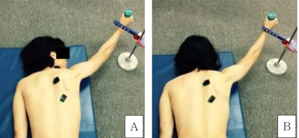
The subjects were placed in the prone position with INR (about 80-90°) and were asked to abduct their shoulder at 120°, in line with the muscle fibers of the lower trapezius using a l-㎏ dumbbell (Reinold et al, 2009). Then, subjects performed the UPAL exercise until they touched a fixed target bar, the height of which was set at the pure frontal plane of each subject (Figure 1A).
UPAL without INR condition
The experimental conditions were the same as the UPAL with INR condition except that UPAL was performed without INR (Figure 1B).
Statistical analysis
All data showed non-normal distributions according to the Kolmogorov-Smirnov test (p<.05). Wilcoxon sign- ed-rank tests were used to compare differences in mus-
Ratio With INRa position Without INR position p value
Middle trapezius/upper trapezius 7.44±5.34b 3.70±3.07 .10
Lower trapezius/upper trapezius 7.81±2.36 4.11±3.68 .02*
aipsilateral neck rotation, bmean±standard deviation, *significant change (p<.05) between with and without ipsilateral neck rotation position.
Table 2. Electromyographic ratio of middle and lower trapezius relative to upper trapezius during unilateral prone arm lift (UPAL) exercises
Muscles With INRa position Without INR position p value
Upper trapezius (%RVCb) 124.88±80.04c 172.05±104.61 .001*
Middle trapezius (%RVC) 476.21±414.99 487.26±451.26 .126
Lower trapezius (%RVC) 527.94±441.78 529.60±453.44 .841
aipsilateral neck rotation, breference voluntary contraction, cmean±standard deviation, *significant change (p<.05) between with and without ipsilateral neck rotation position.
Table 1. Electromyographic data in trapezius during unilateral prone arm lift (UPAL) exercises
A B
Figure 1. UPAL exercises (A: UPAL exercise with INR, B: UPAL without INR).
cle activity between the two conditions (UPAL with INR position vs. UPAL without INR position) for the upper, middle, and lower trapezius. All statistical analyses were performed using SPSS ver. 20.0 (SPSS Inc., Chicago, IL, USA). The significance level was set at α=.05.
Results
EMG activity in the upper trapezius muscle was de- creased significantly in the INR condition during per- formance of the UPAL exercise, compared to without INR condition (p<.05). EMG activity in the middle and lower
trapezius showed no significant difference between the with and without INR conditions (p>.05; Table 1).
The ratios of middle trapezius to upper trapezius activation showed no significant increases between two conditions (p>.05). Lower trapezius to upper tra- pezius activation, showed significant increases in the with INR condition versus in the without INR during the UPAL exercise (p<.05; Table 2).
Discussion
Exercise of the lower trapezius with UPAL is
commonly used to minimize muscle imbalance; i.e., overactivation of the upper trapezius and insufficient activation of the lower trapezius (Ekstrom et al, 2003;
Reinold et al, 2009). In the current study, we inves- tigated the effects of the position with INR on in- hibition of the upper trapezius during a lower tra- pezius exercise. The results of this study showed that the position with INR was significantly effective in decreasing the amplitude of upper trapezius activity, although activity in the middle and lower trapezius was not increased significantly in the INR condition compared to without the INR condition during per- formance of the UPAL exercise. However, the ratio of to upper trapezius activity increased more in the INR versus without INR condition during the UPAL exercise, indicating reduced muscle imbalance.
The INR position is effective in decreasing upper trapezius activity in subjects with upper trapezius tenderness. There is an explanation for this, the first of which is the increased length of the upper trapezius due to INR. In INR position, the upper trapezius is stretched during the UPAL exercise. Regarding the re- sults of previous studies, there has been controversy about the relationship between muscle length and EMG activity (Lunnen et al, 1981; Mohamed et al, 2002). Some researchers have suggested that EMG activity decreased when the hamstring and triceps surae muscles were in a more stretched position (Andriacchi et al, 1984; Heckathorne and Childress, 1981; Lunnen et al, 1981). In contrast, others have suggested that EMG activity did not change con- sistently according to changes in muscle length (Eloranta and Komi, 1981; Mohamed et al, 2002). Our findings support the results of earlier studies on the decreased EMG activity in stretched position of muscle (Lunnen et al, 1981; Mohamed et al, 2002).
Although this association between upper trapezius muscle length and EMG activity do not suggest cause and effect due to cross-sectional study, this result suggest that INR position may impact on re- duction of muscle activity of upper trapezius in sub- jects with upper trapezius tenderness. Further study
would be needed to investigate the relationship be- tween upper trapezius length and muscle activity.
The EMG ratio of the lower trapezius relative to the upper trapezius increased significantly during UPAL with INR. This indicates that the position with INR is helpful for improving the muscle im- balance between the upper trapezius and lower trapezius. A previous study suggested that exercises that improve the activation ratio of the lower to the upper trapezius should be used to reduce muscle im- balance and to improve scapulocostal posture (Reinold et al, 2009). Taping of the upper trapezius improved muscle imbalance between the upper and lower tra- pezius in patients with subacromical impingement syndrome. The ratio of upper to lower trapezius ac- tivation was 3.15 in the without tape condition, com- pared to 3.08 in the with tape condition, during humeral elevation in the scapula plane, indicating re- duced activation of the upper trapezius relative to the lower trapezius. Consistent with these previous re- sults using tape, in our study the ratio of lower to upper trapezius activation was 4.11 in the without INR position and 7.81 in with INR position, indicat- ing increased lower trapezius activity relative to the upper trapezius during the lower trapezius exercise.
Thus, the INR position resulted in inhibition of the upper trapezius and facilitation of the lower trapezius, leading to reduced muscle imbalance.
This study had some limitations. First, we did not assess differences in neck pain intensity between the two conditions, because we focused on muscle im- balance in the trapezius during lower trapezius exercises. Further study is required to investigate the effects of the UPAL exercise with INR position on upper trapezius pain and the pressure pain threshold.
Second, we focused only on trapezius muscle im- balance during UPAL, because muscle imbalance commonly occurs between the upper versus lower trapezius. However, the serratus anterior also partic- ipates in upward rotation during the UPAL exercise.
Also, the levator scapulae can induce muscle im- balance because it can act as a downward rotator
and elevator during the UPAL exercise (Ekstrom et al, 2003). Thus, future studies examining the effects of the UPAL exercise with INR position on muscle imbalance should include the serratus anterior and levator scapulae. Lastly, non-parametric statistic was used because the variables were not normally distributed.
Small sample size and non-parametric statistic may ren- der the result underpowered, thus future study requires a sufficient sample size with power analysis.
Conclusion
We investigated whether the position with INR is an effective position to reduce activation of the upper trapezius during performance of the UPAL exercise, which focuses on achieving lower trapezius activation in subjects with upper trapezius tenderness. The re- sults of this study showed that the INR position is appropriate to decrease upper trapezius activity and reduce muscle imbalance between the upper and lower trapezius muscles. These results may help clinicians to design exercise programs for people with upper trapezius tenderness when inclusion of lower trapezius exercises is appropriate.
References
Andriacchi TP, Anderrson GB, Ortengern R, et al. A study of factors influencing muscle activity about the knee joint. J Orthop Res. 1984;1(3):266-275.
Azevedo DC, de Lima Pires T, de Souza Andrade F, et al. Influence of scapular position on the pres- sure pain threshold of the upper trapezius mus- cle region. Eur J Pain. 2008;12(2):226-232.
Cools AM, Witvrouw EE, Declercq GA, et al.
Scapular muscle recruitment patterns: Trapezius muscle latency with and without impingement symptoms. Am J Sports Med. 2003;31(4):542-549.
Ekstrom RA, Donatelli RA, Soderberg GL. Surface electromyographic analysis of exercises for the
trapezius and serratus anterior muscles. J Orthop Sports Phys Ther. 2003;33(5):247-258.
Eloranta V, Komi PV. Function of the quadriceps femoris muscle under the full range of forces and differing contraction velocities of concentric work. Electromyogr Clin Neurophysiol. 1981;21(4):
419-431.
Fischer AA. Pressure algometry over normal muscles.
Standard values, validity and reproducibility of pressure threshold. Pain. 1987;30(1):115-126.
Gerwin RD, Dommerholt J, Shah JP. An expansion of Simons’ integrated hypothesis of trigger point formation. Curr Pain Headache Rep. 2004;8(6):
468-475.
Hansson GA, Nordander C, Asterland P, et al.
Sensitivity of trapezius electromyography to differ- ences between work tasks-influence of gap defi- nition and normalisation methods. J Electromyogr Kinesiol. 2000;10(2):103-115.
Heckathorne CW, Childress DS. Relationships of the surface electromyogram to the force, length, ve- locity, and contraction rate of the cineplastic human biceps. Am J Phys Med. 1981;60(1):1-19.
Hsu YH, Chen WY, Lin HC, et al. The effects of taping on scapular kinematics and muscle per- formance in baseball players with shoulder im- pingement syndrome. J Electromyogr Kinesiol. 2009;
19(6):1092-1099. http://dx.doi.org/10.1016/j.jelekin.
2008.11.003
Kelley MJ. Anatomic and biomechanical rationale for rehabilitation of the athlete’s shoulder. J Sport Rehabil. 1995;4(2):121-154.
Kendall FP, McCreary EK, Provance PG. Muscles:
Testing and function with posture and pain. 5th ed. Baltimore, Williams & Wilkins, 2005:303, 330.
Lunnen JD, Yack J, LeVeau BF. Relationship be- tween muscle length, muscle activity, and torque of the hamstring muscles. Phys Ther. 1981;61(2):
190-195.
Mohamed O, Perry J, Hislop H. Relationship between wire EMG activity, muscle length, and torque of the hamstrings. Clin Biomech (Bristol, Avon). 2002;
This article was received October 15, 2015, was reviewed October 16, 2015, and was accepted November 17, 2015.
17(8):569-579.
Page P, Frank C, Lardner R. Assessment and Treatment of Muscle Imbalance: The Janda approach. 1st ed.
Champaign, IL, Human Kinetics, 2010:52-53.
Pink MM, Tibone JE. The painful shoulder in the swimming athlete. Orthop Clin North Am. 2000;
31(2):247-261.
Reinold MM, Escamilla RF, Wilk KE. Current con-
cepts in the scientific and clinical rationale behind exercises for glenohumeral and scapulothoracic musculature. J Orthop Sports Phys Ther. 2009;39(2):
105-117. http://dx.doi.org/10.2519/jospt.2009.2835