• 검색 결과가 없습니다.

A Comparison of EMG Activity for the Middle and Lower Trapezius Muscle in the Frontal and Scapular Plane According to Shoulder Abduction Angles

N/A
N/A
Protected

Academic year: 2021

Share "A Comparison of EMG Activity for the Middle and Lower Trapezius Muscle in the Frontal and Scapular Plane According to Shoulder Abduction Angles"

Copied!
7
0
0

로드 중.... (전체 텍스트 보기)

전체 글

(1)

| Abstract |

8)

Purpose: The purpose of this study was to compare muscle activities in the frontal plane and scapular plane of the middle fiber and lower fiber of the trapezius muscle at different shoulder abduction angles.

Methods: Twenty male and female students in their 20s participated in this study. Each subject maintained shoulder abduction at 75˚, 90˚, 125˚, and 160˚ in a standing position and repeated motions three times each in the frontal plane and the scapular plane.

While maintaining the motions for 10 seconds in each posture, surface electromyography (EMG) was used to measure muscle activity of the middle fiber and lower fiber of the trapezius muscle. The collected EMG data were normalized using maximal voluntary isometric contraction (MVIC). Differences in muscle activity of the middle fiber and lower fiber of the trapezius muscles according to the angles at each plane were statistically processed using repeated measured analysis of variance, and an independent t-test was used to examine the differences between the two planes at each angle.

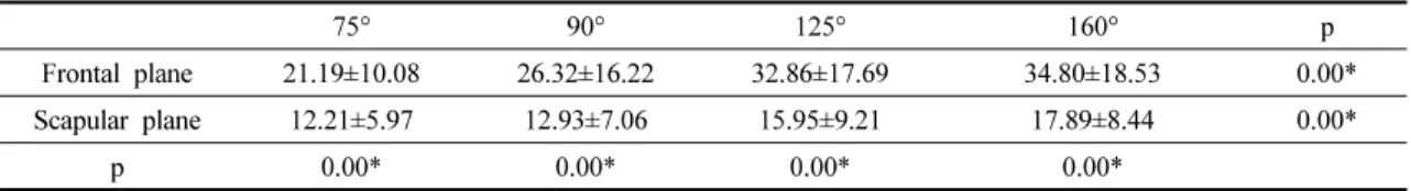
Results: Muscle activity of the middle and lower trapezius during shoulder abduction in the frontal plane and scapular plane significantly increased as the angles increased (p<.05). However, muscle activity of the middle trapezius was significantly lower in the scapular plane than in the frontal plane for all shoulder abduction angles (p<.05).

Conclusion: The results of this study suggest that during shoulder abduction, angles should be different according to the goals, and for training during an acute phase or early phase for functional recovery, it is more efficient to perform the training in the scapular plane than in the frontal plane.

†Corresponding Author : Myoung-Hee Lee ([email protected])

Original Article Open Access

어깨관절의 이마면과 어깨면에서 벌림각도에 따른 중간 등세모근과 아래 등세모근의 근 활성도 비교

김병곤⋅이명희 1†

대구보건대학교 물리치료과, 1 위덕대학교 물리치료학과

A Comparison of EMG Activity for the Middle and Lower Trapezius Muscle in the Frontal and Scapular Plane According to Shoulder Abduction Angles

Byung-Kon Kim⋅Myoung-Hee Lee 1†

Department of Physical Therapy, Daegu Health College

1 Department of Physical Therapy, Ui-duk University Received: August 2, 2016 / Revised: August 22, 2016 / Accepted: August 23, 2016

ⓒ 2016 Journal of Korea Proprioceptive Neuromuscular Facilitation Association

This is an Open Access article distributed under the terms of the Creative Commons Attribution Non-Commercial License

(http://creativecommons.org/licenses/by-nc/3.0) which permits unrestricted non-commercial use, distribution, and reproduction

in any medium, provided the original work is properly cited.

(2)

Key Words: Shoulder abduction, Trapezius muscle, Movement plane, Electromyography

Ⅰ. 서 론

등세모근의 근섬유들은 주행방향이 각각 달라 그 역할도 서로 다르다(Ekstrom et al, 2005; Sahrmann, 2002). 또한 각 근섬유들은 어깨 벌림의 각도에 따라 다른 역할을 하는데 벌림 초기에는 위 등세모근이 어 깨뼈의 상방회전과 상승을 돕고, 벌림 후기에는 아래 등세모근이 어깨뼈를 아랫방향으로 당겨 어깨뼈의 정 렬을 유지하는데 그 역할을 한다(Neumann, 2002).

등세모근 근 섬유중 하나라도 약화되면 길이 장력 관계(length-tension relationship)와 짝힘에 변화가 발생 하며 근 불균형이 나타나고 근 지구력이 감소하여 어 깨뼈-위팔뼈 리듬에 변화를 초래할 수 있다(Ebaugh

& Spinelli, 2010; Moseley et al, 1992). 특히 중간 등세모 근과 아래 등세모근은 어깨가슴관절(scapulothoracic joint) 의 안정화에 중요한 역할을 하는 근육으로 이들 의 소실은 충돌 증후군(subacromial syndrome)과 같은 어깨관절의 기능손상과 어깨관절의 불안정성 증가를 초래한다(De Mey et al, 2009).

중간 등세모근과 아래 등세모근의 근력은 특별한 마비가 없더라도 잘못된 자세나 반복된 동작으로 인 해 감소될 수 있다. 특히 이들의 약화는 척추 뒤굽음증 (kyphosis)이 증가되어 있는 경우에 나타날 수 있으며, 이 근육들의 점진적이고 지속적인 염좌는 위등뼈 부 위에 통증을 유발할 수 있다(Kendall et al, 2005). 어깨 뼈가 벌림되어 있고 앞쪽으로 어깨가 돌출된 사람에 게는 주로 중간 등세모근의 약화가 나타나고, 작은가 슴근(pectoralis minor)의 단축으로 인해 어깨뼈가 앞기 울임(anterior tilt)되거나 어깨뼈가 올림된 사람에게는 아래 등세모근의 약화가 나타난다(Kendall et al, 2005).

어깨관절의 움직임에 대한 평가 및 재활은 신체의 시상면, 이마면, 가로면 즉 일차적인 운동면에서 일어 나는 움직임에 집중되어 왔으나, 사람의 일상적인 생 활에서의 움직임이 이마면과 시상면 사이에 있는 중

간면에서 더 기능적이고 자연스러운 형태로 일어나기 때문에 어깨면에서의 움직임에 대해 논의되기 시작했 다(Cahalan et al, 1991). 이것은 어깨뼈가 이마면에서 30∼45도 전방으로 놓여 있어 위팔뼈의 벌림과 굽힘 은 어깨면을 향하여 위팔뼈가 정렬되었을 때 어깨세 모근(deltoid)과 돌림근띠(rotator cuff)가 팔를 움직이 는데 적절한 길이-장력 관계에 놓이게 되어 어깨면에 서의 벌림 시 위팔뼈의 회전을 요구하지 않게 된다 (Poppen & Walker, 1976). 특히 고유수용성신경근촉진 법이 기능을 강조한 효율적인 치료법임을 고려할 때 어깨관절의 이러한 역학적 특성은 치료 시 반드시 인 지해야 하는 요소이다.

어깨관절의 역학적 축과 길이-장력 관계, 어깨관절 주머니의 관계에 따라 이마면과 어깨면에서의 벌림 시에 근 활성도에의 유의한 차이가 있을 것이라 생각 하며, 따라서 본 연구는 어깨관절의 벌림을 이마면과 어깨면에서 모두 실시하고 그것을 비교함으로써 어깨 관절의 효율적이고 기능적인 훈련을 적용하기 위한 근거를 찾고자 하였다.

Ⅱ. 연구 방법

1. 연구 대상

본 연구의 대상자는 대구에 소재한 D대학교에 재학 중인 20대 성인 21명을 대상으로 실시하였다. 각각의 대상자들에게 연구의 목적과 방법, 절차에 대해 충분 히 설명하였고 대상자들은 실험참여에 동의하였다.

연구대상자 중 팔에 신경학적 질환이 있는 자, 목과

등 또는 어깨관절에 통증이 있는 자, 근골격계 관련

선천적 기형이 있는 자, 수술 경험이 있는 자는 제외하

였다. 연구대상자의 일반적 특성은 Table 1과 같다.

(3)

Male (n=10) Female (n=11) Age (years) 24.00±3.31 22.58±1.16 Height (cm) 173.11±6.07 162.50±5.62 Weight (kg) 69.78±5.63 55.25±8.89

*Mean±SD

Table 1. General charterstics of subjects

2. 측정 도구 및 방법

본 연구에서 등세모근의 중간섬유, 아래섬유의 근 전도 자료수집을 위해서 표면근전도(Telemyo DTS, Noraxon Inc., USA) 를 사용하였다. 수집된 근전도 신호 는 MyoResearch XP를 이용해 대역통과필터(band pass filter, 60∼250㎐)와 노치필터(notch filter, 60㎐)과정을 거쳐 %MVIC를 이용한 정량화(normalization)를 실시 한 후 컴퓨터 파일로 저장하였다. 등세모근의 중간섬 유와 아래섬유의 근 활성도를 측정하기 위해 기존의 선행 연구들을 참여하였고 근육의 전극부착위치를 결 정하였다. 등세모근의 중간섬유는 T4 높이에서 어깨 뼈가시 내측으로 5㎝에 부착하였고, 등세모근의 아래 섬유는 어깨뼈 밑각에서 10㎝ 내측에 55〬 비스듬한 각도 로 부착하였다(Cram et al, 1998). 부착 전에 정확한 데이 터 수집을 위해 면도기로 부착부위의 제모 작업을 하 고, 사포로 각질을 제거한 후 알코올 솜으로 닦았다.

측정 시 어깨관절 벌림을 일정하게 하기 위해 5〬

와 10〬 간격으로 그려진 각도판을 사용하였고, 벌림 각도를 넘어가지 않도록 50㎝ 자를 사용하였다. 그리 고 30〬 수평모음 시 goniometer를 사용하였다.

3. 실험 방법

이마면과 어깨면에서 등세모근의 중간섬유와 아래 섬유의 근 활성도를 측정하기 위해 연구대상자는 기 립자세에서 75〬, 90〬, 125〬, 160〬, 벌림자세를 취하였다 (Kinney et al, 2008). 대상자들은 어깨면에서 어깨관절 벌림 근 활성도를 측정하기 위해 이마면에서 30〬 수평 모음한 자세를 취하였다(Fig. 1).

실험 전 대상자들의 시선은 정면에 정해진 점을 바 라보게 지시하였고, 각 면에서의 벌림 연습을 실시하여 목을 움직이거나, 어깨관절의 안쪽돌림, 그리고 팔꿉 관절의 굽힘이 일어나지 않도록 하였다. 이후 이들은 4가지 각도의 어깨벌림을 한 자세에서 10초간 유지하 였다. 이 때 모든 대상자가 정확한 동작을 수행하도록 하기 위해 각도판을 사용하였는데, 대상자가 팔을 몸통 에 붙이고 서 있을 때 어깨뼈봉우리를 각도판의 중심점 과 일치시킨 후 동작을 수행하도록 하였다. 그리고 팔 의 벌림각도를 통제하기 위해 50㎝ 자를 사용하여 동작 을 유도하였다(Fig. 2). 각 동작은 3번씩 실시하였으며 각도별 동작의 순서는 무작위로 실시하였고, 근피로의 최소화를 위해 다른 각도에서 측정하기 전 30초의 휴식

Fig. 1. The 90 〬 shoulder abduction in scapular plane. Fig. 2. The 50 ㎝ stick for control of shoulder abduction.

(4)

시간을, 면이 변할 때 5분간 휴식시간을 주었다.

각 동작을 수행할 때와 최대 등척성 수축 시 수집된 근전도 신호는 연구대상자가 각 각도에서 팔을 벌림 후 10초간 유지할 때 기록된 근전도 신호에서 처음과 끝의 각 3초를 제외한 중간 범위 4초의 신호를 사용하였 고 3번 측정된 값의 평균을 구하여 자료를 분석하였다.

4. 자료 분석

이마면과 어깨면에서의 어깨관절 벌림 각도에 따 른 중간 등세모근과 아래 등세모근의 근 활성도값을 비교하기 위해 반복 측정 일요인 분산 분석(repeated one-way ANOVA)을 실시하였고, 이마면과 어깨면에 서 평균비교를 위해 독립 t-test(independent t-test)를 실 시하였다. 통계학적인 유의수준 a는 0.05로 하였고 자 료의 통계처리는 상용 통계프로그램인 윈도 SPSS ver.

18.0 프로그램을 사용하였다.

Ⅲ. 연구 결과

1. 이마면과 어깨면에서의 어깨관절 벌림각도에 따른 중간 등세모근의 근 활성도

이마면과 어깨면에서의 어깨관절 벌림각도에 따른

중간 등세모근의 근 활성도 결과는 Table 2와 같다.

중간 등세모근은 160° 벌림 하였을 때 가장 높은 근 활성도를 보였고, 75° 벌림하였을 때 가장 낮은 근 활성도를 보였다. 각 각도별로 이마면에서 나타난 근 활성도 보다 어깨면에서 낮은 근 활성도를 나타내었 다(p<0.05). 또 벌림 각도에 다른 중간 등세모근의 근 활성도는 벌림각이 증가할수록 유의하게 증가하였다 (p<0.05).

2. 이마면과 어깨면에서의 어깨관절 벌림각도에 따 른 아래 등세모근의 근 활성도

이마면과 어깨면에서의 어깨관절 벌림 각도에 따 른 아래 등세모근의 근 활성도는 Table 3와 같다. 아래 등세모근은 160° 벌림하였을 때 가장 높은 근 활성도 를 보였고, 75° 벌림하였을 때 가장 낮은 근 활성도를 보였다. 각 각도별로 어깨면에서 이마면에서 보다 낮 은 근 활성도를 나타내었으나 유의한 차이는 없었다 (p>0.05). 또 벌림각도에 다른 아래 등세모근의 근 활성 도는 벌림각이 증가할수록 유의하게 증가하였다 (p<0.05).

(unit:%MVIC)

75° 90° 125° 160° p

Frontal plane 21.19±10.08 26.32±16.22 32.86±17.69 34.80±18.53 0.00*

Scapular plane 12.21±5.97 12.93±7.06 15.95±9.21 17.89±8.44 0.00*

p 0.00* 0.00* 0.00* 0.00*

*p<0.05

Table 2. The EMG activity of the middle trapezius in the frontal and scapular plane

(unit:%MVIC)

75° 90° 125° 160° P

Frontal plane 13.31±18.59 13.92±11.27 22.24±14.43 30.53±17.06 0.00*

Scapular plane 12.51±8.61 13.19±10.63 21.95±17.43 25.42±14.55 0.00*

P 0.85 0.50 0.95 0.30

*p<0.05

Table 3. The EMG activity of the lower trapezius in the frontal and scapular plane

(5)

Ⅳ. 고 찰

어깨관절에 관한 많은 연구들은 해부학적 면에서 움직임을 정의함에 따라 주로 이마면과 시상면에서 근력을 측정하고 기능을 평가해 왔다. 하지만 어깨뼈 는 이마면의 30°-45° 전방으로 경사지게 놓여있어 해 부학적 움직임은 위팔뼈와 어깨뼈 사이의 움직임이 아니라 몸통과 위팔뼈 사이의 보여지는 움직임을 설 명한 것이라 할 수 있다(Whitcomb et al, 1995). 이러한 이유로 어깨관절에서의 정확한 평가를 위해 어깨면에 서의 팔의 움직임이 거론되기 시작하였다. 어깨면과 같은 면에 위팔뼈(humerus)가 정렬되었을 때 어깨관절 의 역학적 축과 어깨뼈의 역학적 축은 같은 선상에 있게 되며 그 결과 적절한 길이-장력 관계에 놓이게 되어 효율적인 움직임을 가능하게 한다(Poppen &

Walker, 1976). 이러한 팔의 기능적이고 효율적인 움직 임과 관련하여 어깨면이 강조되었으며 어깨관절의 재 활훈련 과정이나 기능을 평가하는데 있어 어깨면의 인지는 필수적인 요소가 되었다(Chae, 1998).

본 연구는 중간 등세모근과 아래 등세모근의 근 활성도를 측정하여 어깨면과 이마면에서의 4개의 어 깨관절 벌림 각도에 따른 변화를 비교해 보았다. 그 결과, 중간 등세모근은 Ekstrom 등(2005)의 연구결과 인 어깨관절 90° 벌림 각도에서 가장 높은 근 활성도를 보인 것과 다르게 어깨관절 160°벌림 각도에서 가장 높은 근 활성도를 보였다. 또한 어깨관절의 벌림 각도 가 증가함에 따라 중간 등세모근의 근 활성도가 증가 하였다.

Ekstrom 등(2003)과 Tucker 등(2010)은 125°, 145°와 같이 90°이상 벌림 되어야 아래 등세모근의 근 활성도 가 증가한다고 보고하였으며, Hislop과 Montgomery (2002) 는 어깨관절 145° 벌림 자세에서, Faria 등(2009) 은 150°에서 180° 사이에서 가장 큰 근 활성도를 보인 다고 하였다. 연구자들 간에 약간의 차이가 있으나 대부분 큰 벌림 각도에서 높은 근 활성도를 보인다고 보고하였다. 본 연구에서는 아래 등세모근은 75°, 90°, 125°, 160°의 순으로 근 활성도가 증가하고, 160° 벌림

각도에서 아래 등세모근이 가장 높은 근 활성도를 보 였다. 이렇게 벌림 각도가 증가함에 따라 아래 등세모 근의 근 활성도가 증가하는 이유로는 첫 번째로 지레 팔의 길이를 들 수 있다(Wise et al, 2004). 등세모근의 근 섬유별 지레 팔을 알아본 연구에서 다른 등세모근 들은 어깨관절이 90°이상 벌림 되면 지레 팔이 현저하 게 줄어드는 반면 아래 등세모근은 벌림 각도가 90°이 상 증가하면서 지레 팔도 함께 증가하였다(Johnson &

Pandyan, 2005). 두 번째로 아래 등세모근은 어깨뼈의 상방회전이 증가함에 따라 아래 등세모근의 근 활성 도가 같이 증가한 것이다(Lee et al, 2011). 마지막으로 어깨관절이 100°이상 벌림 되면 어깨뼈의 후방 경사가 나타난다(Borstad & Ludewig, 2002). 또한 아래 등세모 근의 근 섬유 방향이 145° 방향이기 때문에, 근 섬유의 방향과 일치하는 벌림 각도에서 근 활성도가 많이 나 온다고 하였다(McMahon et al, 1996). 따라서 본 연구에 서 어깨관절 125°, 160° 벌림각도에서 75°, 90°보다 높 은 근 활성도를 보인 것은, 75°, 90°보다 근 섬유의 방향이 유사하기 때문이라고 생각된다.

본 연구에서 중간 등세모근은 어깨면보다 이마면 에서 더 높은 근 활성을 보인다. 따라서 근력증가를 위한 어깨 벌림 운동을 훈련함에 있어 손상이 있는 환자의 경우 그 운동면은 달라져야 할 것이며 효율성 을 고려했을 때 어깨면에서 훈련을 시행하는 것이 좋 을 것으로 생각된다.

본 연구에서는 이마면과 어깨면에서의 근 활성도

를 비교함으로써 어깨관절의 기능을 정확하게 평가하

고 효율적이며 기능적으로 운동을 적용시키기 위한

근거를 찾고자 하였다. 그러나 본 연구의 제한점으로

연구에 참여한 대상자의 수가 적기 때문에 일반화하

기에 어려움이 있었으며 차후 좀 더 많은 대상자를

연구에 참가하도록 하여 일반화 할 수 있도록 해야

할 것이다.

(6)

Ⅴ. 결 론

본 연구는 20대의 정상 성인 남자 10명 여자 11명을 대상으로 어깨관절에서 4개의 벌림 각도에 따른 중간 등세모근과 아래 등세모근의 이마면과 어깨면에서의 근 활성도를 비교하였다.

그 결과, 중간 등세모근과 아래 등세모근 모두 어깨 면에서 낮은 근 활성도를 나타내었으며 벌림각이 증 가함에 따라 근 활성도가 높아지는 것을 확인할 수 있었다. 따라서 어깨관절 벌림 운동 시 목적에 따라 각도를 달리 하여야 하며 급성기나 초기기능회복을 위하여 훈련을 할 때에는 이마면보다 어깨면에서 시 행하는 것이 효율적이라 판단된다.

References

Ballantyne BT, O’Hare SJ, Paschall JL, et al. Electromyographic activity of selected shoulder muscles in commonly used therapeutic exercises. Physical Therapy. 1993;

73(10): 668-677.

Borstad JD, Ludewig PM. Comparison of scapular kinematics between elevation and lowering of the arm in the scapular plane. Clinical Biomechanics. 2002;17(9-10):

650-659.

Cahalan TD, Johnson ME, Chao EY. Shoulder strength analysis using the Cybex Ⅱ isokinetic dynamometer. Clinical Orthopaedics and Related Research. 1991;271:

249-257.

Chae YW. An evaluation for isokinetic strength during shoulder rotation movement in the scapular plane with various abduction. Journal of Korean Society of Physical Therapy. 2000;12(2):95-105.

Chester R, Smith TO, Hooper L, et al. The impact of subacromial impingement syndrome on muscle activity patterns of the shoulder complex: a systematic review of electromyographic studies. BMC Musculoskeletal

Disorders. 2010;11:45.

Cools AM, Witvrouw EE, Declercq GA, et al. Scapular muscle recruitment patterns : Trapewius muscle latency with and without impingement symptoms. American Journal of Sports Medicine. 2003;31(4):542-549.

Cram JR, Kasman GS. Introduction to surface electromyography.

Gaithersburg. Aspen. 1998.

De Mey K, Cagnie B, Danneels LA, et al. Trapezius muscle timing during selected shoulder rehabilitation exercises. Journal of Orthopaedic & Sports Physical Therapy. 2009;39(10):743-752.

Ebaugh DD, Spinelli BA. Scapulothoracic motion and muscle activity during the raising and lowering phases of an overhead reaching task. Journal of Electromyography and Kinesiology. 2010;20(2):

199-205.

Ekstrom RA, Donatelli RA, Soderberg GL. Surface electromyographic analysis of exercises for the trapezius and serratus anterior muscles. Journal of Orthopaedic & Sports Physical Therapy. 2003;33(5):

247-258.

Ekstrom RA, Soderberg GL, Donatelli RA. Normalization procedures using maximum voluntary isometric contractions for the serratus anterior and trapezius muscles during surface EMG analysis. Journal of Electromyography and Kinesiology. 2005;15(4):

418-428.

Faria CD, Teixeira-Salmela LF, Gomes PF. Applicability of the coactivation method in assessing symergies of the scapular stabilizing muscles. Journal of Shoulder and Elbow Surgery. 2009;18(5):764-772.

Hislop HJ, Montgomery J. Daniels and Worthingham’s muscle testing: Techniques of manual examination and performance testing. Philadelphia. Saunders. 2002.

Johnson GR, Pandyan AD. The activity in the three regions

of the trapezius under controlled loading conditions--

an experimental and modelling study. Clinical

(7)

Biomechanics (Bristol, Avon). 2005;20(2):155-161.

Kendall F, McCreary E, Provance P, et al. Muscles: Testing and function, with posture and pain, 5th ed. Baltimore.

Lippincott Williams & Wilkins. 2005.

Kinney E, Wusthoff J, Zyck A, et al. Activation of the trapezius muscle during varied forms of Kendall exercises.

Physical Therapy in Sport. 2008;9(1):3-8.

Lee WH, Ha SM, Park KN, et al. A comparison of EMG activity for middle and lower trapezius muscle according to shoulder abduction angles. Physical Therapy Korea. 2011;18(1):47-56.

McMahon PJ, Jobe FW, Pink MM, et al. Comparative electromyographic analysis of shoulder muscles during planar motions: anterior glenohumeral instability versus normal. Journal of Shoulder and Elbow Surgery. 1996;5(2 Pt 1):118-123.

Moseley JB Jr, Jobe FW, Pink M, et al. EMG analysis of the scapular muscles during a shoulder rehabilitation program. American Journal of Sports Medicine.

1992;20(2):128-134.

Neumann DA. Kinesiology of the musculoskeletal system,

foundations for physical rehabilitation. St. Louis.

Mosby Inc. 2002.

Poppen NK, Walker PS. Normal and abnormal motion of the shoulder. The Journal of Bone & Joint Surgery.

1976;58(2):195-201.

Sahrmann SA. Diagnosis and treatment of movement impairment syndromes. St. Louis. Mosby Inc. 2002.

Tucker WS, Armstrong CW, Gribble PA, et al. Scapular muscle activity in overhead athletes with symptoms of secondary shoulder impingement during closed chain exercises. Archives of Physical Medicine and Rehabilitation. 2010;91(4):550-556.

Whitcomb LJ, Kelley MJ, Leiper CI. A comparison of torque production during dynamic strength testing of shoulder abduction in the coronal plane and the plane of the scapula. Journal of Orthopaedic & Sports Physical Therapy. 1995;21(4):227-232.

Wise MB, Uhl TL, Mattacola CG, et al. The effect of limb support on muscle activation during shoulder exercises. Journal of Shoulder and Elbow Surgery.

2004;13(6):614-620.

수치

Fig. 1. The 90 〬  shoulder abduction in scapular  plane. Fig. 2. The 50 ㎝ stick for control of shoulder abduction.
Table 2. The EMG activity of the middle trapezius in the frontal and scapular plane

참조

관련 문서

In contrast, Chiropractic has shown better result than sports massage in the range of motion of the shoulder joint, Thoracic Scapular rhythm test, and the

The purpose of this study was to examine the effects of football on PAPS in sports club activity for 12 weeks in middle school boys.. The following

The analysis results of differences in tenacity according to the career of winning a prize indicate some differences in desire for victory, fighting spirit,

This study was to do a comparative analysis on kinematic differences and differences in muscle activity between the skilled and the unskilled in windsurfing

In conclusion, Pilates chair exercise is a positive factor in improving skeletal muscle mass among menopausal women's body composition and lower muscles EMG

The results of analysis on the difference of psychological happiness according to frequency of participation of external physical activity of female middle school

The purpose of this study was to investigate the lower extremity muscle strength and student health of male middle school students. Twenty middle school

Fresh samples were immediately tested for antioxidant activity, and antioxidant activity was compared according to gestational age and measured the difference