- 37 -
Ciliary Activity and Electron Microscopic Structure according to the Levels of Respiratory Tract in the Mouse,
Rat and Guinea Pig*
Chae-Seo Rhee, M.D.1, Kang Soo Lee, M.D.2, Ja Bock Yun, M.D.1, Chul Hee Lee, M.D.1 and Yang-Gi Min, M.D.1
ABSTRACT
Few studies have attempted a systematic comparison of ciliary beat frequency (CBF) and ciliary ultrastructure across different species and different levels of the respiratory tract. The aim of this study was to observe the CBF and ciliary ultrastructure of mice, rats and guinea pigs according to varying sites of the respiratory tract. Balb/c mice, Wistar rats and Dunkin-Hartley guinea pigs were used. We measured CBF using a video-computerized analysis technique at the middle of the maxilloturbinal, the nasop- harynx, the upper trachea and the main bronchus in vitro. Ciliary length and the proportion of ciliated epithelium were assessed with scanning electron microscopy (SEM). In the rat, CBF was lower in the main bronchus (9.7±0.4 Hz) than at other sites, but there was no difference in CBF values across different airway sites in mice and guinea pigs. The CBF in the main bronchus was higher in guinea pigs than in rats and mice. SEM showed that the cilia of the rat were significantly shorter in the upper trachea and the main bronchus than in the maxilloturbinal and the nasopharynx. The respiratory epithelia of guinea pigs were more ciliated than those of mice and rats, especially in the upper trachea and the main bronchus. The guinea pig may be a superior experimental animal for ciliary function studies because the guinea pig has a less variable CBF and more uniform distribution of ciliated cells along different levels of the airway. These results provide valuable data relevant to ciliary functional studies using animal models.
KEY WORDS:Ciliary beat frequency·Ciliary ultrastructure·Mouse·Rat·Guinea pig.
INTRODUCTION
The mucosa of the upper respiratory tract is the place where first contact is made with foreign substances such as toxic materials and bacteria. Various interactions be- tween epithelial cells, ciliary movement and constituent of mucus protect the body from invasion by these subs-
tances. Drugs, chemical agents or inflammatory media- tors can influence ciliary movement. To study the effects of these agents on ciliary movement, the respiratory epithelial tissue of rabbits and rats have usually been observed. While ciliary movement is known to differ de- pending on the species of animal and the location of the airway,1-4) no systematic study has been made to date on ciliary movement in relation to species or airway locat- ion. This study was conducted to identify differences in ciliary movement in accordance with species and airway location by examining ciliary movement in the nasal ca- vity, the nasopharynx, the upper trachea and the main bronchus of mice, rats and guinea pigs. The electron- microscopic structure of cilia and the distribution status of ciliated cells were also examined.
MATERIALS AND METHODS Study materials
As experimental animals, 10 Balb/c mice weighing
*Supported by a grant from Seoul National University Hospital research fund, 04-1996-028-0
*Presented at the 2nd Asian Research Symposium in Rhinology, Seoul, Korea, November 1, 1997.
1Department of Otorhinolaryngology-Head and Neck Surgery, Seoul National University College of Medicine, Seoul, 2Depar- tment of Otorhinolaryngology Head and Neck Surgery, Hallym University College of Medicine, Hallym University Sacred He- art Hospital, Anyang, Korea
Address correspondence and reprint requests to Yang-Gi Min, M.D., Department of Otorhinolaryngology Head and Neck Sur- gery, Seoul National University Hospital 28 Yongon-Dong, Ch- ongno-Gu, Seoul 110-744, Korea
Tel:82-2-760-2446, Fax:82-2-744-9945 E-mail:[email protected]
Accepted for publication on February 20, 1999
80-120 g, 10 Wistar rats weighing 200-300 g and 10 Dunkin-Hartley guinea pigs weighing 200-300 g were chosen. All subjects appeared healthy and were purch- ased from the Korean Animal Association.
Harvest of ciliated epithelial cells and measu- rement of ciliary movement
The animals were anesthetized with a peritoneal inj- ection of 40-50 mg/kg sodium pentobarbital of 1-3%.
Through microscopy, a 5×5 mm portion of mucosa was harvested from the middle portion of the maxilloturbinal, the nasopharynx, the upper trachea and the main bronchus of each animal. The harvested tissues were irrigated with normal saline containing ampicillin, placed in Dulbecco’s modified Eagle’s medium-Ham’s nutrient F12 culture medium (DMEM-F12;Gibco BRL, Grand Island, NY) and maintained in a 37℃ CO2 incubator for one hour.
Following this, ciliary beat frequency (CBF) was mea- sured via the video-computerized analysis technique.5) The cells were observed under 1,000 times magnification through an inverting microscope and CBF of each har- vested tissue was recorded with a charged coupled device (CCD) camera (Digistar Xomed, Jacksonville, Florida) at 10 sites for 15 seconds each. The CBF of each tissue was obtained by calculating the mean value of the freq- uencies in the 10 locations. Some of the harvested tissues were fixed in 2.5% glutaraldehyde solution for examin- ation with a scanning electron microscope (SEM).
Scanning electron microscopic examination
After fixation in a glutaraldehyde solution for 24 hours, selected tissue was dehydrated with ethanol and coated with palladium gold. Ciliated epithelial cells were exa- mined with a SEM (H-600, Hitachi Co., Tokyo, Japan) and selected areas were photographed at 3,000 and 6,000 times magnification. For measurement of ciliary length, the cilia was viewed under 6,000 times magnification and 10 of the straightest and most upward growing cilia were selected. The measurement was conducted with fine wi- res and ciliary length was defined as the mean value ob- tained in each tissue (Fig. 1).4) The ratio between the area covered with cilia and the entire area, both viewed under 3,000 times magnification, was determined using a Bio-Imaging analyzer system (BAS-2500, Fuji Photo Film Co., Tokyo, Japan), and in this way the ratio of ci- liated epithelial cells in each tissue was obtained.
Statistical Analysis
Variations in CBF related to different locations in the airway and different species of animals were verified ba- sed on ANOVA and the Kruskal-Wallis test by using the SAS release 6.11 program of the 5% significance level for each.
RESULTS
Table 1 and 2 show the average CBFs and ciliary le- ngths in four different locations in the airways of mice, rats and guinea pigs. The average CBF in the maxillot- urbinal, the nasopharynx, the upper trachea and the main bronchus of mice were 11.5, 11.5, 11.2, and 10.9 Hz, respectively, indicating no location-related difference (p>
0.05). In guinea pigs, CBF did not vary between the ma- xilloturbinal (10.9±0.7 Hz), the nasopharynx (11.8±
0.9 Hz), the upper trachea (12.3±0.8 Hz) and the main bronchus (12.9±0.3 Hz) (p>0.05). Rats, however, ind- icated a lower CBF (9.7±0.4 Hz) in the main bronchus than in the other areas. The CBF in the main bronchus of guinea pigs was higher than in the main bronchus of rats and mice (p<0.01, Table 1).
The average ciliary lengths in the maxilloturbinal, the nasopharynx, the upper trachea and the main bronchus of mice were 3.6, 3.6, 3.5, 3.4 μm respectively, showing no significant difference in accordance with area (p>0.05).
In guinea pigs as well, there was no statistically signifi- cant difference (p>0.05) in the ciliary lengths in these
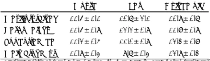
Table 1. Ciliary beat frequencies (Hz) in four different levels of the respiratory tract of the mouse, the rat and the guinea pig
Mouse Rat Guinea pig
Maxilloturbinal 11.5±1.1 10.7±2.1 10.9±0.7 Nasopharynx 11.5±0.9 12.2±0.9 11.8±0.9 Upper trachea 11.2±0.6 11.0±0.9 12.3±0.8 Main bronchus 10.9±0.4 9.7±0.4 12.9±0.3
*Significantly lower compared to the ciliary beat frequencies of the other sites of the rat
†Significantly higher compared to the ciliary beat frequencies in the main bronchi of the mouse and the rat
Table 2. Ciliary lengths (μm) in the respiratory tract of the mo- use, the rat and the guinea pig
Mouse Rat Guinea pig
Maxilloturbinal 3.6±0.1 4.1±0.2 3.8±0.2 Nasopharynx 3.6±0.2 4.2±0.3 3.6±0.2 Upper trachea 3.5±0.3 3.5±0.2 3.7±0.2 Main bronchus 3.4±0.2 3.5±0.1 3.7±0.3
*Significantly shorter compared to the ciliary lengths of the ma- xillo-turbinal and the nasopharynx of the rat
four areas. But in rats, the ciliary lengths in the upper tr- achea (3.5±0.2 μm) and the main bronchus (3.5±0.1 μm) were shorter than those in the maxilloturbinal (4.1
±0.2 μm) and the nasopharynx (4.2±0.3 μm) (p<0.001, Table 2). Table 3 shows the proportion of ciliary epith- elial cell coverage in each airway of the mouse, the rat and the guinea pig. There was no difference in ciliated area in the maxilloturbinal and the nasopharynx of mice,
rats and guinea pigs. The percentage of ciliated area, ho- wever, in the upper trachea (Fig. 2D) and the main bro- nchus of guinea pigs were higher than that in the upper
Table 3. Proportion (%) of the area covered with cilia in four di- fferent levels of the respiratory tract of the mouse, the rat and the guinea pig
Mouse Rat Guinea pig
Maxilloturbinal 87.5±8.4 91.5±4.3 93.5±6.0 Nasopharynx 89.2±6.2 93.4±5.7 92.3±5.7 Upper trachea 33.5±6.7* 38.5±6.7* 88.6±8.3 Main bronchus 35.4±7.3* 35.2±8.3* 87.0±9.2
*Significantly lower compared to proportion of the ciliated area of the upper trachea and the bronchus of the guinea pig
Fig. 2. Scanning electron micrographs of ciliated epithelia of mouse bronchus (A), rat trachea (B), guinea pig maxilloturbinal (C) and guinea pig upper trachea (D). In the rat respiratory cilia the percentage of ciliated area decreases from the maxilloturbinal to- ward the upper trachea. Tracheal and bronchial mucosae of the guinea pig are more ciliated than those of the mouse and the rat.
(original magnification×6,000).
Fig. 1. Measurement of ciliary length on a scanning electron mi- crograph (originial magnification×6,000).
B B B B A
AA A
DD DD CCC
C
trachea and the main bronchus of mice (Fig. 2A) or rats (Fig. 2B). In guinea pigs, proportion of ciliated area re- mained constant in the maxilloturbinal (Fig. 2C), the nasopharynx, the upper trachea (Fig. 2D) and the main bronchus.
DISCUSSION
In a recent study, the CBF in the inferior turbinate, the nasopharynx and the upper trachea of rats and gui- nea pigs, measured by the photoelectric method in vitro, was 12.7, 15.0, 10.5 Hz and 15.7, 16.0, 12.7 Hz, respe- ctively.3) Richelmann and colleagues reported an average CBF of 16.6 Hz in tracheal mucosa of the guinea pigs.6) These measurements vary from those obtained in this study, but it would be unreasonable to compare the res- ults, since ciliary movement in a certain area of the air- way can fluctuate according to different experimental conditions.
Joki et al. examined the ultrastructure and distribution of cilia in the respiratory epithelium of the upper and lower trachea and the subsegmental bronchus of rats and guinea pigs with a SEM.4) According to their study, the average ciliary lengths in the upper trachea of rats and guinea pigs were both 3.9 μm, which is close to the av- erage ciliary length of 3.5 μm found in our study of the upper trachea of rats and guinea pigs. According to the results of several studies, the CBF of rats differs widely at different locations of the airway, which makes it diff- icult to analyze results when rats are used for studying the function of cilia.1)2) The correlation between CBF and the structure of the ciliated epithelium is under some debate. In rats, it has been reported that the CBF decr- eases from the upper airway to the lower airway, which may be due to the differences in ratio of ciliated epith- elium and ciliary ultrastructure at different sites in the airway.3) Morgan et al., however, could not find correl- ation between CBF and ciliary length in rats.2) Meanw- hile, some authors suggested that differences in ciliary length may exist due to variations in regeneration rather than because of differences along the airway.4)
Ciliary length measured through electron microscopy can deviate from the actual length of the cilia due to different angles of view or the direction of the cilia.
Although it would be difficult to eliminate these factors completely, accuracy was maximized in this study by selecting cilia that were straightest and most upwardly directed. In an electron microscopic picture, non-ciliated
cells can be covered by the cilia of ciliated epithelial cells, which makes an exact calculation of ciliated epithelial cell area difficult. In this study, an image analyzer was used to define the area covered by cilia over the entire area of the picture. The ciliated area determined through this method may differ from the actual ciliated area, but the difference would not be significant in comparative analyses since ciliated area was measured under the same conditions throughout the study.
In this study, the CBF of rats was lower with statisti- cal significance in the main bronchus than in the maxil- loturbinal and the nasopharynx. The cilia in the main bronchus of rats were also shorter than those in the max- illoturbinal and the nasopharynx. These findings accor- ded with indicated variations in ciliated epithelium co- verage ratios. The results suggest that the CBF of rats is higher in portions of the airway where the cilia is long and the ratio of ciliated epithelium is high than in areas without these characteristics. Thus, we should be careful to interpret the results of CBF in rats because CBF varies along different parts of the airway and app- ears to depend on ciliary length and the ratio of ciliary epithelial cells.
To date, there has been no study on ciliary movements in different areas of the airway of mice. Nor has there been an examination of the structure with an electron microscope. In mice and guinea pigs, CBF and ciliary length did not appear to differ in different areas of the airway. However, while in guinea pigs the ratio of cil- iated epithelium remained constant along the airway, in the case of mice the ratio was lower in the main bron- chus than in the maxilloturbinal and the nasopharynx.
The finding suggests that there is no correlation between CBF and the ratio of ciliated epithelium in mice, unlike in rats. In this study, SEM finding of the guinea pigs va- ried somewhat from the results obtained by Joki et al.
They reported that the CBF in the upper trachea of gu- inea pigs was the same as in the upper trachea of rats,4) however, in this study the CBF in the upper trachea of guinea pigs was higher than in the same area in rats.
In conclusion, guinea pigs are considered to be a ex- cellent experimental animal model for the study of cili- ary movement because the CBF in these animals remains constant in different locations of the airway and because guinea pigs demonstrate an unbiased distribution of cil- iated epithelium. In rats, the CBF varies along the air- way, which means that studies of drug effects on CBF of airway epithelium must use ciliated epithelial cells
harvested from the same sites. However, in this study, mice indicated a constant CBF along the airway, which suggests that mice may also be used in studies on ciliary movement. In the upper airway there would be few pr- oblems related to differences in CBF and the distribution of ciliated epithelial cells.
REFERENCES
1) Sanderson MJ, Sleigh MA. Ciliary activity of cultured rabbit tr- acheal epithelium: Beat pattern and metachrony. J Cell Sci 1981;
47:331-47.
2) Morgan KT, Jiang XZ, Patterson DL, Gross EA. The nasal muco-
ciliary apparatus. Correlation of structure and function in the rat.
Am Rev Respir Dis 1984;130:275-81.
3) Joki S, Saano V. Ciliary beat frequency at six levels of the respi- ratory tract in cow, dog, guinea-pig, pig, rabbit and rat. Clin Exp Pharmacol Physiol 1994;21:427-34.
4) Joki S, Toskala E, Saano V, Nuutinen J. Ciliary ultrastructure and beating activity in rat and guinea-pig respiratory mucosa. Clin Exp Pharmacol Physiol 1995;22:619-23.
5) Min YG, Yun YS, Rhee CS, Sung MW, Lee KS, Ju MS, et al. Ef- fects of phenylephrine on ciliary beat in human nasal respiratory epithelium: quantitative measurement by video-computerized an- alysis. Laryngoscope 1998;108:418-21.
6) Riechelmann H, Mann W, Maurer J. The influence of Ca2+ ant- agonists on the ciliary activity of the guinea pig trachea. Eur Arch Otorhinolaryngol 1990;248:35-9.