대한소화기학회지 2009;54:220-226 DOI: 10.4166/kjg.2009.54.4.220
접수: 2009년 2월 23일, 승인: 2009년 7월 14일 연락처: 김현수, 501-757, 광주시 동구 학동 8
전남대학교 의과대학 내과학교실 Tel: (062) 220-6296, Fax: (062) 228-1330 E-mail: [email protected]
Correspondence to: Hyun Soo Kim, M.D.
Department of Internal Medicine, Chonnam National Uni- versity Medical School, 8, Hak-dong, Dong-gu, Gwangju 501- 757, Korea
Tel: +82-62-220-6296, Fax: +82-62-228-1330 E-mail: [email protected]
위선종 환자에서 대장내시경 선별 검사가 필요한가?
전남대학교 의과대학 내과학교실
박선영ㆍ김현수ㆍ윤경원ㆍ조성범ㆍ이완식ㆍ박창환ㆍ주영은ㆍ최성규ㆍ류종선
Prevalence of Colorectal Adenoma Is Increased in Patients with Gastric Adenoma
Seon Young Park, M.D., Hyun Soo Kim, M.D., Kyung Won Yoon, M.D., Sung Bum Cho, M.D., Wan Sik Lee, M.D., Chang Hwan Park, M.D., Young Eun Joo, M.D., Sung-Kyu Choi, M.D., and Jong-Sun Rew, M.D.
Department of Internal Medicine, Chonnam National University Medical School, Gwangju, Korea
Background/Aims: It has been reported that patients with gastric cancer may be at increased risk of synchronous or metachronous colorectal cancer. However, the incidence of colorectal adenoma in patients with gastric adenoma has not been discussed earlier. The aims of this study were to investigate the incidence of colorectal adenoma and to evaluate the necessity of colonoscopic surveillance in patients with gastric adenoma. Methods: We per- formed colonoscopy in 221 patients with gastric adenoma between January 2002 and June 2008. As a control group, 387 consecutive patients without gastric adenoma on gastroscopy who underwent colonoscopy were included. We retrospectively examined the endoscopic and colonoscopic findings as well as the clinicopathologic features. Results: Colorectal adenoma were diagnosed in 57.5% (127/221) of the gastric adenoma group and 38.0% (147/387) of the control group (p<0.001). Univariate analysis demonstrated that gender, age, past history of diabetes, and past history of gastric adenoma were associated with the risk of colorectal adenoma. Multivariate analysis demonstrated that gender (male, aOR 2.31, 95% CI 1.61-3.31), age (≥50 years, aOR 2.47, 95% CI 1.53-4.01), past history of diabetes (aOR 2.35, 95% CI 1.32-4.20), and presence of gastric adenoma (aOR 1.63, 95% CI 1.13-2.36) appeared to be independent risk factors for colorectal adenoma. Conclusions: The risk of col- orectal adenoma increases significantly in patients with gastric adenoma. We suggest that colonoscopic surveillance may be necessary in patients with gastric adenoma. (Korean J Gastroenterol 2009;54:220-226)
Key Words: Gastric adenoma; Colorectal adenoma; Colonoscopy
서 론
대장암은 미국에서 두 번째로 높은 사망률을 보이는 암으 로1 국내에서도 발생 빈도가 증가하고 있어서 암으로 인한
전체 사망 원인 중 4위에 해당한다.2 대장암 치료는 제한적 이어서 전구 병변인 선종을 미리 발견하고 제거하는 것이 중요하다. 이러한 대장의 선종과 암의 선별 검사로 대장내 시경검사를 언제 실시할 것인가에 대해서는 각 나라마다 다
박선영 외 8인. 위선종 환자에서 대장내시경 선별 검사가 필요한가? 221
소 차이를 보이지만, 우리나라의 경우 2001년 11월에 제정 된 대장암 조기검진 지침에 따르면 무증상 성인의 경우 50 세부터 대장내시경검사를 권고하고 있다.3,4 최근, 상부위장 관 내시경검사의 보편화로 위암의 조기 진단율이 높아지며, 특히 조기 위암으로 수술 받은 환자들의 90% 이상에서 장 기 생존이 가능하여 위암과 관련된 다발 종양의 발생이 높 았으며 그 중에서도 특히 대장암과 병발하는 경우가 많다.5 또한 대장암 환자에서 다발 종양의 발생을 분석한 결과에서 도 위암이 가장 많았다.6 그러나, 위선종에서 대장 병변이 얼마나 동반되어 있는지에 대해서는 연구가 거의 없는 실정 으로, 이런 환자에서 추가로 대장내시경검사가 필요한지에 관한 연구는 아직 없다. 이에 저자 등은 상부위장관 내시경 으로 위선종을 진단한 환자에서 대장 병변, 특히 대장 종양 이 얼마나 동반되었는지에 대해 알아보고 대장내시경 선별 검사가 필요한지에 대해 알아보고자 하였다.
대상 및 방법
1. 대상
2002년 1월부터 2008년 6월까지 전남대학교병원에 내원 하여 상부위장관 내시경검사에서 위선종7,8을 진단받고, 대 장내시경을 시행한 221명의 환자를 ‘위선종군’으로 하였다.
‘대조군’은 2002년 1월부터 2008년 6월까지 상부위장관 내 시경검사에서 위 종양이 발견되지 않았고 대장내시경검사 를 같이 시행한 387명을 무작위로 선정하였다. 위선종군과 대조군에서 이전에 위나 장 수술을 받은 경우, 염증성 장질 환, 가족 대장용종증의 과거력, 대장내시경검사에서 전 장 을 관찰하지 못하거나 기술적으로 실패한 경우는 이번 연구 에서 제외하였다.
2. 방법
위선종과 위 선암종은 생검이나 내시경 점막 절제술 및 점막하 절제술, 또는 수술을 통하여 조직학적으로 진단하였 다.8 위 선암의 경우에는 기저에 형성이상을 동반한 경우만 포함시켰다. 대장내시경검사에서 용종의 유무, 크기, 위치 및 육안 형태를 확인하고, 대장 용종은 생검이나 내시경 점 막 절제술을 통해 조직학적으로 확진하였으며, 여러 개의 용종이 있는 경우 병리적인 특성이 가장 심한 용종을 분석 에 포함시켰다. 대장 종양(colorectal neoplasm)은 대장 선종 과 대장암을 합쳐서 정의하였으며, 정상 소견과 함께 과형 용종 및 염증성 용종은 비종양 병변(non-neoplastic lesion, without colorectal neoplasm)으로 분류하였다. 진행 선종은 크 기가 1 cm가 넘거나 융모 선종 및 고등급 이형성, 또는 암 이 동반된 경우로 정의하였다.9 모든 위선종군과 건강 대조
군의 연령, 성별, 키, 몸무게, 체질량지수, 위내시경검사 소 견과 병리 소견, 대장내시경검사 소견과 병리 소견은 후향 조사하였다. 이번 연구는 전남대학교병원 임상연구윤리위 원회의 심의를 통과하였다.
3. 통계분석
모든 통계분석은 SPSS 14.0 for Windows를 이용하였다.
범주형 변수의 분석은 Chi-square를, 연속변수의 분석은 independent t-test를 이용하였다. 연령과 성별을 보정하기 위 해 공변량분석(ANCOVA)을 시행하였고, 대장선종과 관련된 인자를 분석하기 위해 가능한 혼란변수의 통제를 위한 다변 량 분석을 실시하였으며, 종속형 변수를 이분형 변수로 하 여 다중 로지스틱 회귀분석을 시행하였다. 모든 통계 유의 성은 p<0.05인 경우로 하였다.
결 과
1. 위선종군과 대조군의 일반 특성
위선종군의 평균 연령은 64.4±8.9세로 대조군의 58.3±11.8 세보다 높았다(p<0.001). 남녀비는 위선종군에서 2.9:1이었 으며, 대조군에서 1.1:1로 위선종군에서 남자의 비율이 더 높았다(p<0.001). 두 군 간의 신장과 몸무게의 유의한 차이 는 없었으나, 대조군의 체질량지수(24.1±3.3 kg/m2)가 위선 종군의 체질량지수(23.5±3.1 kg/m2)보다 높았다(p=0.032). 두 군 간의 당뇨병과 고혈압의 유병률 차이는 없었다(Table 1).
위선종군의 위상피병변의 병리 진단은 저도 형성이상 (low-grade dysplasia)이 56.6% (125/221), 고도 형성이상(high- grade dysplasia)이 21.3% (47/221), 선암종이 22.2% (49/221) 를 차지하였다.
2. 대장 종양의 위험인자에 관한 분석
전체 608명의 환자 중에서 대장 종양은 274명(45.1%)이었 고, 비종양성 병변은 334명(54.9%)이었다. 대장 종양에서 대 장암은 38명(6.3%), 진행성 선종은 103명(16.7%)에서 발견되 었다. 위선종군에서 대장 종양의 발생빈도는 57.5% (127/
221)였으며, 대조군의 대장 종양의 발생빈도는 38.0% (147/
387)이었다(p<0.001). 위선종군을 저도 형성이상군, 고도 형 성이상군, 선암종군으로 나누었을 때 대조군에 비해 저도 이형성군(70/125 vs. 94/221, p<0.001)과 선암종군(34/49 vs.
94/221, p<0.001)에서 대장 종양의 발생 빈도가 더 높았으 나, 고도 이형성군(23/47 vs. 94/221, p=0.146)에서는 차이가 없었다. 그 외 단변량 분석에서 대장 종양과 관련된 인자로 는 성별, 나이, 당뇨병 여부였다. 대장 종양이 있는 군에서 남자는 71.5% (196/274)였으나 대장 종양이 없는 군에서 남
222 The Korean Journal of Gastroenterology: Vol. 54, No. 4, 2009
Table 1. Baseline Characteristics of the Subjects with or without Gastric Adenoma Characteristics Subjects with gastric
adenoma (n=221)
Subjects without gastric
adenoma (n=387) p-value p-value† Sex
Male 164 (74.2%) 201 (51.9%) <0.001 Female 57 (25.8%) 186 (48.1%)
Age* 64.4±8.9 58.3±11.8 <0.001
Height (cm)* 163.3±10.9 163.1±8.4 0.844 0.817
Body weight (kg)* 62.4±9.8 64.4±11.1 0.065 0.981
BMI (kg/m2)* 23.5±3.1 24.1±3.3 0.032 0.041
Diabetes 22/216 (10.2%) 40/367 (10.9%) 0.787 0.372 Hypertension 53/216 (24.5%) 85/367 (23.2%) 0.706 0.054 Subjects with colon adenoma 127/221 (57.5%) 94/221 (42.5%) <0.001 0.014 Subjects with advanced colon adenoma‡ 44/221 (19.9%) 59/387 (15.2%) 0.140 0.151 Subjects with colon cancer 18/221 (8.1%) 20/387 (5.2%) 0.145 0.406
* Values are mean±SD.
†Significance by ANCOVA (co-variance) test adjusted for age and sex.
‡Advanced adenomas were defined as polyps with size>10 mm, villous components, high grade dysplasia, or carcinoma.
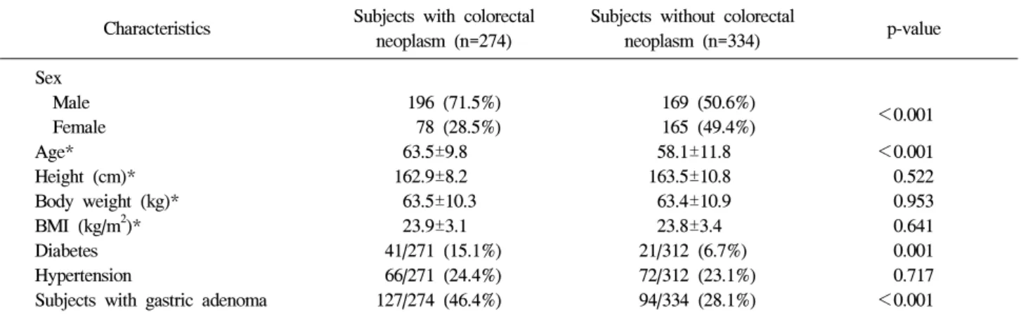
Table 2. Baseline Characteristics of the Subjects with or without Colorectal Neoplasm Characteristics Subjects with colorectal
neoplasm (n=274)
Subjects without colorectal
neoplasm (n=334) p-value Sex
Male 196 (71.5%) 169 (50.6%) <0.001 Female 78 (28.5%) 165 (49.4%)
Age* 63.5±9.8 58.1±11.8 <0.001
Height (cm)* 162.9±8.2 163.5±10.8 0.522
Body weight (kg)* 63.5±10.3 63.4±10.9 0.953
BMI (kg/m2)* 23.9±3.1 23.8±3.4 0.641
Diabetes 41/271 (15.1%) 21/312 (6.7%) 0.001
Hypertension 66/271 (24.4%) 72/312 (23.1%) 0.717 Subjects with gastric adenoma 127/274 (46.4%) 94/334 (28.1%) <0.001
*Values are mean±SD.
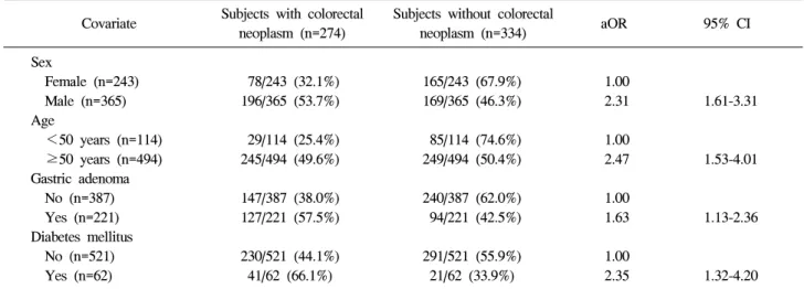
자는 50.6% (169/334)였다(p<0.001). 대장 종양이 있는 군에 서 당뇨병이 동반된 경우는 15.1% (41/271)였으나, 대장 종 양이 없는 군에서 당뇨병이 동반된 경우는 6.7% (21/312)였 다(p<0.001) (Table 2). 이를 다변량 분석을 하였을 때 남성, 50세 이상, 당뇨병이 있을 때, 위선종군에서 대장 종양의 빈 도가 의의 있게 높았다(Table 3).
3. 위선종군과 대조군의 대장 종양의 특징
위선종군과 대조군 간의 대장 종양의 최대 크기는 각각 10.8±10.1 mm, 11.2±8.2 mm로 두 군 간의 차이는 없었다 (p=0.748). 위선종군에서 대장 종양의 전체 개수가 3.6±10.1 로 대조군의 2.1±1.7로 높았으나 통계적인 의의는 없었다 (p=0.099). 그 외 두 군 간의 대장 종양의 위치분포 차이는
없었다(p=0.853) (Table 4). 진행 선종의 분포는 위선종군에 서 19.9% (44/221), 대조군에서 15.2% (59/387)였고(p=0.140) 대장암의 분포는 위선종군에서 8.1% (18/221), 대조군에서 5.2% (20/387)였다(p=0.145) (Table 1).
고 찰
대장 용종의 유병률은 서구에서 40대 이상의 인구 중 5-10%의 빈도를 보이고, 에스자결장경검사에서 5-10%, 대장 내시경검사에서 25% 이상 보고되고 있다. 나이가 많을수록 용종의 수가 증가하고 세포 이형성의 정도가 심해질 뿐 아 니라, 큰 용종의 빈도가 높으며,10 이번 연구에서도 50세 이 상에서 대장 종양의 빈도는 49.6%로 50세 미만에서의
Park SY, et al. Prevalence of Colorectal Adenoma Is Increased in Patients with Gastric Adenoma 223
Table 3. Multivariate Analysis of Risk Factor for Colorectal Neoplasm Covariate Subjects with colorectal
neoplasm (n=274)
Subjects without colorectal
neoplasm (n=334) aOR 95% CI Sex
Female (n=243) 78/243 (32.1%) 165/243 (67.9%) 1.00
Male (n=365) 196/365 (53.7%) 169/365 (46.3%) 2.31 1.61-3.31 Age
<50 years (n=114) 29/114 (25.4%) 85/114 (74.6%) 1.00
≥50 years (n=494) 245/494 (49.6%) 249/494 (50.4%) 2.47 1.53-4.01 Gastric adenoma
No (n=387) 147/387 (38.0%) 240/387 (62.0%) 1.00
Yes (n=221) 127/221 (57.5%) 94/221 (42.5%) 1.63 1.13-2.36 Diabetes mellitus
No (n=521) 230/521 (44.1%) 291/521 (55.9%) 1.00
Yes (n=62) 41/62 (66.1%) 21/62 (33.9%) 2.35 1.32-4.20
Table 4. Characteristics of Colorectal Neoplasm in the Subjects with or without Gastric Adenoma Variables Subjects with gastric
adenoma (n=127)
Subjects without gastric
adenoma (n=147) p-value
Maximal size 10.8±10.1 mm 11.2±8.2 mm 0.748
Total number 3.6±10.1 2.1±1.7 0.099
Location (n=266) 0.853
Ascending colon 26.0% (32/123) 31.5% (45/143) 0.328 Transverse colon 16.3% (20/123) 12.6% (18/143) 0.393 Descending colon 8.9% (11/123) 8.4% (12/143) 0.873 Sigmoid colon 30.9% (38/123) 30.1% (43/143) 0.884
Rectum 17.9% (22/123) 17.5% (25/143) 0.931
Histology
Non-advanced adenoma 64.8% (81/125) 58.2% (82/141)
0.267 Advanced adenoma* 35.2% (44/125) 41.8% (59/141)
Colon cancer 14.4% (18/125) 14.4% (20/141) 0.892
*Advanced adenomas were defined as polyps with size>10 mm, villous components, high grade dysplasia, or carcinoma.
25.4%보다 더 높았다. 성별에 따른 분석을 살펴보면, 일반 적으로 성별은 선종의 발생에 무관하다는 보고도 있으나, 서양의 다른 연구에서 여자보다 남자에서 약 1.3-1.7배 정도 대장 종양의 유병률이 높았으며, 이번 연구에서는 남자가 여자에 비해 2.3배 대장 종양의 유병률이 높았다.11,12 그 외 대장 종양의 발생 요인으로 유전적인 감수성과 지방질 음 식, 섬유소 부족, 비만, 흡연 등의 환경 요인이 중요하다.13-17 환경 요인 중에 특히 고혈당, 고인슐린혈증 및 인슐린 저항 성 등이 대장암 발생과 연관이 있으며, 당뇨병 및 인슐린 저 항성과 관련이 많은 비만도 대장암과 연관이 있다.18,19 대장 선종 환자를 대상으로 한 연구에서도 대장 선종의 발생과 체질량지수, 고혈당, 그리고 복부 비만과 연관성이 있었다.20 이번 연구에서 당뇨병이 있는 환자에서 대장 종양의 빈도는 66.1% (41/62)로 당뇨병이 없는 환자의 대장 종양의 빈도인 44.1% (230/521)보다 더 높았다. 이번 연구에서 타 연구보다
전반적인 대장 종양의 빈도가 더 높았으며, 이는 대상 환자 가 위선종군을 포함하였고, 증상이 있거나 대장 종양에 관 심을 갖고 3차 병원에 내원하여 검사를 시행한 환자를 포함 하였기에, 기존 연구에 비해 대상환자의 연령과 기저질환의 차이가 있기 때문으로 생각한다.
이번 연구에서 대장 종양의 빈도는 위선종이 있는 환자에 서 증가하였다. 서양의 한 보고에서 41명의 위선종을 가지 고 있는 환자가 나이를 보정한 101명의 대조군에 비해 대장 용종, 대장 선종, 대장암의 빈도가 더 높음을 보고하였다.21 국내의 한 연구에서는 위암 환자에서 대장선종과 대장암의 빈도는 각각 22.9%, 9.5%였으며, 대조군에서 대장선종과 대 장암의 빈도는 각각 29.0%, 0.7%로 위암 환자에서 대장암의 빈도가 증가하였다.22 동아시아의 경우 다른 장기에 비해 발 생빈도가 비교적 높은 위암에서 다발 원발 악성 종양의 빈 도가 높으며, 특히 대장암과 병발한 경우가 많다.23 실제로
224 대한소화기학회지: 제54권 제4호, 2009
이번 연구에서 대장 종양의 빈도와 연관이 있는 인자를 알 기 위해 시행한 단변량 분석에서 위선종의 유무 외에 나이, 성별, 당뇨병 유무가 관련이 있었으며, 이를 다변량 로지스 틱 회귀분석을 하였을 때도 위선종을 포함한 모든 인자들에 서 대장 종양의 발생 빈도와 연관이 있었다. 이제까지의 연 구들에서 위암과 대장암의 연관성은 형태, 유전적인 연관성 으로 설명하고 있다. 위암은 Helicobacter pylori (H. pylori) 감염과 관련되어 장상피화생에서 발생하고,24 대장암은 선 종-암종 연속성에 따라 선종을 대장암의 전구병변으로 생각 한다. 그 외 H. pylori 감염이 위암과 관련이 있는 것처럼 대 장암과 관련이 있다는 몇몇 연구 결과들도 있다.25,26 위암과 대장암의 유전적인 연관성은 여러 연구에서 밝혀졌듯이 p53, APC, DCC, K-ras, hMLH1과 hMLH2, Microsatellite instability (MSI)와 같은 동일한 유전자가 위암과 대장암에서 중요한 역할을 한다고 알려져 있다.27-32 이와 유사하게 APC/MCC 유전자(5q21), Topoisomerase II alpha, Klf4 (epith- elial zinc-finger transcription factor), COX-2 발현 등이 위암의 전구병변과 대장암의 전구병변에서 같이 관여하고 있다.33-37 이번 연구에서 위선종을 동반한 경우 대장 종양의 발생 빈도가 높았다. 아직까지 대장내시경검사가 상부위장관 내 시경검사에 비해 보편화되지 못하였고 대장암의 발생이 점 차 증가하고 있는 시점에서 상부위장관 내시경검사에서 위 선종을 발견한 경우 적극적으로 대장내시경검사를 시행하 여 대장암의 발생에 관심을 두는 것이 대장 선종뿐만 아니 라 대장암을 조기에 발견하는 데 도움이 될 것이다.
이번 연구의 제한점은 의무기록을 통한 후향 연구로 연구 대상자 수, 특히 여자 환자 수가 상대적으로 적었고, 위선종 군과 대조군의 연령 차이로 연구 대상 선발에 선택 오차가 있겠다. 하지만 연령과 성별을 보정한 통계 분석에서 위선 종군에서 대장 종양의 빈도가 높았다. 이에 위선종을 갖고 있는 환자에서 대장내시경검사를 시행해야 하는지 임상적 인 유용성을 갖기 위해서는 향후 좀 더 많은 수의 증례 수 를 가진 대규모 전향 연구가 필요하리라 생각한다. 그리고, 위선종으로 내시경 점막 절제술, 내시경 점막하 절제술 및 수술을 시행한 환자의 추적관찰 시 항상 대장 종양에 대한 발생 가능성에 관심을 갖고 이에 대한 감시 체계의 구축이 필요할 것으로 생각한다.
요 약
목적: 위암과 관련된 다발 종양의 발생 중에서 대장 선암 종의 빈도가 증가한다는 몇몇 보고들이 있다. 그러나, 위선 종 환자에서 대장 선종의 위험성과 대장내시경검사가 필요 한지는 잘 알려져 있지 않다. 이에 저자 등은 위선종 환자에 서 대장종양의 빈도를 알아보고, 대장내시경검사의 선별 검
사가 필요한지에 대해 알아보고자 하였다. 대상 및 방법:
2002년 1월부터 2008년 6월까지 전남대학교병원에 내원하 여 위선종 및 위암을 진단하고 내시경 점막 절제술 및 점막 하 절제술을 시행받았고, 대장내시경검사를 시행한 221명의 환자를 위선종군으로 하였다. 대조군은 상부위장관 내시경 검사에서 위종양이 발견되지 않았고 대장내시경검사를 시 행한 387명을 대상으로 하였다. 위선종군과 대조군의 기저 임상 특성과 내시경검사 소견 및 병리 소견은 후향 조사하 였다. 결과: 위선종군에서는 저도 선종이 56.6% (125/221)로 가장 많았다. 위선종군에서 대장 선종의 발생 빈도는 57.5%
(127/221)였으며, 대조군에서 대장 선종의 발생빈도는 38.0%
(147/387)였다(p<0.001). 단변량 분석에서 대장 종양과 관련 된 인자로는 성별, 나이, 당뇨병 여부였다. 이를 다변량 분 석을 하였을 때 성별(남성, aOR 2.31, 95% CI 1.61-3.31), 나 이(50세 이상, aOR 2.47, 95% CI 1.53-4.01), 당뇨병 유무(당 뇨병, aOR 2.35, 95% CI 1.32-4.20), 위선종 유무(위선종, aOR 1.63, 95% CI 1.13-2.36)가 대장 종양의 발생 빈도에 의 의 있게 영향을 주었다. 결론: 이번 연구에서 대장 선종은 위선종 환자에서 유의하게 증가하였다. 따라서 위선종 환자 의 추적관찰 시 대장 종양에 대한 발생 가능성에 관심을 갖 고 이에 대한 감시 체계의 구축이 필요할 것으로 생각한다.
색인단어: 위선종, 대장 종양, 대장내시경
참고문헌
1. Ries LA, Wingo PA, Miller DS, et al. The annual report to the nation on the status of cancer, 1973-1997, with a special section on colorectal cancer. Cancer 2000;88:2398-2424.
2. 대한민국보건복지부. 한국인 암등록 조사자료 분석보고 서. 2003.
3. Jung SY. 대장암 조기검진을 위한 권고안. Korean J Gas- trointest Endosc 2002;24:317-320.
4. Winawer S, Fletcher R, Rex D, et al. Colorectal cancer scre- ening and surveillance: clinical guidelines and rationale-up- date based on new evidence. Gastroenterology 2003;124:544- 560.
5. Yoshino K, Asanuma F, Hanatani Y, Otani Y, Kumai K, Ishibiki K. Multiple primary cancers in the stomach and an- other organ: frequency and the effects on prognosis. Jpn J Clin Oncol 1985;15:183-190.
6. Maruyama H, Hasuike Y, Furukawa J, et al. Multiple color- ectal carcinomas and colorectal carcinoma associated with ex- tracolonic malignancies. Surg Today 1992;22:99-104.
7. Kang HJ, Park DY, Kim KH, Song GA, Lauwers GY.
Pathologic diagnosis of gastric epithelial neoplasia. Korean J
박선영 외 8인. 위선종 환자에서 대장내시경 선별 검사가 필요한가? 225
Gastroenterol 2008;52:273-280.
8. Kim WH, Park CK, Kim YB, et al. A standardized pathology report for gastric cancer. Korean J Pathol 2006;39:106- 113.
9. Chang HJ, Park CK, Kim WH, et al. A standardized pathol- ogy report for colorectal cancer. Korean J Pathol 2006;2006:
193-203.
10. Ansher AF, Lewis JH, Fleischer DE, et al. Hyperplastic co- lonic polyps as a marker for adenomatous colonic polyps.
Am J Gastroenterol 1989;84:113-117.
11. Stevens T, Burke CA. Colonoscopy screening in the elderly:
when to stop? Am J Gastroenterol 2003;98:1881-1885.
12. Rex DK, Khan AM, Shah P, Newton J, Cummings OW.
Screening colonoscopy in asymptomatic average-risk African Americans. Gastrointest Endosc 2000;51:524-527.
13. Sandler RS, Lyles CM, Peipins LA, McAuliffe CA, Woosley JT, Kupper LL. Diet and risk of colorectal adenomas: macro- nutrients, cholesterol, and fiber. J Natl Cancer Inst 1993;85:
884-891.
14. Bayerdorffer E, Mannes GA, Ochsenkuhn T, Kopcke W, Wiebecke B, Paumgartner G. Increased risk of ‘high-risk' colorectal adenomas in overweight men. Gastroenterology 1993;104:137-144.
15. Giovannucci E, Colditz GA, Stampfer MJ, et al. A pro- spective study of cigarette smoking and risk of colorectal ad- enoma and colorectal cancer in U.S. women. J Natl Cancer Inst 1994;86:192-199.
16. Cannon-Albright LA, Skolnick MH, Bishop DT, Lee RG, Burt RW. Common inheritance of susceptibility to colonic adenomatous polyps and associated colorectal cancers. N Engl J Med 1988;319:533-537.
17. Grady WM. Genetic testing for high-risk colon cancer pa- tients. Gastroenterology 2003;124:1574-1594.
18. Calle EE, Thun MJ. Obesity and cancer. Oncogene 2004;23:
6365-6378.
19. Kaaks R, Toniolo P, Akhmedkhanov A, et al. Serum C-pep- tide, insulin-like growth factor (IGF)-I, IGF-binding proteins, and colorectal cancer risk in women. J Natl Cancer Inst 2000;92:1592-1600.
20. Chung YW, Han DS, Park YK, et al. Association of obesity, serum glucose and lipids with the risk of advanced colorectal adenoma and cancer: a case-control study in Korea. Dig Liver Dis 2006;38:668-672.
21. Cappell MS, Fiest TC. A multicenter, multiyear, case-con- trolled study of the risk of colonic polyps in patients with gastric polyps. Are gastric adenomas a new indication for surveillance colonoscopy? J Clin Gastroenterol 1995;21:198- 202.
22. Oh SY, Park DI, Yoo TW, et al. Is gastric cancer a new in- dication for surveillance colonoscopy? Colon cancer is in- creased in gastric cancer patients. Korean J Gastroenterol 2006;47:191-197.
23. Okamoto N, Morio S, Inoue R, Akiyama K. The risk of a second primary cancer occurring in five-year survivors of an initial cancer. Jpn J Clin Oncol 1987;17:205-213.
24. Correa P. Human gastric carcinogenesis: a multistep and mul- tifactorial process--First American Cancer Society Award Lec- ture on Cancer Epidemiology and Prevention. Cancer Res 1992;52:6735-6340.
25. Thomas JE, Gibson GR, Darboe MK, Dale A, Weaver LT.
Isolation of Helicobacter pylori from human faeces. Lancet 1992;340:1194-1195.
26. Saunders KE, Shen Z, Dewhirst FE, Paster BJ, Dangler CA, Fox JG. Novel intestinal Helicobacter species isolated from cotton-top tamarins (Saguinus oedipus) with chronic colitis. J Clin Microbiol 1999;37:146-151.
27. Laurent-Puig P, Olschwang S, Delattre O, et al. Survival and acquired genetic alterations in colorectal cancer. Gastroenter- ology 1992;102:1136-1141.
28. Uchino S, Noguchi M, Ochiai A, Saito T, Kobayashi M, Hirohashi S. p53 mutation in gastric cancer: a genetic model for carcinogenesis is common to gastric and colorectal cancer.
Int J Cancer 1993;54:759-764.
29. Nakatsuru S, Yanagisawa A, Ichii S, et al. Somatic mutation of the APC gene in gastric cancer: frequent mutations in very well differentiated adenocarcinoma and signet-ring cell car- cinoma. Hum Mol Genet 1992;1:559-563.
30. Uchino S, Tsuda H, Noguchi M, et al. Frequent loss of heter- ozygosity at the DCC locus in gastric cancer. Cancer Res 1992;52:3099-3102.
31. Vogelstein B, Fearon ER, Hamilton SR, et al. Genetic alter- ations during colorectal-tumor development. N Engl J Med 1988;319:525-532.
32. Ohtani H, Yashiro M, Onoda N, et al. Synchronous multiple primary gastrointestinal cancer exhibits frequent microsatellite instability. Int J Cancer 2000;86:678-683.
33. Sanz-Ortega J, Sanz-Esponera J, Caldes T, Gomez de la Concha E, Sobel ME, Merino MJ. LOH at the APC/MCC gene (5Q21) in gastric cancer and preneoplastic lesions.
Prognostic implications. Pathol Res Pract 1996;192:1206- 1210.
34. Fogt F, Nikulasson ST, Holden JA, et al. Topoisomerase II alpha expression in normal, inflammatory, and neoplastic con- ditions of the gastric and colonic mucosa. Mod Pathol 1997;
10:296-302.
226 The Korean Journal of Gastroenterology: Vol. 54, No. 4, 2009
35. Katz JP, Perreault N, Goldstein BG, et al. Loss of Klf4 in mice causes altered proliferation and differentiation and pre- cancerous changes in the adult stomach. Gastroenterology 2005;128:935-945.
36. Katz JP, Perreault N, Goldstein BG, et al. The zinc-finger transcription factor Klf4 is required for terminal differ-
entiation of goblet cells in the colon. Development 2002;
129:2619-2628.
37. Konturek PC, Kania J, Burnat G, Hahn EG, Konturek SJ.
Prostaglandins as mediators of COX-2 derived carcinogenesis in gastrointestinal tract. J Physiol Pharmacol 2005;56:57-73.