Korean J Gastroenterol Vol. 58 No. 1, 58-60 DOI: 10.4166/kjg.2011.58.1.58
IMAGE OF THE MONTH
Korean J Gastroenterol, Vol. 58 No. 1, July 2011 www.kjg.or.kr
폐 침범이 의심되는 자가면역 췌장염
방승민, 박정엽
연세대학교 의과대학 소화기내과학교실, 소화기병연구소
Suspected Pulmonary Involvement of Autoimmune Pancreatitis
Seungmin Bang and Jeong Youp Park
Division of Gastroenterology, Department of Internal Medicine, Yonsei Institute of Gastroenterology, Yonsei University College of Medicine, Seoul, Korea
CC This is an open access article distributed under the terms of the Creative Commons Attribution Non-Commercial License (http://creativecommons.org/licenses/
by-nc/3.0) which permits unrestricted non-commercial use, distribution, and reproduction in any medium, provided the original work is properly cited.
교신저자: 박정엽, 120-752, 서울시 서대문구 성산로 250, 연세대학교 의과대학 소화기내과학교실, 소화기병연구소
Correspondence to: Jeong Youp Park, Division of Gastroenterology, Department of Internal Medicine, Yonsei Institute of Gastroenterology, Yonsei University College of Medicine, 250, Seongsan-ro, Seodaemun-gu, Seoul 120-752, Korea. Tel: +82-2-2228-1937, Fax: +82-2-2227-7900, E-mail: [email protected]
Financial support: None. Conflict of interest: None.
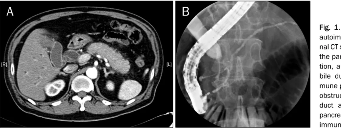
Fig. 1. Abdominal CT and ERCP of autoimmune pancreatitis. (A) Abdomi- nal CT showed diffuse enlargement of the pancreas, peripancreatic infiltra- tion, and dilatation of the common bile duct, compatible with autoim- mune pancreatitis. (B) ERCP showed obstruction of the distal common bile duct and diffuse slightly irregular pancreatic duct, compatible with auto- immune pancreatitis.
증례: 자가면역 췌장염으로 추적 관찰 중인 55세 남자 환자 가 CT 검사에서 폐에 이상 소견을 보여 내원하였다. 환자는 내원 2년 전에 복시(diplopia)로 스테로이드 치료를 받은 적이 있었으며 6개월 전 황달을 주소로 내원하여 시행한 검사상 자가면역 췌장염과 그로 인한 담도 폐색으로 진단되었다. 플 라스틱 스텐트를 이용한 담도 배액술과 스테로이드 치료를 받 았고, 스테로이드 치료 후 혈당 상승 소견 보여 당뇨 치료를 시작했다. 6개월 전 시행한 ANA와 같은 자가면역질환 표지 자들은 모두 음성이었고 혈청 IgG는 1,108 mg/dL (정상수치 700-1,600)로 정상이었으나 IgG4는 1,610 mg/L (정상수치 39.2-864.0)로 증가되어 있었다. 복부 CT상 췌장의 미만성 종
대와 췌장주위 침윤 소견을 보였고 원위부 담도는 폐색된 소 견을 보였다 (Fig. 1A). ERCP 검사에서도 CT와 마찬가지로 원위부 담도 폐색으로 인한 간내담관과 근위부 담도의 확장 소견을 보였고, 췌관은 전반적으로 불규칙한 모양을 보였다 (Fig. 1B). 이와 같은 소견을 바탕으로 자가면역 췌장염을 의 심하고 스테로이드 치료를 한 결과, 증상이 호전 양상을 보이 고 IgG4 수치가 정상화되어 자가면역 췌장염으로 진단하고 스테로이드 용량을 감량하였다.
내원 2개월 전 시행한 추적 복부 CT 검사에서 췌장에는 이상 없었으나 흉부에 이상 소견을 보였다. 내원 당시 시행한 말초혈액검사에서 백혈구 9,750/mm3, 혈색소 14.8 g/dL, 혈
Bang SM and Park JY. Suspected Pulmonary Involvement of Autoimmune Pancreatitis
59
Vol. 58 No. 1, July 2011
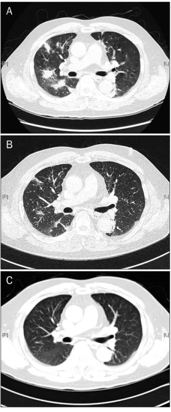
Fig. 2. Chest CT of pulmonary involvement of autoimmune pancreatitis. (A) Chest CT showed multifocal consolidation with fibrosis and emphysematous change at both lungs. (B) Chest CT 1 month after azathioprine therapy showed the improvement of consolidation. (C) Chest CT 3 months after azathioprine therapy showed that previously noted consolidation disappeared completely.
소판 364,000/mm3이었다. 혈액 검사상 BUN 23 mg/dL, 크 레아티닌 1.0 mg/dL, 총 단백 8.4 g/dL, 알부민 5.0 g/dL, 총 빌리루빈 0.6 mg/dL, AST/ALT 23/18 IU/L, ALP 58 IU/L로 모두 정상이었다. 혈청 IgG는 1,306 mg/dL로 정상이 었으나 IgG4는 1,450.0 mg/L로 증가된 소견을 보였다. 흉부 CT 검사상 양쪽 폐의 여러 곳에 폐기종과 섬유화를 동반한 부정형의 결절(consolidation) 소견이 나타났다(Fig. 2A). 자 가면역 췌장염의 재발 그리고 폐 침범을 의심하여 폐 조직 검사 후 스테로이드 치료를 시행하려 하였으나 환자는 침습적 검사를 거부하였을 뿐만 아니라 과거 스테로이드 치료 동안 혈당이 과도하게 상승하여 힘들었다는 이유로 스테로이드 치 료도 거부하여 azathioprine 50 mg 경구 투여로 면역 억제 치료를 시작하였다. 치료 1달 후 시행한 흉부 CT 검사상 폐결 절은 호전되는 양상을 보였고(Fig. 2B), 치료 3달 후 시행한 혈청 IgG4 검사는 783.0 mg/L로 정상 소견을 보였으며 흉부 CT 검사에서 그전에 보이던 결절들은 사라졌다(Fig. 2C). 현 재 azathioprine 투약 중단 4개월 째 재발 소견 없이 추적 관찰 중이다.
진단: 자가면역 췌장염의 폐침범 의심
자가면역 췌장염은 폐쇄성 황달, 불규칙한 췌관의 좁아짐, 췌장의 부어오름을 특징으로 하는 췌장염의 일종이며 스테로 이드 치료에 반응을 잘하는 특징을 가지고 있다. 또한 혈청 IgG4의 상승과 자가면역표지자가 양성 소견을 보이기도 하며 조직 검사에서는 형질 세포(plasma cell)의 침윤이 관찰된다.
때때로 췌장 이외의 장기에도 문제를 일으키기도 하는데 침 샘, 후복막강, 담관, 폐, 신장 등을 침범하기도 한다.1-5
2004년 Taniguchi 등6은 자가면역 췌장염을 가지고 있는 환자에서 발생한 간질성 폐렴을 보고한바 있다. Tsushima 등7은 19명의 폐를 침범한 자가면역 췌장염 환자를 분석한 결과 혈청 IgG와 IgG4의 상승을 보였으며 폐문부의 림프절 비대(hilar lymphadenopathy), 소결절(nodule), 그물 모양의 음영(reticular shadow)과 같은 소견을 보였다고 보고하였다.
국내에서도 Yoo 등8이 조직 검사를 통해 IgG4 양성 형질 세 포가 폐에 침윤되어 있음을 확인한 후 폐를 침범한 자가면역 췌장염 2예를 보고한 바 있었다. 이번 증례의 경우 림프절 비 대 소견은 보이지 않았으며 폐기종과 함께 섬유화를 동반한 다발성 결절 소견을 보였고 혈청 IgG4 상승으로 자가면역 췌 장염의 폐침범을 의심하였다. 종괴나 결절의 모양으로 나타나 기 때문에 다른 폐 질환과의 감별 진단이 중요하므로 이 증례 에서도 확실한 진단을 위해서는 조직 검사가 필요했으나, 폐 조직 검사의 난이도와 민감도를 고려할 때 면역 억제 치료에 대한 치료 반응을 통해 임상적으로 자가면역 췌장염의 폐침범 진단을 내릴 수 있다고 생각된다.7
대부분의 증례에서 자가면역 췌장염의 폐침범은 스테로이 드 치료에 좋은 반응을 보였다. 이 증례의 경우 환자가 스테로
60
방승민, 박정엽. 폐 침범이 의심되는 자가면역 췌장염The Korean Journal of Gastroenterology
이드 치료 중 혈당 조절이 잘 되지 않아 힘들어 했기 때문에 스테로이드 대신 azathioprine을 이용한 면역억제치료를 시 도하였고 좋은 치료 반응을 보였다. 자가면역췌장염의 스테로 이드 치료 후 재발이 발생하는 경우 또는 스테로이드를 줄일 수 없는 경우 azathioprine 치료의 효과는 이미 보고된 바 있 지만, 폐와 같이 타장기를 침범한 경우에도 반응이 좋은지는 알려진 바가 없다. 또한 azathioprine 자체가 부작용으로 췌 장염을 유발할 수 있고 아직까지 어느 정도의 용량으로 얼마 나 투여해야 하는 지에 대하여도 정해진 바가 없다.9
자가면역 췌장염으로 추적 중에는 폐침범 여부도 같이 확 인해야 하며 폐의 종괴나 결절로 내원한 환자에서는 IgG4 연 관 질환일 가능성에 대하여 검토가 필요하다. 자가면역 췌장 염의 폐침범에 대한 진단과 치료, 예후를 알기 위해 더 많은 증례를 대상으로 하는 연구가 필요할 것으로 판단된다.
REFERENCES
1. Hamano H, Kawa S, Horiuchi A, et al. High serum IgG4 concen- trations in patients with sclerosing pancreatitis. N Engl J Med 2001;344:732-738.
2. Hamano H, Kawa S, Ochi Y, et al. Hydronephrosis associated with retroperitoneal fibrosis and sclerosing pancreatitis. Lancet
2002;359:1403-1404.
3. Kim KP, Kim MH, Song MH, Lee SS, Seo DW, Lee SK. Autoi- mmune chronic pancreatitis. Am J Gastroenterol 2004;99:
1605-1616.
4. Yoshida K, Toki F, Takeuchi T, Watanabe S, Shiratori K, Hayashi N. Chronic pancreatitis caused by an autoimmune abnormality.
Proposal of the concept of autoimmune pancreatitis. Dig Dis Sci 1995;40:1561-1568.
5. Zen Y, Harada K, Sasaki M, et al. IgG4-related sclerosing chol- angitis with and without hepatic inflammatory pseudotumor, and sclerosing pancreatitis-associated sclerosing cholangitis:
do they belong to a spectrum of sclerosing pancreatitis? Am J Surg Pathol 2004;28:1193-1203.
6. Taniguchi T, Ko M, Seko S, et al. Interstitial pneumonia asso- ciated with autoimmune pancreatitis. Gut 2004;53:770.
7. Tsushima K, Tanabe T, Yamamoto H, et al. Pulmonary involve- ment of autoimmune pancreatitis. Eur J Clin Invest 2009;39:
714-722.
8. Yoo JW, Roh JH, Lim CM, et al. Two cases of pulmonary involve- ment of immunoglobulin G4 related autoimmune disease.
Tuberc Respir Dis 2009;67:359-363.
9. Sandanayake NS, Church NI, Chapman MH, et al. Presentation and management of post-treatment relapse in autoimmune pancreatitis/immunoglobulin G4-associated cholangitis. Clin Gastroenterol Hepatol 2009;7:1089-1096.