105
ISSN 1975-4612 Copyright ⓒ 2008 Korean Society of Echocardiography www.kse-jcu.org
ST-segment elevation is usually resulted from total occlusion of the epicardial coronary arteries like acute myocardial infarction. However, it is important to remember that acute myocardial infarction is not the only cause of ST-segment elevation.1)It can be seen in patients with acute pericarditis and
myocarditis, Brugada syndrome, left bundle branch block and also in normal variants. Here we report a very rare case with ST-segment elevation from cancer invasion to the right ventricle and interventricular septum.
A 45 year-old female with recurred breast cancer was pre-
Received:August 25, 2008 Accepted:August 26, 2008
Address for Correspondence:Jae-Hyeong Park, MD, PhD, Division of Cardiology, Department of Internal Medicine, College of Medicine, Chungnam National University, Chungnam National University Hospital, 33 Munhwa-ro, Jung-gu, Daejeon 301-721, Korea Tel: +82-42-259-8237, Fax: +82-42-259-8238, E-mail: [email protected]
A
A R Ra ar re e C Ca au us se e o of f S ST T- -S Se eg gm me en nt t E El le ev va at tiio on n
K
Kyyee TTaaeekk AAhhnn,, MMDD,, JJaaee--HHyyeeoonngg PPaarrkk,, MMDD,, PPhhDD,, JJaaee--HHwwaann LLeeee,, MMDD,, PPhhDD,, S
Sii WWaann CChhooii,, MMDD,, PPhhDD,, JJiinn--OOkk JJeeoonngg,, MMDD,, PPhhDD aanndd IInn--WWhhaann SSeeoonngg,, MMDD,, PPhhDD
Division of Cardiology, Department of Internal Medicine, College of Medicine, Chungnam National University, Chungnam National University Hospital, Daejeon, Korea
K
KEEYY WWOORRDDSS: ST-elevation·Malignancy.
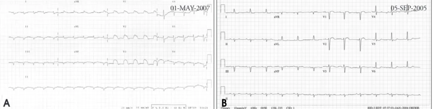
Fig. 1. A: Initial electrocardiogram showed low voltage, Q waves in inferior leads and ST-segment elevation in the leads V1 to V3. B: Previous electrocardiogram checked twenty months ago revealed no ST-segment elevation in the V1 to V3.
Fig. 2. A: The echocardiogram revealed external mass (white arrow) at the anterior side of the right ventricle. The mass invaded to the right ventricle and apical septum. B: The computerized tomography demonstrated external mass (black arrow) of the left chest wall and the mass invade to the right ventricle and apical portion of the interventricular septum.
01-MAY-2007 05-SEP-2005
A
A B
B
J Cardiovasc Ultrasound 2008;16(3):105-106
I
IMMAAGGEESS IINN CCAARRDDIIOOVVAASSCCUULLAARR UULLTTRRAASSOOUUNNDD
106
sented with oliguria and poor oral intake to the emergency room. She underwent left radical mastectomy for her breast cancer for eleven years ago. Despite of several times of chemo- therapy and radiotherapy, her breast cancer recurred from the left chest wall after five years. Initial blood pressure was 72/50 mmHg and the electrocardiogram showed low voltage, Q waves in inferior leads and ST-segment elevation in the leads V1 to V3 (Fig. 1A). It was totally different from previous electrocardiogram (Fig. 1B). We performed echocardiogram and checked cardiac biomarkers. The echocardiogram revealed mildly decreased left ventricular systolic function and external mass at the anterior side of the right ventricle. The mass invaded to the right ventricle and apical septum (Fig. 2A).
The result of cardiac biomarkers was within normal range.
The computerized tomography demonstrated external mass of the left chest wall and the mass invaded to the right ventricle and apical portion of the interventricular septum (Fig. 2B).
We speculated cancer invasion to the myocardium as the cause of ST-segment elevation. The nonmyocardial cancer tissue might cause marked conduction delay of endocardial action potential to the epicardium and it produce ST-segment elevations.
R
Reeffeerreenncceess
1. Wang k, Asinger RW, Marriott HJ. ST-segment elevation in conditions other than acute myocardial infarction. N Engl J Med 2007;356:47-54.
Journal of Cardiovascular Ultrasound 16|September 2008