ORIGINAL ARTICLE
담도 협착이 의심되는 병변에서 원발 부위에 따라 동시에 시행한 초음파 내시경 유도하 조직 검사와 내시경 역행성 췌담도 조영술 유도하 조직 검사 간의 비교
여승재1,2, 조창민1,2, 정민규1,3, 서안나4, 배한익4
경북대학교 의과대학 내과학교실1, 칠곡경북대학교병원 담도췌장암센터2, 경북대학교병원 소화기내과3, 경북대학교 의과대학 병리학교실4
Comparison of the Diagnostic Performances of Same-session Endoscopic Ultrasound- and Endoscopic Retrograde Cholangiopancreatography-guided Tissue Sampling for Suspected Biliary Strictures at Different Primary Tumor Sites
Seong Jae Yeo1,2, Chang Min Cho1,2, Min Kyu Jung1,3, An Na Seo4 and Han Ik Bae4
Department of Internal Medicine, School of Medicine, Kyungpook National University1, Center for Pancreatobiliary Tumors, Kyungpook National University Chilgok Hospital2, Division of Gastroenterology, Kyungpook National University Hospital3, Department of Pathology, School of Medicine, Kyungpook National University4, Daegu, Korea
Background/Aims: Determining the cause of suspected biliary stricture is often challenging in clinical practice. We aimed to compare the diagnostic yields of endoscopic ultrasound-guided tissue sampling (EUS-TS) and endoscopic retrograde cholangiopancreatog- raphy-guided tissue sampling (ERCP-TS) in patients with suspected biliary stricture at different primary lesions.
Methods: We enrolled patients who underwent same-session EUS- and ERCP-TS for the evaluation of suspected biliary stricture.
Forceps biopsy and/or brush cytology of intraductal lesions and fine-needle aspiration for solid mass lesions were performed during ERCP and EUS, respectively.
Results: One hundred and twenty-five patients treated at our institution between January 2011 and September 2016, were initially considered for the study. However, 32 patients were excluded due to loss of follow-up (n=8) and ERCP-TS on the pancreatic duct (n=20) or periampullary lesions (n=4). Of the 93 patients included, 86 had a malignant tumor including cholangiocarcinoma (n=39), pancre- atic cancer (n=37), and other malignancies (n=10). Seven patients had benign lesions. EUS-TS had higher rate of overall diagnostic accuracy than ERCP-TS (82.8% vs. 60.2%, p=0.001), and this was especially true for patients with a pancreatic lesion (84.4% vs.
51.1%, p=0.003).
Conclusions: EUS-TS was found to be superior to ERCP-TS for evaluating suspected biliary strictures, especially those caused by pancre- atic lesions. (Korean J Gastroenterol 2019;73:213-218)
Key Words: Endosonography; Cholangiopancreatography, endoscopic retrograde; Diagnosis; Stricture
Received December 3, 2018. Revised February 25, 2019. Accepted February 27, 2019.
CC This is an open access article distributed under the terms of the Creative Commons Attribution Non-Commercial License (http://creativecommons.org/licenses/
by-nc/4.0) which permits unrestricted non-commercial use, distribution, and reproduction in any medium, provided the original work is properly cited.
Copyright © 2019. Korean Society of Gastroenterology.
교신저자: 조창민, 41404, 대구시 북구 호국로 807, 칠곡경북대학교병원 담도췌장암센터
Correspondence to: Chang Min Cho, Center for Pancreatobiliary Tumors, Kyungpook National University Chilgok Hospital, 807 Hoguk-ro, Buk-gu, Daegu 41404, Korea.
Tel: +82-53-200-2608, Fax: +82-53-200-2028, E-mail: cmcho@knu.ac.kr, ORCID: https://orcid.org/0000-0002-9903-1282 Financial support: None. Conflict of interest: None.
INTRODUCTION
Evaluation of biliary stricture is often challenging in clinical
practice, and endoscopic ultrasound (EUS) and endoscopic retrograde cholangiopancreatography (ERCP) are comple- mentary procedures used to evaluate suspected biliary
stricture. Pathologic confirmation of malignancy before surgical resection or neoadjuvant therapy is important in such patients.
The commonest methods of tissue sampling are ERCP with forceps biopsy and/or brush cytology and EUS-guided fine-nee- dle aspiration.
Several authors have reported that the diagnostic yield of ERCP-guided tissue sampling (ERCP-TS) ranges from 35 to 70% with little improvement in diagnostic yield when brush cytology and forceps biopsy are performed simultaneously.1-6 Meanwhile, previous reports for EUS-guided tissue sampling (EUS-TS) using fine-needle aspiration or biopsy provide higher diagnostic yields for solid pancreatic masses, and EUS-TS has been increasingly used to overcome the moderate diagnostic yield of ERCP-TS.7-9 Furthermore, in patients with suspected biliary stricture, the diagnostic yield of EUS-TS is reportedly superior to that of ERCP-TS when tissue samples are obtained from lesions in the pancreas, lymph node (LN), and others that cause biliary obstruction.10-13
Although EUS-TS has a better diagnostic yield for suspected biliary strictures than ERCP-TS, there is some controversy as to which technique is better for sampling different primary tumor sites. The aim of this study was to compare the diag- nostic yields of EUS- and ERCP-TS in patients with suspected biliary stricture at different primary tumor sites.
SUBJECTS AND METHODS
1. Patients
Retrospective analysis was performed on consecutive pa- tients referred for the evaluation of suspected biliary stricture from January 2011 to September 2016. Patients that under- went same-session EUS and ERCP examinations were consid- ered for enrollment. We excluded the followings; 1) those that underwent EUS or ERCP, 2) those that underwent both EUS and ERCP on the same day but without tissue sampling, and 3) those that underwent EUS and ERCP on different days.
2. EUS- and ERCP-TS procedures
EUS was first performed using a curvilinear echoendoscope (GF-UCT240 or GF-UCT260; Olympus Medical Systems, Tokyo, Japan). EUS-TS targeted any solid lesions in the pancreas, bile duct, gallbladder, or LN and periampullary lesions. All EUS-TS procedures were performed without on-site cytopatho- logic assessment. Specimen were expressed on 1 to 2 slides
for alcohol-fixation with Papanicolaou smear. In each case, an additional specimen was placed in a 10% formalin contain- er for subsequent histologic analysis.
ERCP was followed by EUS, if clinically indicated, and per- formed by the same endoscopist. During ERCP, selective bili- ary cannulation and cholangiography were performed to identify the level of the bile duct stricture. ERCP-TS was then performed using brush cytology (BC-15C; Olympus, Tokyo, Japan) without stricture dilation before tissue sampling.
Cytology brushings were obtained using 10 to-and-fro move- ments across strictures, and 2 glass slides were then smeared with brushes, which were then fixed in 95%
alcohol. Brush tips were cut and placed in 10% formalin for analysis. However, brush cytology was not performed in all patients. An intraductal biliary forceps was then introduced to the distal end of the stricture under fluoroscopy. Biopsies were performed in triplicate and additional biopsies were conducted when no adequate specimen was obtained in any of the three initial trials. EUS- and ERCP-TS procedures were performed by two experienced endoscopists (C. C. and J. M.) who conducted more than 300 EUS- and ERCP-related pro- cedures per year.
A pathologist without prior knowledge of the sampling tech- nique evaluated cytopathologic specimens. Based on histo- pathologic results, we compared diagnostic accuracies by tis- sue sampling method and lesion location. Tissue samples were classified as: 1) malignant, 2) suspicious for malignancy, 3) atypical, 4) benign, and 5) inadequate for diagnosis or non-diagnostic.
3. Definitions and outcomes
We considered tissue samples as malignant when the path- ologist concluded "suspicious for malignancy" or “malignant”.
Failure to collect a specimen by EUS- or ERCP-TS was consid- ered “non-diagnostic”. The final diagnostic standard of malig- nancy was defined as the followings; 1) a malignant histo- pathology based a surgical specimen, 2) a malignant cytopa- thology from EUS- and/or ERCP-TS, or 3) clinical follow-up for more than 12 months with findings consistent with malignancy, such as clinical progression. When no sign of malignancy was noted during follow-up, the lesion was considered benign.
Hyperbilirubinemia was defined as a total bilirubin level of
≥2.5 mg/dL.
The primary endpoints of this study were the overall diag-
Table 1. Patient Baseline Characteristics and Final Diagnoses Total (n=93)
Sex (male:female) 62:31
Age (years) 65.8 (45-85)
Hyperbilirubinemiaa
Yes 74 (79.6)
No 19 (20.4)
EUS-TS
Target size (mm)
Long axis 26.5±12.2
Short axis 18.9±9.1
Needle size
19 G 2 (2.2)
20 G 2 (2.2)
22 G 52 (55.9)
25 G 37 (39.7)
ERCP-TS
Sampling method
Forceps biopsy 92 (98.9)
Brushing cytology 4 (4.3)
Final diagnosis
Benign 7 (7.5)
AIP 4 (4.3)
CBD stricture 2 (2.2)
Chronic pancreatitis 1 (1.1)
Malignancy 86 (92.5)
Cholangiocarcinoma 39 (52.0)
Pancreatic cancer 37 (39.8)
GB cancer 6 (6.5)
HCC 1 (1.1)
Malignant IPMN 1 (1.1)
Malignant lymphoma 2 (2.32)
Values are presented as the mean±standard deviation, median (range), or n (%).
EUS-TS, endoscopic ultrasound-guided tissue sampling; ERCP-TS, endoscopic retrograde cholangiopancreatography-guided tissue sampling; AIP, autoimmune pancreatitis; CBD, common bile duct; GB, gallbladder; G, gauge; HCC, hepatocellular carcinoma; IPMN, intraductal papillary mucinous neoplasm.
aHyperbilirubinemia was defined as a total bilirubin level of ≥2.5 mg/dL.
nostic accuracies of EUS- and ERCP-TS and differences between these at different primary tumor sites. The secondary endpoints were specimen adequacies of EUS- and ERCP-TS for different target lesions. This study was approved by the Institutional Review Board of Kyungpook National University Chilgok Hospital, Daegu, Korea (IRB No. 2017-01-028).
4. Statistical analysis
The Chi-squared (χ2) or Fisher’s exact test were used to determine differences between categorical variables, and the Student’s t-test was used to determine differences between continuous variables. McNemar’s test was used to compare diagnostic yields and to determine the significances of differ- ences between EUS- and ERCP-TS. Results are presented as means±SDs, and p-value of <0.05 was considered statistically significant. The analysis was conducted using SPSS version 23.0 (IBM Corp., Armonk, NY, USA) and Medcalc (version 18.11; http://www.medcalc.org).
RESULTS
One hundred and twenty-five patients with suspected bili- ary stricture treated at our institution between January 2011 and September 2016 were considered as the study enrollment. Of these, 32 patients were excluded due to; 1) follow-up loss (n=8), 2) ERCP-TS being performed on the pan- creatic duct (n=20) or a periampullary lesion (n=4). Finally, 93 patients were included in the study. No complications, such as pancreatitis, perforation, or hemorrhage, requiring treatment occurred.
Patient baseline characteristics and final diagnoses are summarized in Table 1. The 93 study subjects (62 males and 31 females; overall median age, 67 years) underwent same-session EUS- and ERCP-TS. Seventy-four patients had hyperbilirubinemia at time of the procedure. Mean solid lesion size targeted by EUS-TS was 26.5±12.2 mm (longest diame- ter) by 18.9±9.1 mm (shortest diameter). Needle sizes were 19 gauge (G) for 2 lesions, 20G for 2, 22G for 57, and 25G for 37. For ERCP-TS, forceps biopsy was used for 92 lesions and brush cytology for 4.
Malignant tumors were noted in 86 patients; final diag- noses were confirmed in 33 patients through surgery.
Forty-nine patients were diagnosed with a malignancy by EUS- or ERCP-TS. Malignancy was not detected in two patients us-
ing either technique, who were diagnosed by long-term clinical follow-up. Malignancy was also identified in one patient by liver biopsy and in another patient by ultrasound-guided lymph node biopsy.
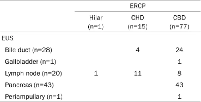
Sites of specimen acquisitions by procedure are summar- ized in Table 2. For ERCP, 1 sample was collected from the hilar area, 15 samples from common hepatic duct (CHD), and 77 from common bile duct (CBD). For EUS, 28 samples were extracted from bile duct, 1 from gallbladder, 20 from lymph
Table 2. Site of Specimen Acquisition by Procedure Type ERCP Hilar
(n=1)
CHD (n=15)
CBD (n=77) EUS
Bile duct (n=28) 4 24
Gallbladder (n=1) 1
Lymph node (n=20) 1 11 8
Pancreas (n=43) 43
Periampullary (n=1) 1
ERCP, endoscopic retrograde cholangiopancreatography; CHD, common hepatic duct; CBD, common bile duct; EUS, endoscopic ultrasound.
Table 3. Cytopathological Classifications Determined by EUS- and ERCP-guided Tissue Sampling
EUS ERCP
Malignancy 70 (75.3) 49 (52.7)
Suspicious for malignancy 2 (2.2) 5 (5.4)
Atypical 2 (2.2) 5 (5.4)
Benign 16 (17.2) 29 (31.1)
Inadequate specimen 3 (3.1) 5 (5.4)
Values are presented as n (%).
EUS, endoscopic ultrasound; ERCP, endoscopic retrograde cholangiopancreatography.
Table 4. Diagnostic Performances for EUS- and ERCP-guided Tissue Sampling by Primary Tumor Site
Overall (n=93) Bile duct (n=41) Pancreas (n=43) Others (n=9)
EUS (%) ERCP (%) p-value EUS (%) ERCP (%) p-value EUS (%) ERCP (%) p-value EUS (%) ERCP (%) p-value
Sensitivity 89.5 65.1 0.001 86.8 78.9 0.334 90.2 56.1 0.001 100.0 42.9 0.002
Specificity 100.0 100.0 - 100.0 100.0 - 100.0 100.0 - 100.0 100.0 -
Accuracy 90.3 67.7 0.001 87.8 80.5 0.581 90.7 58.1 0.002 100.0 77.8 0.125
PPV 100.0 100.0 - 100.0 100.0 - 100.0 100.0 - 100.0 100.0 -
NPV 43.8 18.9 0.001 37.5 27.3 0.646 33.3 10.0 0.001 100.0 33.3 0.001
EUS, endoscopic ultrasound; ERCP, endoscopic retrograde cholangiopancreatography; PPV, positive predictive value; NPV, negative predictive value.
Table 5. Diagnostic Results according to Procedures
EUS-TS positive EUS-TS negative
ERCP-TS positive 47 (50.5) 9 (9.7)
ERCP-TS negative 30 (32.3) 7 (7.5)
Values are presented as n (%).
EUS-TS, endoscopic ultrasound-guided tissue sampling; ERCP-TS, endoscopic retrograde cholangiopancreatography-guided tissue sampling.
node, 43 from pancreas, and one from periampullary lesion.
EUS-TS resulted in the detections of malignancy and sus- pected malignancy in 70 and two patients, respectively, and ERCP-TS resulted in the detections of malignancy and sus- pected malignancy in 49 and five patients, respectively.
Inadequate specimens were obtained from three patients dur- ing EUS-TS and from five patients during ERCP-TS (Table 3).
No significant difference was noted between the two modal- ities in terms of specimen inadequacy (EUS-TS: 3.1% vs.
ERCP-TS: 5.4%, p=0.687).
The sensitivities, specificities, accuracies, positive pre-
dictive values, and negative predictive values of EUS- and ERCP-TS are present in Table 4. The overall diagnostic accu- racy of EUS-TS was superior to that of ERCP-TS (EUS-TS:
90.3% vs. ERCP-TS: 67.7%, p=0.001), and this was especially true for patients with a pancreatic lesion (EUS-TS: 90.7% vs.
ERCP-TS: 58.1%, p=0.002). However, no significant differ- ences were observed for the diagnostic accuracies between EUS- and ERCP-TS for bile duct lesions (EUS-TS: 87.8% vs.
ERCP-TS: 80.5%, p=0.581) or at other primary sites (EUS-TS:
100% vs. ERCP-TS: 77.8%, p=0.125). Overall diagnostic accu- racies of EUS- and/or ERCP-TS were around 92.5%.
Table 5 details cases diagnosed by EUS- and ERCP-TS. As shown in the table, 47 cases were diagnosed when both pro- cedures were used, 30 cases were diagnosed by EUS-TS, 9 cases were diagnosed by ERCP-TS, and seven cases were not diagnosed by either procedure. Of the 30 cases diagnosed by EUS-TS alone, eight were CBD lesions, 18 were pancreatic lesions, and four were other lesions. Of the nine cases diag- nosed by ERCP-TS, five were CBD lesions and four were pan- creatic lesions.
DISCUSSION
It is difficult to determine the exact cause of a suspected biliary stricture. Preoperative histopathological confirmation and the institution of neoadjuvant therapy are important for
determining optimal treatment modalities for this condition.
ERCP-TS is conventionally used in these cases, but its re- ported diagnostic accuracy ranges from 35 to 70%; though this can be slightly increased by performing forceps biopsy and brush cytology.1-6 Recently, EUS-TS was shown to have better diagnostic accuracy, that is, between 85% and 93%.
Furthermore, EUS-TS is known to be superior to ERCP-TS in patients with a biliary stricture caused by lesions involving the pancreas and LN.7,14-17 However, few studies have com- pared the diagnostic yields of these two methods, particularly in patients that undergo initial EUS-TS followed by ERCP-TS at the same time. Previous studies on this topic are limited by incomplete protocols and small cohort sizes, and little in- formation is available on the diagnostic yields of EUS-TS and ERCP-TS for different primary lesion types and locations.
The merits of our study are as follows: 1) all 93 patients included underwent ERCP-TS after EUS-TS, and thus, the ex- perimental protocol did not affect outcomes. Two previous studies reported diagnostic accuracies for EUS-TS and ERCP-TS performed during same sessions as ~20% and
~60%, respectively.18,19 Thus, it appears the experimental pro- tocols influence results. And 2) we found results were un- affected by bias secondary to primary lesions. Stenosis caused by biliary or pancreatic lesions occurred in 41 (44.1%) and 43 patients (46.2%), respectively, and thus, the number of patients with a pancreatic or biliary lesion were similar.
Previous studies have suggested that pancreatic lesion-in- duced strictures are more likely to be diagnosed by EUS-TS than by evidence of narrowing caused by bile ducts or other etiologies.7,14,19 Therefore, if a primary lesion has an asym- metric influence on the pancreas or biliary tract, diagnostic outcome are likely to be affected.
In the present study, the diagnostic accuracy of EUS-TS (84.4%) for biliary strictures caused by pancreatic lesions was significantly greater than that of ERCP-TS (51.1%) because EUS-TS directly targets these lesions, whereas ERCP-TS tar- gets sites of bile duct compression.20 The diagnostic accu- racies of EUS-TS and ERCP-TS for identifying biliary stricture secondary to a biliary lesion were similar (80.5% vs. 73.2%, respectively). However, EUS-TS detected a bile duct lesion in 11 patients, in which ERCP-TS failed to detect a lesion.
We enrolled patients that underwent initial EUS-TS followed by ERCP-TS during same sessions, because biliary stents in- serted during ERCP to decompress biliary obstruction can neg-
atively affect the diagnostic accuracy of future EUS-TS procedures.21,22 Therefore, when both procedures are per- formed during same sessions, initial EUS-TS improves diag- nostic yield and relieves obstruction. A small number of studies have reported that same-session EUS-TS and ERCP-TS did not increase complication rates, although procedure times were obviously extended.18,20,23 We concur with these findings as no patient developed a cardiopulmonary complication in the present study. Nevertheless, careful monitoring and caution are required to safely perform EUS-TS and ERCP-TS during same sessions.
Several limitations of our study warrant mention: 1) the study is inherently limited by its retrospective single-center study design. 2) Not all patients underwent brush cytology and forceps biopsy during ERCP-TS. 3) Cholangioscopy is known to improve the diagnostic accuracy of ERCP-TS, but was not performed, and this might have reduced diagnostic accuracy. 4) EUS-TS was performed using different kinds of needles, which may have affected diagnostic accuracies and outcomes. 5) EUS-TS was performed on an LN for CHD and hilar lesions, and thus, it cannot be assumed our results well-reflect diagnostic results for primary lesions. Moreover, a benign finding may indicate that the procedure had been correctly performed for reactive lymphadenopathy secondary to cholangitis. Although EUS-TS returned a benign finding, the final diagnosis was malignancy for all. However, when EUS-TS of an LN returned a finding of benignity, this did suggest a higher probability of a benign final diagnosis. And 6) all proce- dures were performed by two expert endoscopists, and thus, techniques or practices employed might not accurately reflect those used at other centers. A large-scale prospective multi- center study that adopts the aforementioned diagnostic meth- ods with or without cholangioscopy is required to address these concerns.
In conclusion, we compared the diagnostic accuracies of EUS-TS and ERCP-TS in patients with a suspected biliary stricture. Overall, EUS-TS was observed to be superior to ERCP-TS, especially in cases of pancreatic lesion-induced bili- ary obstruction. However, the use of EUS-TS alone as a diag- nostic aid was limited in such cases. As shown by our results, in patients with pancreatic lesions, nine cases (21%) were diagnosed only by ERCP-TS, and thus, a combination of EUS-TS and ERCP-TS is likely to be more effective than EUS-TS alone for the diagnosis of pancreatic lesions. In cases of sus-
pected malignant primary biliary obstruction, the diagnostic accuracy EUS-TS was found to be non-significantly better than that of ERCP-TS. Furthermore, the present study suggests EUS-TS may be useful in cases of suspected chol- angiocarcinoma, though it is not routinely performed in pa- tients with obstruction caused by bile duct cancer. Therefore, we recommend that both, EUS-TS and ERCP-TS be performed in patients with suspected biliary stricture to improve the diag- nostic accuracy.
REFERENCES
1. Glasbrenner B, Ardan M, Boeck W, Preclik G, Möller P, Adler G.
Prospective evaluation of brush cytology of biliary strictures dur- ing endoscopic retrograde cholangiopancreatography. Endoscopy 1999;31:712-717.
2. Pugliese V, Conio M, Nicolò G, Saccomanno S, Gatteschi B.
Endoscopic retrograde forceps biopsy and brush cytology of bili- ary strictures: a prospective study. Gastrointest Endosc 1995;42:
520-526.
3. Jailwala J, Fogel EL, Sherman S, et al. Triple-tissue sampling at ERCP in malignant biliary obstruction. Gastrointest Endosc 2000;51(4 Pt 1):383-390.
4. Ponchon T, Gagnon P, Berger F, et al. Value of endobiliary brush cytology and biopsies for the diagnosis of malignant bile duct stenosis: results of a prospective study. Gastrointest Endosc 1995;42:565-572.
5. Stewart CJ, Mills PR, Carter R, et al. Brush cytology in the assess- ment of pancreatico-biliary strictures: a review of 406 cases. J Clin Pathol 2001;54:449-455.
6. Schoefl R, Haefner M, Wrba F, et al. Forceps biopsy and brush cy- tology during endoscopic retrograde cholangiopancreatography for the diagnosis of biliary stenoses. Scand J Gastroenterol 1997;32:363-368.
7. Hewitt MJ, McPhail MJ, Possamai L, Dhar A, Vlavianos P, Monahan KJ. EUS-guided FNA for diagnosis of solid pancreatic neoplasms: a meta-analysis. Gastrointest Endosc 2012;75:
319-331.
8. Jeong H, Park CS, Kim KB, et al. Predictors of malignancies in pa- tients with inconclusive or negative results of endoscopic ultra- sound-guided fine-needle aspiration for solid pancreatic masses. Korean J Gastroenterol 2018;71:153-161.
9. Nam HS, Kang DH. Endoscopic ultrasound-guided biliary drainage. Korean J Gastroenterol 2017;69:164-171.
10. DeWitt J, Misra VL, Leblanc JK, McHenry L, Sherman S. EUS-guid-
ed FNA of proximal biliary strictures after negative ERCP brush cytology results. Gastrointest Endosc 2006;64:325-333.
11. Eloubeidi MA, Chen VK, Jhala NC, et al. Endoscopic ultra- sound-guided fine needle aspiration biopsy of suspected cholangiocarcinoma. Clin Gastroenterol Hepatol 2004;2:209-213.
12. Mohamadnejad M, DeWitt JM, Sherman S, et al. Role of EUS for preoperative evaluation of cholangiocarcinoma: a large sin- gle-center experience. Gastrointest Endosc 2011;73:71-78.
13. Fritscher-Ravens A, Broering DC, Knoefel WT, et al. EUS-guided fine-needle aspiration of suspected hilar cholangiocarcinoma in potentially operable patients with negative brush cytology. Am J Gastroenterol 2004;99:45-51.
14. Gress F, Gottlieb K, Sherman S, Lehman G. Endoscopic ultra- sonography-guided fine-needle aspiration biopsy of suspected pancreatic cancer. Ann Intern Med 2001;134:459-464.
15. Puli SR, Bechtold ML, Buxbaum JL, Eloubeidi MA. How good is en- doscopic ultrasound-guided fine-needle aspiration in diagnos- ing the correct etiology for a solid pancreatic mass?: a meta-anal- ysis and systematic review. Pancreas 2013;42:20-26.
16. Tummala P, Munigala S, Eloubeidi MA, Agarwal B. Patients with obstructive jaundice and biliary stricture ± mass lesion on imag- ing: prevalence of malignancy and potential role of EUS-FNA. J Clin Gastroenterol 2013;47:532-537.
17. Turner BG, Cizginer S, Agarwal D, Yang J, Pitman MB, Brugge WR.
Diagnosis of pancreatic neoplasia with EUS and FNA: a report of accuracy. Gastrointest Endosc 2010;71:91-98.
18. Oppong K, Raine D, Nayar M, Wadehra V, Ramakrishnan S, Charnley RM. EUS-FNA versus biliary brushings and assessment of simultaneous performance in jaundiced patients with sus- pected malignant obstruction. JOP 2010;11:560-567.
19. Rösch T, Hofrichter K, Frimberger E, et al. ERCP or EUS for tissue diagnosis of biliary strictures? A prospective comparative study.
Gastrointest Endosc 2004;60:390-396.
20. Weilert F, Bhat YM, Binmoeller KF, et al. EUS-FNA is superior to ERCP-based tissue sampling in suspected malignant biliary ob- struction: results of a prospective, single-blind, comparative study. Gastrointest Endosc 2014;80:97-104.
21. Gress F, Michael H, Gelrud D, et al. EUS-guided fine-needle aspira- tion of the pancreas: evaluation of pancreatitis as a complication.
Gastrointest Endosc 2002;56:864-867.
22. Agarwal B, Abu-Hamda E, Molke KL, Correa AM, Ho L. Endoscopic ultrasound-guided fine needle aspiration and multidetector spi- ral CT in the diagnosis of pancreatic cancer. Am J Gastroenterol 2004;99:844-850.
23. Ross WA, Wasan SM, Evans DB, et al. Combined EUS with FNA and ERCP for the evaluation of patients with obstructive jaundice from presumed pancreatic malignancy. Gastrointest Endosc 2008;68:461-466.