1352
대한안과학회지 2019년 제 60 권 제 12 호 J Korean Ophthalmol Soc 2019;60(12):1352-1355 ISSN 0378-6471 (Print)⋅ISSN 2092-9374 (Online)
https://doi.org/10.3341/jkos.2019.60.12.1352
Case Report
항암 치료 후 발생한 빈혈망막병증
Anemic Retinopathy after Chemotherapy
이승찬⋅오백록
Seung Chan Lee, MD, Baek Lok Oh, MD
서울대학교 의과대학 서울대학교병원 안과학교실
Department of Ophthalmology, Seoul National University Hospital, Seoul National University College of Medicine, Seoul, Korea
Purpose: To report a case of anemic retinopathy after chemotherapy.
Case summary: A 32-year-old male presented with visual disturbance in both eyes. He had been diagnosed with testicular can- cer and had undergone right orchiectomy 4 months prior. He completed adjuvant chemotherapy 2 weeks before presentation.
His best-corrected visual acuities were 20/35 in both eyes. Fundus examination revealed multiple flame-shaped hemorrhages around the posterior pole, and boat-shaped hemorrhages on the macula in both eyes. Laboratory results showed that he had re- duced hemoglobin (5.5 g/dL) and platelet counts (77,000/µL). After transfusion, visual acuities were improved and retinal hemor- rhages were resolved along with normalization of the hematological conditions.
Conclusions: Many ocular and medical conditions can cause bilateral retinal hemorrhages. This case emphasizes that compre- hensive history evaluation, systemic evaluation, and laboratory findings, as well as a detailed fundus examination, are important in the diagnosis of patients with bilateral retinal hemorrhages.
J Korean Ophthalmol Soc 2019;60(12):1352-1355
Keywords: Anemia, Retinal hemorrhage, Thrombocytopenia
■Received: 2019. 7. 26. ■ Revised: 2019. 9. 19.
■Accepted: 2019. 11. 29.
■Address reprint requests to Baek Lok Oh, MD
Department of Ophthalmology, Seoul National University Hospital, #101 Daehak-ro, Jongno-gu, Seoul 03080, Korea Tel: 82-2-2072-3740, Fax: 82-2-741-3187
E-mail: [email protected]
*Conflicts of Interest: The authors have no conflicts to disclose.
ⓒ2019 The Korean Ophthalmological Society
This is an Open Access article distributed under the terms of the Creative Commons Attribution Non-Commercial License (http://creativecommons.org/licenses/by-nc/3.0/) which permits unrestricted non-commercial use, distribution, and reproduction in any medium, provided the original work is properly cited.
빈혈망막병증은 빈혈환자의 28%에서 발생할 수 있다고 보고된 바 있다.1 빈혈의 정도가 심할수록 망막병증의 발생 가능성이 올라가며, 혈색소 수치가 6 g/dL보다 낮은 경우 고위험군으로 알려져 있다. 빈혈과 함께 저혈소판증이 동 반될 경우 발병 가능성은 더욱 높아진다.2 고환암에 대해 항암 치료를 마친지 2주 뒤 심한 빈혈이 있었던 환자에서 양안에 다발성 망막출혈이 발견되었고, 빈혈을 교정하면서 안저 소견 또한 함께 호전된 증례를 경험하였기에 문헌 고
찰과 더불어 보고하는 바이다.
증례보고
32세 남자 환자가 3일 전부터 시작된 양안 시야장애를 주소로 내원하였다. 처음에는 눈앞에 뭔가 끼어있고 시야 가 약간 붉게 보이는 증상이 좌안에 있었다가 2일 뒤 우안 에도 동일한 증상이 발생하였다고 호소하였다. 환자는 과 거력상 고혈압, 당뇨는 없었고 내원 4개월 전 우측 고환암 에 대해 고환절제술을 시행받았다. 수술 후 혈액종양내과 에서 Bleomycin, Etoposide, Cisplatin regimen (BEP)으로 보조항암요법을 진행하였으며, 마지막 항암 치료는 내원 14일 전 완료하였다.
내원 시 최대교정시력은 양안 모두 20/35이었으며 전안 부검진상 특이 소견은 없었다. 안저 검진상 양안 시신경과 주요 혈관궁을 따라서 다발성 화염상 출혈이 관찰되었고,
1353 - 이승찬⋅오백록 : 항암 치료 후 발생한 빈혈망막병증 1예 -
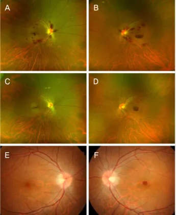
A B
C D
E F
Figure 1. Fundus photographs at the initial presentation (A,
B), at 1 month follow-up visit (C, D) and at 3 months fol- low-up visit (E, F). Fundus examination revealed multiple flame-shaped hemorrhages at the posterior pole in both eyes and premacular boat-shaped hemorrhage in the left eye (A, B).Retinal hemorrhages almost resolved in the right eye and small residual premacular hemorrhage remained in the left eye (E, F).
A
B
C
D
E
F
Figure 2. Spectral domain-optical coherence tomography
(SD-OCT) images at the initial presentation (A, B), at 1 month follow-up visit (C, D) and at 3 months follow-up visit (E, F).Vertical macular scans of SD-OCT demonstrated premacular sub-internal limiting membrane (ILM) hemorrhage and multi- ple intra-retinal nerve fiber layer hemorrhage in both eyes.
There was no definite outer retinal abnormality except optical shadowing effect due to hemorrhage (A, B). Vertical scans of SD-OCT demonstrated a small amount of sub-ILM hemor- rhage on the macula in both eyes (E, F).
황반부에는 배 모양 출혈(boat shaped hemorrhage)이 관찰 되었다(Fig. 1A, B). 광학단층촬영에서는 양안 황반부 내경 계막하출혈과 황반주변부 망막신경섬유층의 출혈이 확인 되었다. 출혈로 인한 광학적 그림자 효과 이외에 망막외층 에서는 뚜렷한 이상 소견이 발견되지 않았다(Fig. 2A, B).
환자의 혈압, 체온은 정상 범위였으며 혈당수치 또한 정상 이었다. 혈액검사상 혈색소 5.5 g/dL, 혈소판 숫자 77,000/µL 가 확인되었다. 환자의 병력, 망막출혈의 양상 및 혈액검사 결과를 바탕으로 양안의 빈혈망막병증으로 진단하였다.
환자는 혈액종양내과에서 수차례 적혈구 및 혈소판 수혈 을 받았고, 안과에서는 특별한 치료 없이 1달 간격으로 외 래에서 경과 관찰하였다. 수혈 후 혈액학적 이상은 교정이 되었으며 망막출혈은 내원 시마다 점점 줄어드는 양상이었다 (Fig. 1, 2). 증상 발생 후 4개월째인 마지막 외래 내원시 교 정시력 양안 모두 20/25이었으며, 안저 검진상 망막출혈은 모두 흡수되었다. 광학단층촬영에서도 양안 모두 정상 소 견이었다. 혈액검사에서 혈색소 11.3 g/dL, 혈소판 114,000/µL 가 확인되었다.
고 찰
빈혈은 망막의 산소결핍을 유발하게 되는데 이로 인해
1354
- 대한안과학회지 2019년 제 60 권 제 12 호 -
망막신경섬유층의 허혈이 발생하여 흔히 환자에게서 면화 반을 관찰할 수 있다. 또한 저산소 상태로 인해 망막혈관이 확장되고, 빈혈에 의한 저단백혈증으로 인해 경벽 투과성 이 높아져 이로 인해 망막의 부종과 출혈이 발생하게 된다.
저혈소판증은 응고장애를 일으켜 망막혈관의 출혈 경향을 더욱 높이게 된다.3 이외에도 정맥저류, 망막혈관연축, 혈액 점도 증가 등도 빈혈망막병증을 일으킬 수 있는 기전으로 생각되고 있다.4
양안에 다발성 망막출혈이 발생하였을 때 감별해야 하는 질환은 여러 가지가 있으며 흔한 질환으로는 고혈압망막병 증, 당뇨망막병증 등이 있다. 상대적으로 드문 원인으로 혈 액 이상(blood dyscrasia)에 의한 망막병증, 낫형적혈구 망 막병증, 백혈병 망막병증, 전자간증 관련 망막병증, 급성 세 균심내막염, 루푸스 혈관병증, 일산화탄소 중독 등이 있다.
위 질환별로 망막출혈의 양상 및 동반되는 망막 병변이 다 소 차이가 있기 때문에 안저 소견을 자세히 관찰하는 것이 감별 진단에 중요하다. 증례의 환자에서는 미세혈관류, 면 화반, 삼출물, 로트반점, 시신경부종 등의 소견은 동반되지 않은 채 망막출혈만 관찰되었다. 출혈은 망막후극부에만 국한되어 있었고 주요 혈관들 자체에는 명확한 변화가 관 찰되지 않았다. 안저 소견만으로는 여러 질환을 명확히 구 분하기 힘든 경우가 많기 때문에 환자의 병력과 동반되는 전신증상 및 검사실 소견을 자세히 파악하는 것이 매우 중 요하다. 증례의 환자는 최근 항암 치료력이 있고 혈액검사 상 심한 빈혈이 동반되어 있었기에 빈혈망막병증으로 진단 을 내릴 수 있었다. BEP regimen은 고환암에서 표준 항암 치료로 사용되며 24-33%에서 빈혈을 유발할 수 있다고 알 려져 있다.5
치료로는 혈액종양내과와 소통하면서 수혈을 통해서 빈
혈과 혈소판감소증을 교정하는 것이 필요하다. 대부분의 경우에서 원인질환을 치료하면 망막출혈이 저절로 흡수되 어 호전되는 것을 기대할 수 있다.6 황반부의 망막앞출혈이 있고 양이 많은 경우에는 유리체절제술이나 야그레이저를 통한 막절개술 통해서 혈액을 배출시킬 수 있다.7
양안에 망막출혈이 있는 환자를 대할 때 안저 소견만으 로 명확하게 감별하기 힘든 경우가 많다. 따라서 저자들은 본 증례를 통하여 주의 깊은 안저 검진 외에도 환자의 병력 및 동반되는 전신증상, 검사실 소견 등을 자세히 파악하는 것이 중요하다는 것을 환기시키고자 하는 바이다.
REFERENCES
1) Carraro MC, Rossetti L, Gerli GC. Prevalence of retinopathy in pa- tients with anemia or thrombocytopenia. Eur J Haematol 2001;67:238-44.
2) Duke-Elder S, Dobree J. The blood diseases. In: Duke-Elder Sm, ed. System of ophthalmology (diseases of the retina). St. Louis: CV Mosby Co.; 1967;373-407.
3) Rubenstein RA, Yanoff M, Albert DM. Thrombocytopenia, ane- mia, and retinal hemorrhage. Am J Ophthalmol 1968;65:435-9.
4) Foulds W. The ocular manifestations of blood diseases. Trans Ophthalmol Soc U K 1963;83:345-67.
5) Kawai K, Ando S, Hinotsu S, et al. Completion and toxicity of in- duction chemotherapy for metastatic testicular cancer: an updated evaluation of Japanese patients. Jpn J Clin Oncol 2006;36:425-31.
6) Kacer B, Hattenbach LO, Hörle S, et al. Central retinal vein occlu- sion and nonarteritic ischemic optic neuropathy in 2 patients with mild iron deficiency anemia. Ophthalmologica 2001;215:128-31.
7) Celebi S, Kükner A. Photodisruptive Nd: YAG laser in the man- agement of premacular subhyaloid hemorrhage. Eur J Ophthalmol 2001;11:281-6.
1355 - 이승찬⋅오백록 : 항암 치료 후 발생한 빈혈망막병증 1예 -
= 국문초록 =
항암 치료 후 발생한 빈혈망막병증
목적: 항암 치료 후 발생한 빈혈망막병증 1예를 보고하고자 한다.
증례요약: 32세 남자 환자가 3일 전부터 시작된 시야이상을 호소하며 내원하였다. 내원 4개월 전 우측 고환암에 대해 고환절제술을 시행받았으며 이후 보조항암요법을 진행하여 2주 전에 치료를 마쳤다. 내원시 양안 경도의 시력저하를 보였으며, 안저검진상 양안 후극부에 다발성 화염상 출혈과 함께 황반부 배 모양 출혈이 관찰되었다. 혈액검사상 중증 빈혈(혈색소 5.5 g/dL)과 함께 경도의 혈소 판감소증(77,000/µL)이 확인되었고 이를 바탕으로 양안의 빈혈성 망막병증으로 진단하였다. 적혈구 및 혈소판 수혈 후 혈액검사수치 는 거의 정상화되었으며, 이와 함께 시력호전 및 안저검진상 망막출혈이 소실되는 것을 확인할 수 있었다.
결론: 양안에 망막출혈을 일으킬 수 있는 여러 질환들은 안저 소견만으로 명확하게 감별하기 힘든 경우가 많다. 주의 깊은 안저 검진 뿐 아니라 환자의 병력, 동반되는 전신증상 및 검사실 소견 등을 자세히 파악하는 것 역시 중요하다.
<대한안과학회지 2019;60(12):1352-1355>
이승찬 / Seung Chan Lee
서울대학교 의과대학 서울대학교병원 안과학교실 Department of Ophthalmology, Seoul National University Hospital, Seoul National University College of Medicine