CONTENTS
Ⅰ. INTRODUCTION
Ⅱ. MATERIALS AND METHODS
Ⅲ. RESULTS
Ⅳ. DISCUSSION
Ⅴ. CONCLUSIONS REFERENCES KOREAN ABSTRACT
Ⅰ. INTRODUCTION
Stress is the nonspecific response of the body to any demand and has been believed to be the cause of various forms of diseases for a long time.
1)Controllable stress triggers the stabilization and facilitation of neuronal networks involved in the generation of appropriate patterns of appraisal and coping, whereas uncontrollable stress favours the extinction of inappropriate patterns and the reorganisation of neuronal connections underlying certain inappropriate behaviors. Both, controllable and uncontrollable stress-reaction- processes are therefore essential prerequisites of, and inherent challenges to, the development and adaptation of an
individual in an ever changing external world but may also lead to psychodevelopmental failures and psychosomatic diseases.
2,3)Apoptosis is a process of genetically programmed alterations of cell structure that lead to failure of proliferation and differentiation, and eventual cell death.
4)Generally, stress-induced apoptosis may occur by high glucose
5), increased glucocorticoid
6)and direct nerve involvement
7). And glucocorticoid and excitatory mechanism involving N-methyl-D- aspartate receptors are important in nerve cell apoptosis or atropy related to stress.
8)A brain stem plays a important part in chronic pain involving wind-up, central sensitiazation, and neuroplasticity. The most of pain disorders in the orofacial area such as temoporomandibular disorder, trigerminal neuralgia, mucosal erosion or ulcer are mediated by trigeminal nervous system.
21)As stress-induced nerve cell change may lead to functional disturbances of nerve and diseases including orofacial pain, the present study was performed to observe the ultrastructural changes of brain nerve cells of rats under restraint stress in order to inquire the relationship between stress and pathologies of central nervous system.
Ultrastructural Changes of the Rat Brain Stem under Restraint Stress
Seung-Ho Baek
1, D.M.D.,M.S.D., Dong-Sik Lee
1, D.M.D.,M.S.D., Yang-Hyun Chun
1, D.M.D.,M.S.D.,Ph.D., Jin-Yong Lee
2, D.M.D.,M.S.D.,Ph.D.,
Jung-Pyo Hong
1, D.M.D.,M.S.D.,Ph.D.
Department of Oral Diagnosis & Oral Medicine
1, Dept. of Oral Microbiology
2,
College of Dentistry, Kyung Hee University
Ⅱ. MATERIALS AND METHODS 1. Experimental animals
Sprague-Dawley rats (8-week-old, 323-367 g) were purchased from Dae-Han Experimental Animal Research Center, Seoul, Korea. They were maintained at 20-23℃ and fed ad libitum on a normal laboratory diet. The rats were divided into 2 groups: 1) Normal control group; 2) Restraint stress group : the rats were placed in the stress cage throughout the period of experiment. All the animals were then sacrificed at day 0, 1, 3, 5, and 7 day of the experiment and the brain stems were excised immediately and fixed in the glutaradehye in phosphate buffer.
2. Electron Microscopy
The excisional tissues of the brain stems were rinsed in 0.1M cacodylate buffer 3 times 10 min each, postfixed in 1% osmium tetroxide for for 90 min, and rinsed in 0.1M malate buffer 3 times 5 min each. They were prestained with 1% uranyl acetate for 90 min and washed in 0.1M malate buffer 3 times 5 min each, and dehydrated through an ascending series of ethanol concentration (50% to
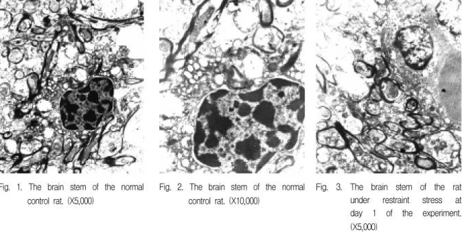
Fig. 1. The brain stem of the normal control rat. (X5,000)
Fig. 2. The brain stem of the normal control rat. (X10,000)
Fig. 3. The brain stem of the rat under restraint stress at day 1 of the experiment.
(X5,000)
100%, 15 min each). They were then placed in 100%
ethanol and propylene oxide (1:1) for 45 min, propylene oxide for 45 min, and propylene oxide and epon (1:1) for 1 hr. After then, they were placed in epon in a vacuum oven overnight, embedded with fresh epon which was polymerized at 60 ℃ for 3 days. The embedded tissues were cut with a diamond knife 50nm thick and stained with uranyl acetate and lead citrate. The tissues were observed under the transmission electron microscope (JEOL Ltd., Japan)
Ⅲ. RESULTS The brain stem of the normal group
1. Nuclei and many dendrites were observed in a normal shape.(Fig. 1,2)
The brain stem of the restraint stress group
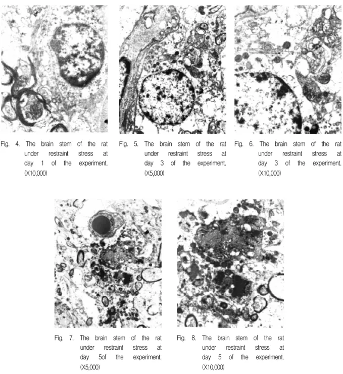
1. Many small-sized mitochondria appeared at day 5 and 7 of the experiment.
2. Spaces around the nucleus were prominent at day 3 and increased with time up to day 7.(Fig.
3-10)
Ⅳ. DISCUSSION
Glucocorticoids are major protectors during and after stress. Centrally, glucocorticoids counter- balance and regulate three neurochemical systems active during stress: the noradrenergic system, the serotonin system and the GABA benzodiazepin system.
9)Stress-regulating circuit is functionally
affected by corticosteroids in adult rats and may imply that human disorders associated with corticosteroid imbalance are allied to a changed circuitry in the brain.
10)Steroid hormones are lipophilic molecules derived from cholesterol and synthesized in the adrenal cortex, the testes, and the ovary and placenta, which can cause the atrophy or apoptosis of Fig. 4. The brain stem of the rat
under restraint stress at day 1 of the experiment.
(X10,000)
Fig. 5. The brain stem of the rat under restraint stress at day 3 of the experiment.
(X5,000)
Fig. 6. The brain stem of the rat under restraint stress at day 3 of the experiment.
(X10,000)
Fig. 7. The brain stem of the rat under restraint stress at day 5of the experiment.
(X5,000)
Fig. 8. The brain stem of the rat under restraint stress at day 5 of the experiment.
(X10,000)
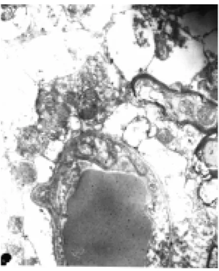
Fig. 9. The brain stem of the rat under restraint stress at day 7 of the experiment. (X5,000)
neuronal cell under stress condition.
11)For example, glucocorticoid can be toxic to neurons, and thus may be important in neurodegenerative diseases including Alzheimer's disease.
12)Adrenal glucocorticoids are thought to be responsible for the damage of nerves because of their adrenal glucocorticoids's ability to com- promise energy metabolism and make neurons more vulnerable to glutamate excitotoxicity. Additional mechanisms by which stress or glucocorticoids could damage the hippocampus are considered in the context of recent evidence that stress regulates neurotrophic factor expression in the brain.
13)Stress induced corticosterone secretion and that excitatory mechanism involving N-methyl- D-aspartate receptors play a major role in driving the atrophy.
8)The other evidences for possible mechanisms of causing neuronal cellular damages are as follows:
activated microglia produce molecules including nitric oxide and tumor necrosis factor-alpha which can be toxic to neurons
14), stress-induced rise in serum anti-brain autoantibody levels occured in the rat
15).
Mitochondria and Ca
2+play an important role in the apoptosis triggered by many stimuli. Mito- chondria integrate death signals through Bcl-2
Fig. 10. The brain stem of the rat under restraint stress at day 7 of the experiment. (X10,000)
family members and coordinate caspase activation through the release of cytochrome c as a result of the outer mitochondrial membrane becoming permeable.
16)Molecular targets for Ca
2+are now being identified and include signal transduction intermediates, endonuclease(s) and proteases, and the enzymes involved in the maintenance of phospholipid asymmetry in the plasma membrane.
17)In addition, the transmembrane receptors p75 and Fas can trigger and in some cases are required for programmed cell death of the neurons that express them, through signalling pathways that are regulated by a variety of cytoplasmic effectors.
18)Examination of a cut surface of any part of the central nervous system reveals that it consists of gray matter and white matter. The white matter contains only axons of nerve cells plus the associated neuroglial cells and blood vessels.
Tracts, grouped axons, do not stand out as
delineated bundles of fibers. In contrast to the white
matter, the nerve tissue of gray matter contains cell
bodies, fibers (both axons and dendrites), and the
associated neurological cells. The gray matter is
the site for synapses. The nucleus means a cluster
or group of neuronal cell bodies plus fibers and
neuroglia.
19)The medulla, pons, and midbrain are collectively referred to as the brain stem. Their organization is closely, but not entirely, associated with the cranial nerves. Histologically, the brainstem contains numerous islands of gray matter surrounded by white matter. Some of this white matter is composed of more or less distinct tracts. In other cases, the definition between white matter and gray matter, as in the reticular substance, is not very clear. Many of the nuclei in the brain stem contain cell bodies of motorneurons of the cranial nerves.
These motor nuclei are counterparts of the anterior horns of the spinal cord.
19)The small-diameter primary afferents inner- vating the temporomandibular joint and masticatory muscles project into the brain and terminate centrally in the trigeminal brain stem sensory nuclear complex, where they release excitatory neurochemicals, such as excitatory amino acids and neuropeptides. The trigeminal brain stem complex is subdivided into the main or principal sensory nucleus and the spinal tract nucleus, which comprises 3 subnuclei - oralis, interpolaris, and caudalis.
20,21)Subnucleus caudalis (medullary dorsal horn) is a principal brain stem relay site of V nociceptive information. It composed of a laminated structure resembling the dorsal horn of the spinal cord, which is the integral part of spinal nociceptive processing.
Also, by analogy with spinal nociceptive afferents, the small-diameter afferents carryig nociceptive information from the various craniofacial tissues, including the TMJ an masticatory muscles, predominantly terminate in the superficial laminae (I and II) of subnucleus caudalis, as well as in its deeper laminae V and VI. Craniofacial noxious stimulation of deep tissues also evokes reflex autonomic changes (eg, in blood pressure and respiration) as well as reflex increases in muscle activity, and many of these reflex effects also are dependent on a relay in subnucleus caudalis, since they can be markedly reduced by caudalis lesions.
20,21)
It was shown that the strong emotional immo-
bilization stress in rats resulted in increased blood-brain barrier permeability and besides in brain parenchymal vessels damages accompanied by haemorrhages and the loss of some nerve cells.
The earliest and strongest brain vessel disruptions under emotional stress were found in the oral part of the brain stem reticular formation.
22)The ultrastructure of rat's pterygopalatine ganglionic neurocytes under immobilization stress was investigated by Kuder T. The rough endoplasmic reticulum occurring in large amounts, was placed predominantly on the circumference of cells. The smooth reticulum, conversely, occupied mainly the perinuclear part of cytoplasm. The Golgi apparatus was much developed in comparison to the control group. Increase in number of lysosomes and lipophuscin was observed. In all experimental groups changes within mitochondria were noticed (atrophy of cristae and matrix, presence of myelin bodies and swellings.
23)It was reported that the hippocampus of diabetic rats is extremely susceptible to additional stressful events, which in turn can lead to irreversible hippocampal damage.
24)Folan JC et al. reported that the drug- and saline-induced alterations of neural connectivity may reflect stress-induced general changes demonstrating the plasticity of the paraganglionic cell population.
25)In the present study, we examined the ultrastructural changes of brain stem nerve cells of the rats under restraint stress to inquire the relationship between stress and pathologies of central nervous system. In the normal group, nuclei and many dendrites were observed in a normal shape. In the restraint stress group, many small-sized mitochondria appeared at day 5 and 7 of the experiment. And Spaces around the nucleus were prominent at day 3 and increased with time up to day 7.
It is likely that degeneration of the brain stem can be induced by stress. It also appears that stress-induced nerve cell damage may lead to functional disturbances of nerve and diseases.
Additional histological study needs to be extended
to observe the histological changes in various tissue types, so it may be able to determine whether the stress can affect a particular type of tissue. A further study is also required to identify the underlying mechanisms of the morphological changes of the nerve cells in the perspective of apoptosis and stress-related protein.
V. CONCLUSIONS
Stress and other psychological factors are believed to play an important role in the major health problems and also closely related with disorders and diseases of the orofacial tissue.
However the mechanisms by which stress induce these types of diseases including temporoman- dibular disorders and burning mouth syndrome has yet to be elucidated. The present study was performed to observe the ultrastructural changes of brain nerve cells of rats under restraint stress to inquire the relationship between stress and central nervous system.
Eighteen Sprague-Dawley rats (8-weeks old, 323-367 g/bw) were used for the experiment and the rats were divided into 2 groups: 1) Normal control group; 2) Restraint stress group : the rats were placed in the stress cage throughout the period of experiment. All the animals were then sacrificed at day 0, 1, 3, 5, and 7 of the experiment and the brain stems were excised immediately and fixed in the glutaradehye in phosphate buffer. The brain stem samples were subjected to transmission electron microscopy. The results were as follows:
1. In the normal control group, nuclei and many dendrites were observed in a normal shape.
2. In the restraint stress group, many small-sized mitochondria appeared at day 5 and 7 of the experiment.
3. In the restraint stress group, spaces around the nucleus were prominent at day 3 and increased with time up to day 7.
It is likely that stress may cause degenerative
cellular changes. It also appears that stress-induced nerve cell damage may lead to functional disturbances of nerve and diseases. Afterthere additional histological and molecular study is required to determine whether the stress can affect a particular type of tissue and identify the underlying mechanisms of the morphological changes of the nerve cells in the perspective of apoptosis and stress-related protein.
REFERENCES
1. Selye H : Selye's guide to stress research. Vol. I., Van Nostrand Reinhold Ltd., Canada, 1980.
2. Rothenberger A, Huther G : The role of psychosocial stress in childhood for structural and functional brain development:neurobiological basis of developmental
psychopathology. Prax Kinderpsychol
Kinderpsychiatr, 46(9):623-44,1997,
3. Belova TI, Sudakov KV : Morphofunctional changes in brain neurons during emotional stress. Vestn Akad Med Nauk SSSR, (2):11-3, 1990.
4. Contran RS, Kumar V, Robbins SL and Schoen FJ : Robbins pathologic basis of disease. 5th Ed, W.B.
Saunders Co, Philadelphia, 1994.
5. Baumgartner-Parzer SM, Wagner L, Pettermann M, Grillari J, Gessl A and Waldhausl W : High-glucose-triggered apoptosis in cultured endothelial cells. Diabetes, 14:1323-7, 1995.
6. Concordet J.P. and Ferry A. : Physiological pro- grammed cell death in thymocytes is induced by physical stress (exercise). Am J Physiol, 265:C626-9, 1993.
7. Saito H. : The relationship between the sympathetic nerves and immunocytes in the spleen. Kaibogaku Zasshi, 66(1):8-19, 1991.
8. Magarinos AM, McEwen BS : Stress-induced atro- phy of apical dendrites of hippocampal CA3c neurons:
Involvement of glucocorticoid secretion and excita- tory amino acid receptors. Neurosci, 69 (1): 89〡98、
1995。
9. McEwen BS : Stress and hippocampus. An update on current knowledge. Presse Med, 14;20(37):1801-6, 1991.
10. Mulders WH, Meek J, Hafmans TG, Cools AR :
Plasticity in the stress-regulating circuit: decreased
input from the bed nucleus of the stria terminalis to
the hypothalamic paraventricular nucleus in Wistar
rats following adrenalectomy. Eur J Neurosci, 9(11):2462-71, 1997.
11. Beato M, Klug J : Steroid hormone receptors: an update. Hum Reprod Update, 6(3):225-36, 2000.
12. Drew PD, Chavis JA : Inhibition of microglial cell activation by cortisol. Brain Res Bull, 15;52(5):391-6, 2000.
13. Smith MA : Hippocampal vulnerability to stress and aging: possible role of neurotrophic factors. Behav Brain Res, 78(1):25-36, 1996.
14. Peterson PK, Hu S, Anderson WR, Chao CC : Nitric oxide production and neurotoxicity mediated by activated microglia from human versus mouse brain.
J Infect Dis, 170(2):457-60, 1994.
15. Andrejevic S, Bukilica M, Dimitrijevic M, Laban O, Radulovic J, Kovacevic-Jovanovic V, Stanojevic S, Vasiljevic T, Markovic BM. : Stress-induced rise in serum anti-brain autoantibody levels in the rat. Int J Neurosci. 89(3-4):153-64, 1997.
16. Desagher S, Martinou JC : Mitochondria as the central control point of apoptosis. Trends Cell Biol, 10(9):369-77, 2000.
17. McConkey DJ : The role of calcium in the regulation of apoptosis. Scanning Microsc, 10(3):777-93;
discussion 793-4, 1996.
18. Raoul C, Pettmann B, Henderson CE : Active killing of neurons during development and following stress:
a role for p75(NTR) and Fas? Curr Opin Neurobiol, 10(1):111-7, 2000.
19. Ross MH, Reith EJ, Romrell LJ : Histology. A Text and Atlas. 2nd. Ed. Williams & Wilkins, Baltimore, USA, 1989.
20. Sessle BJ, Hu JW : Mechanisms of pain arising from articular tissues. Can J Physiol Pharmacol, 69(5):617- 26, 1991.
21. Okeson JP : Bell's orofacial pain. 5th ed. Quintessence Publishing Co., Inc., Chicago, USA, 1995.
22. Belova TI, Sudakov KV : Morphofunctional changes in brain neurons during emotional stress. Vestn Akad Med Nauk SSSR, (2):11-3, 1990.
23. Folan JC, Johansson O, Heym CJ : Paraganglionic cell response to chronic imipramine and handling stress:
an ultrastructural study. Neural Transm Gen Sect 79(3):169-81, 1990.
24. Reagan LP, Magarinos AM, McEwen BS : Neurolo- gical changes induced by stress in streptozotocin diabetic rats. Ann N Y Acad Sci, 893:126-37, 1999.
25. Folan JC, Johansson O, Heym C : Paraganglionic cell response to chronic imipramine and handling stress:
an ultrastructural study. J Neural Transm Gen Sect, 79(3):169-81, 1990.
Corresponding Author : Jung-Pyo Hong, Professor,
Department of Oral Diagnosis & Oral Medicine, School
of Dentistry, Kyung Hee University, 1 Hoegi-Dong,
Dongdaemun-Ku, Seoul 130-701, Korea
국문초록
구속스트레스에 의한 백서 뇌세포의 미세구조 변화